Introduction
Multiple myeloma (MM) is a type of plasma cell
malignancy, which has a relatively high incidence rate among
hematological malignancies. MM accounts for ~2% of all
cancer-associated fatalities (1)
and is exhibiting an increasing trend. Currently, the pathogenesis
of MM is hypothesized to involve a progressive process in which
monoclonal gammopathy of undetermined significance (MGUS) develops,
later progresses to MM in select cases, and eventually evolves into
extramedullary myeloma (plasma cell leukemia) in a proportion of
patients. Mutations in multiple genes in patient bone marrow plasma
cells have been observed throughout this process (2). MicroRNAs (miRNA) are a class of small
non-coding RNA molecules ~22 nucleotides in length. As a member of
the miRNA family, miRNA-21 (miR-21) is closely associated with
tumors and may regulate the expression of sprouty homolog 2 (SPRY2)
(3–4). SPRY2 is a specific signaling pathway
inhibitor and a member of the Sprouty (SPRY) family. SPRY proteins
have four subtypes, SPRY1, 2, 3 and 4. Human SPRY2 consists of 315
amino acid residues (35 kDa), with cysteine-rich residues 178–282
located on the C terminus. Due to its distinct biological effects,
SPRY2 has become a focus of intense research. Lao et al
(5) found that the inhibitory
effect of SPRY2 on the receptor tyrosine kinase signaling pathway
is significantly increased compared with that of other subtypes. It
has been reported (5–8) that miR-21 may regulate the expression
of the inhibitory factor SPRY2 of the mitogen-activated protein
kinase/extracellular signal-regulated kinase (MAPK/ERK) signaling
pathway. In the present study, the expression of miR-21 and SPRY2
in myeloma cells was investigated and the results lay the
foundation for the identification of the association of miR-21 and
SPRY2 expression with the pathogenesis, progression and malignant
characteristics of myeloma, as well as for the clarification of the
transcriptional regulatory mechanism of miR-21 in myeloma
cells.
Materials and methods
Clinical samples
A total of 30 serum samples were obtained from
patients with MM, including 16 with κ-type myeloma, 12 with λ-type
myeloma and two with non-secretory myeloma. According to the
International Staging System (ISS), 13 patients had stage I
myeloma, eight had stage II myeloma, and nine had stage III
myeloma. The patients in the MM group included 17 males and 13
females, aged between 34 and 86 years, with a mean age of 61.7±12.1
years. There were 15 MGUS serum samples from nine male and six
female patients, aged between 35 and 85 years, with a mean age of
60.7±13.2 years. All samples were obtained from outpatients and
inpatients in the First Affiliated Hospital of China Medical
University (Shenyang, China) between July 2010 and October 2012. MM
and MGUS were diagnosed using the 2012 MM diagnostic criteria of
the International Myeloma Working Group as a reference (9). There were 20 serum samples from the
normal control group (NC group), including 12 male and eight female
outpatients without any detectable bone marrow abnormalities that
were aged between 35 and 82 years, with a mean age of 59.8±10.6
years. Patients with autoimmune diseases and malignant tumors that
may have affected miR-21 expression were excluded from the present
study. The study was approved by the ethics committee of the First
Affiliated Hospital of China Medical University. Written informed
consent was obtained from the patients or their families.
Cell lines and cell culture
U-266, KM3 and RPMI 8226 human myeloma cell lines
were provided by the Cell Biology Laboratory of China Medical
University (Shenyang, China). The U-266, KM3 and RPMI 8226 myeloma
cell lines were cultured and passaged in RPMI-1640 medium (Hyclone,
Thermo Fisher Scientific, Logan, UT, USA) containing 10% fetal
bovine serum in a humidified incubator at 37°C with 5%
CO2. The cells at the logarithmic growth phase were
harvested for the subsequent experiments when the cells reached 80%
confluence.
Detection of miR-21 expression
Venous blood was collected from all subjects in
morning fasting-state and 5 ml of blood was collected and
centrifuged at 950 × g for 10 min prior to use. Reverse
transcription-quantitative polymerase chain reaction (RT-qPCR) of
miR-21 was performed according to the methods used by Chen et
al (10). Serum (200
μl) was used to extract total RNA (1 μg) and
preserved at −80°C. Total RNA was subsequently subjected to cDNA
synthesis using a ReverTra Ace Kit (Promega Corp., Madison, WI,
USA). The PCR conditions were as follows: 10 min denaturation at
95°C, followed by 40 two-step cycles of 15 sec at 95°C, 15 sec at
55°C and 20 sec at 72°C; and the reaction mixture was 20μl.
The average Ct value of miR-21 was recorded according to the
comparative threshold method outlined by Schmittgen and Livak et
al (11), represented by
2−ΔΔCt. ΔΔCt = Experimental group (Cttarget
gene−Cthousekeeping gene) − control
group(Cttarget gene−Cthousekeeping gene). The
experiments were repeated three times. The following primers
(Shanghai GenePharma Co.,Ltd) were used: U6 forward,
5′-ATTGGAACGATACAGAGAAGATT-3′ and reverse,
5′-GGAACGCTTCACGAATTTG-3′; miR-21 forward,
5′-ACGTTGTGTAGCTTATCAGACTG-3′ and reverse,
5′-AATGGTTGTTCTCCACACTCTC-3′.
Detection of protein expression using
western blot analysis
Cells were harvested in the logarithmic growth phase
at 80% confluence. The cells were lysed in radioimmunoprecipitation
analysis lysis buffer (Beyotime Institute of Biotechnology,
Shanghai, China) supplemented with protease inhibitors to extract
the total cellular protein. The protein concentration was
quantified using a bicinchoninic acid protein assay kit
(Sigma-Aldrich, St. Louis, MO, USA). The sample was added to 5X
loading buffer (Beyotime Institute of Biotechnology), boiled at
100°C for 5 min, and cooled prior to loading. The samples were
subject to 10% SDS-polyacrylamide gel electrophoresis, transferred
onto nitrocellulose membranes (Takara Bio Inc., Otsu, Japan) and
blocked in 5% skimmed milk, whilst agitated for 1 h on a shaker.
Following the addition of primary antibodies Sgol (1:100; Shanghai
Ximei Chemical Co., Ltd, Shanghai, China) and monoclonal GAPDH
(1:5,000; Abbkine, Inc., Redlands, CA, USA), the membranes were
sealed within a bag and incubated on the shaker at a speed of 60
rpm at 4°C overnight. The membrane was then washed with
phosphate-buffered saline with Tween-20 (PBST) three times for 10
min each time. Following the addition of horseradish
peroxidase-labeled goat anti-mouse immunoglobulin M antibody
(Luoyang Baitaike Biotechnology Co., Ltd, Luoyong, China; 1:5,000),
the membrane was incubated at room temperature for 1 h and then
washed with PBST three times, followed by visualization using
enhanced chemiluminescence and gel imaging using a gel
documentation system (Bio-Rad Laboratories, Inc., Hercules, CA,
USA). The quantification of target proteins was accomplished by
calculating the relative band intensity in gray-scale images of the
proteins (miR-21/GAPDH or SPRY2/GAPDH).
Transfection of miR-21
mimics/inhibitor
U-266 cells were subject to Lipofectamine™
2000-mediated G418 transfection (Baiao Biotech Inc., Changchun,
China). The cells in the logarithmic growth phase cultured in
complete medium were seeded into 12-well plates at a density of
1×10⁵ cells/ml. The cells were divided into four groups:
miR-21 mimic group [transfected with miR-21 mimics (Biomics
Biotechnologies Co., Ltd, Nantong, China)], miR-21 inhibitor group
[transfected with miR-21 inhibitor (Biomics Biotechnologies Co.,
Ltd)], untreated group (non-transfected cells) and siRNA NC group
[transfected with siRNA negative control (Biomics Biotechnologies
Co.,Ltd)]. Each group included three wells of cells. The DNA
concentration for transfection was 100 nmol/l. The cells were
switched to normal medium after 6 h of transfection and Dulbecco’s
modified Eagle’s medium/serum containing G418 was added 24 h later
to obtain clones after selection for two weeks. Observation under a
fluorescence microscope (BX51; Olympus, Tokyo, Japan) confirmed a
transfection efficiency exceeding 90%. The clones were cultured
continuously to collect cells for RT-qPCR quantification.
Statistical analysis
Data were analyzed using SPSS 17.0 statistical
software (SPSS Inc., Chicago, IL, USA). Measurement data were
expressed as the mean ± standard deviation and the difference of
means between the two groups were assessed using the small sample
t-test. P<0.05 was considered to indicate a statistically
significant difference.
Results
miR-21 expression levels in the different
groups
The relative serum miR-21 level of each group was
determined using RT-qPCR. The miR-21 level in the circulating serum
of the MM group was significantly increased compared with the MGUS
and NC groups (P<0.01). The difference in the expression level
between the MGUS group and NC group was not statistically
significant (P>0.05). According to ISS staging, the circulating
level of miR-21 of stage I MM patients was significantly lower than
those of stage II and stage III patients and the differences were
statistically significant (P<0.05; Table I).
 | Table ICirculating miR-21 in all groups |
Table I
Circulating miR-21 in all groups
| Variables | n | miR-21 |
|---|
| Group |
| MM | 30 | 2.38±0.32b |
| MGUS | 15 | 0.77±0.20 |
| Normal control | 20 | 0.43±0.13 |
| ISS installments | | |
| I | 13 | 0.86±0.19a |
| II | 9 | 2.79±0.64 |
| III | 12 | 2.88±0.52 |
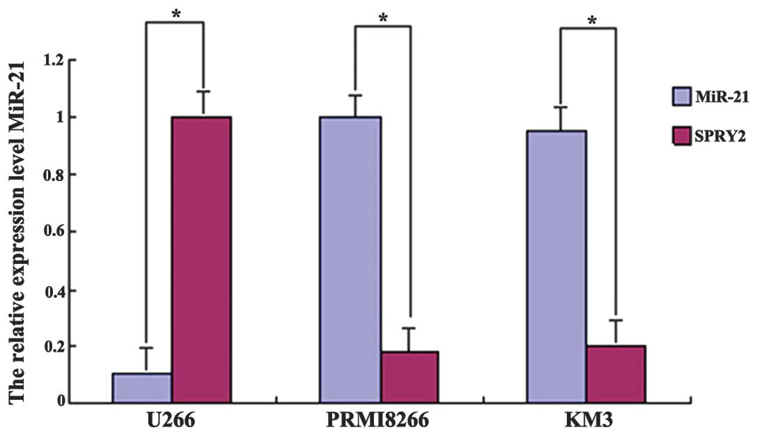
RT-qPCR detection of miR-21 and SPRY2
gene expression in MM cell lines
miR-21 and SPRY2 were expressed in the MM cell
lines. In the RPMI 8226 and KM3 cell lines, miR-21 was highly
expressed and SPRY2 was expressed at a low level. However, in the
U-266 cell line, in which a low level of miR-21 was detected, SPRY2
was expressed at a high level. The differences were statistically
significant (P<0.01; Fig.
1).
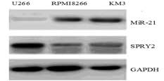
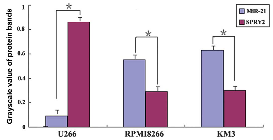
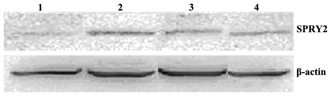
Western blot analysis detection of miR-21
and SPRY2 expression in MM cell lines
Western blot analysis revealed that in the MM cell
lines (RPMI 8226 and KM3) with a high level of endogenous miR-21
expression, SPRY2 was expressed at a significantly lower level. By
contrast, in the U-266 cell line with low endogenous miR-21
expression, SPRY2 was expressed at a significantly higher level
(Fig. 2). The differences in the
band intensity of the SPRY2 and miR-21 protein between the
different cell lines were statistically significant (P<0.01;
Fig. 3).
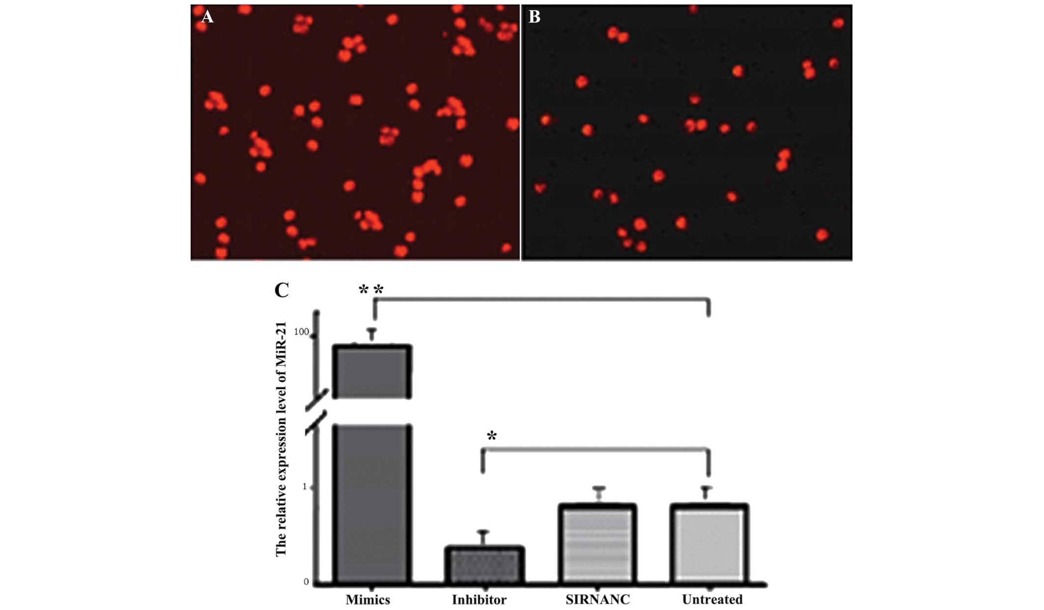
Effect of mimics and inhibitor
transfection on miR-21 expression in U-266 cells
U-266 cells were transfected with a
fluorescence-labeled miR-21 mimic (Fig. 4A) and inhibitor (Fig. 4B) using liposome and the
observation under a fluorescence microscope confirmed a
transfection efficiency exceeding 90%. RT-qPCR was used to detect
miR-21 expression levels in different groups. The results revealed
that the miR-21 level was 98.6±14.2 in the mimic-transfected cells,
representing an increase of 120.2-fold compared with that of the
non-transfected cells (0.82±0.13); the difference was statistically
significant (P<0.001). The expression level was 0.37±0.06 in the
inhibitor-transfected cells, a significant decrease of 61.9%
compared with the non-transfected cells (P<0.05), whereas no
significant difference in miR-21 expression was found between the
siRNA NC cells and the non-transfected cells (Fig. 4C).
Western blot analysis detection of SPRY2
in the miR-21-transfected U-266 cells
After U-266 cells were transfected with miR-21
mimics (50 nM) and inhibitors (100 nM), the band intensity of SPRY2
protein in the miR-21 mimic-transfected cells was significantly
lower than that of inhibitor-transfected, siRNA-transfected and
untreated cells and the differences were statistically significant
(P<0.01; Fig. 5).
Discussion
MM is an immune system lymphocyte-derived cancer.
Due to the annual increases in MM incidence with the aging
population, MM has become the second-most common type of
hematological malignancy (12–22).
The severe humoral immune deficiency and bone damage caused by the
excessive production of abnormal plasma cells in the bone marrow
and monoclonal immunoglobulin lead to high rates of MM-associated
mortality. Currently, the mechanism of MM pathogenesis remains to
be elucidated, treatment outcomes exhibit large individual
differences and the available prognostic indicators cannot
appropriately reflect the complexity of the disease. Therefore,
in-depth studies on the mechanism of pathogenesis and prognostic
factors may be the key for the development of an effective solution
to the clinical problems.
An ideal tumor biomarker should be detected easily
and non-invasively. miR-21, an miRNA molecule, which is abnormally
expressed in a variety of malignant tissues and has a key role in
the regulation of tumorigenesis and progression, is a current area
of intense research interest. miR-21 overexpression is closely
associated with cancer cell proliferation, metastasis, and disease
prognosis of MM, non-Hodgkin’s lymphoma, leukemia and various other
non-hematological solid tumors (14–16)
miR-21 may regulate the expression of SPRY2. Structurally, the
SPRY2 protein (3–5) has a highly conserved, cysteine-rich
C-terminus, which may be positioned in the target region of the
activated cell membrane, while its N-terminus is highly mutable.
Several studies (5,17–19)
have demonstrated that the expression of SPRY2 was downregulated or
inhibited in prostate cancer, breast cancer and malignant glioma,
leading to the deregulation and overactivation of MAPK/ERK
signaling in tumor cells. Therefore, SPRY2 is considered to be an
oncogene, which inhibits MAPK/ERK signaling. The findings of the
present study provided a basis for the further investigation of
SPRY2 function, its molecular mechanisms in MM and its clinical
significance.
The serum miR-21 levels in each group were
determined using RT-qPCR and it was observed that miR-21 was
over-expressed in patients with MM. Serum circulating miR-21 levels
in the MM group were significantly higher than those in the MGUS
group and NC group (P<0.01), indicating a close correlation of
miR-21 with the pathogenesis and progression of MM, which is
consistent with findings previously reported in the literature
(20–22). A study (23) reported that the expression of
miR-21 is significantly higher in myeloma cells than in the plasma
cells of MGUS and NC groups, suggesting a positive correlation
between circulating miR-21 levels and the tumor cell load in
patients. As the high level of serum circulating miR-21 in MM
patients is likely to be due to myeloma cells, the serum
circulating miR-21 level may be used to monitor MM progression and
to evaluate therapeutic performance. The level of circulating
miR-21 in the MGUS group was higher than in the NC group; however,
the difference was not statistically significant (P>0.05); a
larger sample size is required for further investigation.
The present results revealed low or absent
expression of SPRY2 in the RPMI 8266 and KM3 MM cell lines, which
may be associated with the inhibition of SPRY2 expression by the
high level of miR-21 in MM cells. Using SPRY2 over-expression
(24), certain studies have
reported that SPRY2 partially inhibits the activation of the
MAPK/ERK signaling pathway and inhibits interleukin 6
(IL-6)-induced MM cell proliferation, suggesting that SPRY2 has a
role in tumor suppression in MM cells through the inhibition of the
activation of MAPK/ERK signaling (5,17–19).
The downregulation of miR-21 by inhibitor transfection may increase
the expression of SPRY2 in the U-266 MM cell line, suggesting that
miR-21 may regulate SPRY2 expression.
Furthermore, using western blotting, it was
identified that the expression level of SPRY2 was significantly
lower in the RPMI 8226 and KM3 MM cell lines, which have high
endogenous miR-21 expression levels, whereas the level of SPRY2 was
significantly higher in the U-266 cell line with low endogenous
miR-21 expression. The negative correlation between miR-21 and
SPRY2 was further confirmed by the observation that the expression
of SPRY2 significantly decreased in the stably-transfected MM cell
line overexpressing exogenous miR-21.
In conclusion, the present study preliminarily
confirmed that miR-21 is likely a negative regulatory factor
causing downregulation of SPRY2 in MM. miR-21 is closely associated
with the pathogenesis, progression and prognosis of MM, and thus
may be used as an indicator of poor prognosis in MM patients. With
the development and application of miRNA antisense oligonucleotide
technology, miR-21 may become a biomarker for MM diagnosis and a
novel target for the treatment of drug resistance.
References
|
1
|
Hatzimichael E, Dasoula A, Benetatos L,
Syed N, Dranitsaris G, Crook T and Bourantas K: Study of specific
genetic and epigenetic variables in multiple myeloma. Leuk
Lymphoma. 51:2270–2274. 2010. View Article : Google Scholar : PubMed/NCBI
|
|
2
|
Pichiorri F, Suh SS, Ladetto M, Kuehl M,
Palumbo T, Drandi D, Taccioli C, Zanesi N, Alder H, Hagan JP, et
al: MicroRNAs regulate critical genes associated with multiple
myeloma pathogenesis. Proc Natl Acad Sci USA. 105:12885–12890.
2008. View Article : Google Scholar : PubMed/NCBI
|
|
3
|
Edwin F, Anderson K, Ying C and Patel TB:
Intermolecular interactions of sprouty proteins and their
implications in development and disease. Mol Pharmacol. 76:679–691.
2009. View Article : Google Scholar : PubMed/NCBI
|
|
4
|
Feng YH, Wu CL, Shiau AL, Lee JC, Chang
JG, Lu PJ, Tung CL, Feng LY, Huang WT and Tsao CJ:
MicroRNA-21-mediated regulation of Sprouty2 protein expression
enhances the cytotoxic effect of 5-fluorouracil and metformin in
colon cancer cells. Int J Mol Med. 29:920–926. 2012.PubMed/NCBI
|
|
5
|
Lao DH, Chandramouli S, Yusoff P, et al: A
Src homology 3-binding sequence on the C terminus of Sprouty2 is
necessary for inhibition of the Ras/ERK pathway downstream of
fibroblast growth factor receptor stimulation. J Biol Chem.
281:29993–30000. 2006. View Article : Google Scholar : PubMed/NCBI
|
|
6
|
Sayed D, Rane S, Lypowy J, He M, Chen IY,
Vashistha H, Yan L, Malhotra A, Vatner D and Abdellatif M:
MicroRNA-21 targets Sprouty2 and promotes cellular outgrowths. Mol
Biol Cell. 19:3272–3282. 2008. View Article : Google Scholar : PubMed/NCBI
|
|
7
|
Leone E, Morelli E, Di Martino MT, Amodio
N, Foresta U, Gullà A, Rossi M, Neri A, Giordano A, Munshi NC, et
al: Targeting miR-21 inhibits in vitro and in vivo multiple myeloma
cell growth. Clin Cancer Res. 19:2096–2106. 2013. View Article : Google Scholar : PubMed/NCBI
|
|
8
|
Shah AA, Leidinger P, Blin N and Meese E:
miRNA: small molecules as potential novel biomarkers in cancer.
Curr Med Chem. 17:4427–4432. 2010. View Article : Google Scholar : PubMed/NCBI
|
|
9
|
Terpos E, Morgan G, Dimopoulos MA, et al:
International Myeloma Working Group recommendations for the
treatment of multiple myeloma-related bone disease. J Clin Oncol.
31:2347–2357. 2013. View Article : Google Scholar : PubMed/NCBI
|
|
10
|
Chen C, Ridzon DA, Broomer AJ, Zhou Z, Lee
DH, Nguyen JT, Barbsin M, Xu NL, Mahuvakar VR, Andersen MR, et al:
Real-time quantification of microRNAs by stem-loop RT-PCR. Nucleic
Acids Res. 33:e1792005. View Article : Google Scholar : PubMed/NCBI
|
|
11
|
Schmittgen TD and Livak KJ: Analyzing
real-time PCR data by the comparative C(T) method. Nat Protoc.
3:1101–1108. 2008. View Article : Google Scholar : PubMed/NCBI
|
|
12
|
International myeloma working group:
Criteria for the classification of monoclonal gammopathies,
multiple myeloma and related disorders: a report of the
International Myeloma Working Group. Br J Haematol. 12l:749–757.
2003.
|
|
13
|
Cavo M, Rajkumar SV, Palumbo A, et al:
International Myeloma Working Group consensus approach to the
treatment of multiple myeloma patients who are candidates for
autologous stem cell transplantation. Blood. 117:6063–6073. 2011.
View Article : Google Scholar : PubMed/NCBI
|
|
14
|
Liu C, Yu J, Yu S, Lavker RM, Cai L, Liu
W, Yang K, He X and Chen S: MicroRNA-21 acts as an oncomir through
multiple targets in human hepatocellular carcinoma. J Hepatol.
53:98–107. 2010. View Article : Google Scholar : PubMed/NCBI
|
|
15
|
Xu J, Wu C, Che X, et al: Circulating
microRNAs, miR-21, miR-122 and miR-223, in patients with
hepatocellular carcinoma or chronic hepatitis. Mol Carcinog.
50:136–142. 2011. View
Article : Google Scholar : PubMed/NCBI
|
|
16
|
Pichiorri F, Suh SS, Ladetto M, Kuehl M,
Palumbo T, Drandi D, Taccioli C, Zanesi N, Alder H, Hagan JP, et
al: MicroRNAs regulate critical genes associated with multiple
myeloma pathogenesis. Proc Natl Acad Sci USA. 105:12885–12890.
2008. View Article : Google Scholar : PubMed/NCBI
|
|
17
|
Cabrita MA and Christofori G: Sprouty
proteins, masterminds of receptor tyrosine kinase signaling.
Angiogenesis. 11:53–62. 2008. View Article : Google Scholar : PubMed/NCBI
|
|
18
|
Lito P, Mets BD, Kleff S, O’Reilly S,
Maher VM and McCormick JJ: Evidence that sprouty 2 is necessary for
sarcoma formation by H-Ras oncogene-transformed human fibroblasts.
J. Biol Chem. 283:2002–2009. 2008. View Article : Google Scholar
|
|
19
|
Asangani IA, Rasheed SA, Nikolova DA,
Leupold JH, Colburn NH, Post S and Allgayer H: MicroRNA-21(miR-21)
post-transcriptionally downregulates tumor suppressor Pdcd4 and
stimulates invasion, intravasation and metastasis in colorectal
cancer. Oncogene. 27:2128–2136. 2008. View Article : Google Scholar
|
|
20
|
Wang X, Li C, Ju S, et al: Myeloma cell
adhesion to bone marrow stromal cells confers drug resistance by
microRNA-21 up-regulation. Leuk Lymphoma. 52:1991–1998. 2011.
View Article : Google Scholar : PubMed/NCBI
|
|
21
|
Löffler D, Brocke-Heidrich K, Pfeifer G,
Stocsits C, Hackermüller J, Kretzschmar AK, Burger R, Gramatzki M,
Blumert C, Bauer K, et al: Interleukin-6 dependent survival of
multiple myeloma cells involves the Stat3-mediated induction of
microRNA-21 through a highly conserved enhancer. Blood.
110:1330–1333. 2007. View Article : Google Scholar : PubMed/NCBI
|
|
22
|
Hu HY, Li KP, Wang XJ, Liu Y, Lu ZG, Dong
RH, Guo HB and Zhang MX: Set9, NF-kappaB and microRNA-21 mediate
berberine-induced apoptosis of human multiple myeloma cells. Acta
Pharmacol Sin. 34:157–166. 2013. View Article : Google Scholar
|
|
23
|
Xiong Q, Zhong Q, Zhang J, Yang M, Li C,
Zheng P, Bi LJ and Ge F: Identification of novel miR-21 target
proteins in multiple myeloma cells by quantitative proteomics. J
Proteome Res. 11:2078–2090. 2012. View Article : Google Scholar : PubMed/NCBI
|
|
24
|
Feng YH, Wu CL, Tsao CJ, et al:
Deregulated expression of sprouty2 and microRNA-21 in human colon
cancer: Correlation with the clinical stage of the disease. Cancer
Biol Ther. 11:111–121. 2011. View Article : Google Scholar
|