Introduction
Osteoporosis is a chronic, metabolic and systemic
skeletal disease characterized by low bone mineral density (BMD)
and micro-architectural deterioration, resulting in increased bone
fragility and fracture risk (1,2). It
is estimated that >200 million individuals worldwide suffer from
osteoporosis and the prevalence is continuing to increase with the
growing elderly population (3).
The incidence of osteoporosis is 2–4 times higher in females than
that in males due to a sharp decrease in ovarian estrogen
production, which causes rapid bone loss during the first decade
following the menopause (4). Bone
fracture, which is the most serious consequence of osteoporosis, is
associated with high economic costs and substantial morbidity and
mortality; therefore, the prevention and treatment of this
condition are of great importance (5). Current drug treatments for the
prevention and treatment of post-menopausal osteoporosis include
estrogen, selective estrogen receptor modulators, calcitonin and
bisphosphonates (4,6,7).
Although these agents are effective in preventing bone loss, they
are not the ideal treatments due to their adverse side effects on
the breast and the gastrointestinal and cardiovascular systems, as
well as increasing the risk of endometrial or ovarian cancer
(4,8–12).
Novel drugs based on medicinal herbs and natural products, and
which possess fewer side effects, are urgently required (13). Traditional Chinese Medicines have
been widely used in the prevention and treatment of post-menopausal
osteoporosis, and as these medicines are prepared from medicinal
plants, are a source of numerous bioactive compounds and are
preferred by patients, they are more suitable for long-term use
compared with chemically synthesized medicines (14).
Heng-Gu-Gu-Shang-Yu-He-Ji (OsteoKing) is a
formulation composed of numerous types of medicinal herbs
(Pericarpium Citri reticulatae, Carthamus tinctorius L., Radix
notoginseng, Eucommia ulmoides Oliv., Radix ginseng, Radix
Astragali Mongolici and Carapax trionycis) based on a concoction
originating from Yunnan Province in China and has been used for
>100 years. It has a notable effect in the treatment of bone
diseases, particularly for femoral head necrosis, prolapse of the
lumbar intervertebral disc and osteoarthritis, and was approved by
the Chinese State Food and Drug Administration in 2002 (15,16).
Previous studies by our group demonstrated that OsteoKing is able
to elevate the gene expression of core binding factor α 1 and
vascular endothelial growth factor, and improve the
micro-architecture in the necrotic femoral head of rabbits
(17–20). Clinical studies have demonstrated
that OsteoKing has an effect in preventing fracture and treating
ischemic necrosis of the femoral head in humans (21–23).
However, no studies have been performed thus far to investigate
whether OsteoKing has any anti-osteoporotic activity. The present
study was conducted to investigate the effects of OsteoKing on an
osteoporosis model of ovariectomized (OVX) rabbits.
Materials and methods
Drugs and reagents
The OsteoKing concoction was prepared according to
the Chinese Pharmacopeia (China Pharmacopeia Committee, 2002) and
was supplied by Crystal Natural Pharmaceutical Co. (Kunming,
China). Briefly, Pericarpium Citri reticulatae (10 g),
Carthamus tinctorius L. (15 g), Radix Notoginseng (30
g), Eucommia ulmoides Oliv. (30 g), Radix Ginseng (20
g), Radix Astragali mongolici (40 g) and Carapax
Trionycis (10 g) were ground into a coarse powder and immersed
in 10X (10 l/kg) distilled water for 12 h at room temperature, and
then boiled using a distillation apparatus for 1 h. This process
was repeated twice and for the second and third extraction, the
residue from the previous extraction was filtered and the same
extraction procedures were applied. Thereafter, the combined
extracts were filtrated and evaporated using a rotary evaporator at
50°C to a relative density of 1.03–1.04 g/cm3,
centrifuged for 30 min at 1,450 × g and the supernatant obtained
was centrifuged once again after standing for 12 h. Subsequently,
the pH was adjusted to 4.0–6.0 using NaOH (Shaihai Experiment Co.,
Shanghai, China), distilled water was added to a total volume of
1,000 ml and the product was filtrated prior to usage. Nilestriol
was purchased from Shanghai New Hualian Pharmaceutical Co. Ltd.
(Shanghai, China). Rabbit enzyme-linked immunosorbent assay (ELISA)
kits for measurement of the serum concentrations of osteocalcin
(OC), procollagen type I N-terminal peptide (PINP),
tartrate-resistant acid phosphatase 5b (TRAP5b), cross linked
N-telopeptide of type I collagen (NTX) with a sensitivity of 0.3
ng/ml, 0.2 ng/ml, 19.5 μU/ml and 0.78 pmol/ml, respectively,
were purchased from Wuhan Huamei Bioengineering Co. (Wuhan, China),
and the intra-assay and inter-assay coefficients of variability of
all the ELISA kits were <8 and 10%, respectively.
Animals
A total of 101 female New Zealand white rabbits aged
6 months were obtained from the Animal Center of Kunming Medical
University (Kunming, China). Their body weight ranged between 2.5
and 3.0 kg. The animals were housed at a constant temperature
(20–25°C), humidity (40–70%) and light-dark cycle (12/12 h). Tap
water was available ad libitum, while standard rabbit chow
was restricted to 50 g per day. All experiments were conducted
under the National Institutes of Health Guide for the Care and Use
of Laboratory Animals and approved by the Ethics Committee (Animal
Care and Use Committee) of Kunming Medical University. All efforts
were made to minimize the pain and suffering of the animals.
Experimental protocol
Following a two-week acclimation period, the animals
were randomly allocated into an OVX model group (OVX group, n=76)
and a sham-surgery group (sham group, n=25). Animals of the OVX
group underwent a bilateral ovariectomy as previously described
under general anesthesia with an intravenous injection of sodium
pentobarbital (30 mg/kg; Shanghai Westang Bio-tech Co., Ltd.,
Shanghai, China), while the sham surgery group was subjected to a
procedure involving exposure of the ovaries without excision
(24). Post-operatively, all
animals (Animal Center of Kunming Medical University, Kunming,
China) fasted for 12 h and sodium benzylpenicillin (0.3 million
IU/kg; Wuhan Dahua Pharmaceutical Co., Ltd., Wuhan, China) was
administered via an intramuscular injection for five days to
prevent infection (25). However,
one rabbit from the sham surgery group and two rabbits from the OVX
group died within 150 days following surgery. To characterize the
experimental animal model, six animals from each group were
selected randomly 150 days following OVX to determine the BMD of
their vertebrae, serum biochemical parameters, mechanical
properties and micro-architecture of the lumbar vertebra. Once the
osteoporotic rabbit model was established, 16 rabbits from the sham
group were randomly selected to continue with the study, and 64
rabbits from the OVX group were randomly divided into four groups:
Model group (Model), OVX with nilestriol group (nilestriol), OVX
with 300 mg/kg OsteoKing group (OsteoKing 300) and OVX with 600
mg/kg OsteoKing (OsteoKing 600) group, containing 16 rabbits each.
OsteoKing was administered orally once every other day with the
dose at 300 mg/kg approximating the clinical application dose for
humans (19,20). Nilestriol (Shanghai Hualian
Pharmaceutical Co., Ltd., Shanghai, China) was administered orally
at a dose of 0.5 mg/kg once weekly (26,27).
Rabbits of the sham group and model group were treated with
deionized water. All animals were weighed and the doses were
adjusted weekly. At 60 days and 120 days after treatment, six
randomly selected rabbits from each group were sacrificed and the
effects of OsteoKing or nilestriol on the BMD of the vertebrae,
serum biochemical parameters and mechanical properties, histology,
and micro-architecture of the lumbar vertebra were recorded.
BMD analysis
The rabbits were anesthetized with an intravenous
injection of sodium pentobarbital (30 mg/kg) and the BMD of the
vertebrae was measured in vivo using dual-energy X-ray
absorptiometry (DXA; Lunar Prodigy Advance; GE Lunar, Madison, WI,
USA) as described previously (22)
Specific software for small animals (GE Medical Systems, enCORE
2004 software; cersion 8.80.001) was used. BMD measurements were
performed following 150 days bilateral ovariectomy (0 days
treatment) and 60, 120 days treatment, respectively.
Mechanical assessment
The second lumbar vertebra was harvested at days 0,
60 and 120 of treatment, frozen at −20°C prior to the assay and the
mechanical properties were measured as described previously
(25,28). The bones were thawed at room
temperature prior to the mechanical assessments and moisture levels
were retained with the use of a moist gauze soaked in 0.9% NaCl
solution (The Third Chemical Reagent Factory, Tianjin, China)
throughout the entire assessment period. The vertebrae were
prepared by cutting off the end plates from the vertebral body to
create parallel planar surfaces using a diamond wafer saw (VT1200,
Leica Microsystems GmbH, Wetzlar,, Germany). The vertebral samples
were then placed centrally between two parallel steel plates
attached to a materials-testing machine (Instron System 5565;
Instron, Norwood, MA, USA) and assessed along the longitudinal axis
at a constant compressive speed of 1 mm/min (25,29).
The specimens were loaded until the specimen succumbed to the
strain/weight and the mechanical parameters (maximum load,
displacement, stiffness and energy absorption capacity) were
calculated from the load-displacement curves. Briefly, the maximum
load (N) was considered as the maximum force on the curve;
furthermore, displacement (mm) and the ultimate deformity prior to
failure and stiffness (N/mm) were determined from the slope of the
linear portion, and the area under the load-displacement curve was
defined as the energy absorption capacity (mJ) (28,30).
Biochemical analysis of serum
Blood samples were collected from the central ear
artery 150 days following bilateral ovariectomy (day 0 of
treatment) and at days 60 and 120 of treatment, respectively, after
an overnight fast, consistently between 09:00 and 11:00 A.M. The
serum was promptly separated and stored at −80°C prior to the assay
(31). The serum calcium
(Ca2+) and inorganic phosphorus (P) levels were
determined using a biochemical automatic analyzer (Hitachi 7080;
Hitachi Ltd., Tokyo, Japan), and the serum concentrations of OC,
PINP, TRAP5b and NTX were measured using rabbit ELISA kits. All
samples were run in the same assay unless an individual value
required repeating.
Histopathological evaluation
The sections of the third lumbar vertebrae, which
were harvested 60 or 120 days following treatment, were prepared as
described previously (32,33). Briefly, samples were fixed in 10%
neutral formal-saline (Day Ning Chemical Reagent Co., Ltd., Jining,
Shandong, China) for five days, dehydrated in a graded ethanol
series, and embedded in paraffin following decalcification in 10%
ethylene diamine tetraacetic acid (Kunming Pegatron Yang Technology
Co., Ltd., Kunming, China) for 30 days. Subsequently, the blocks
were cut into 5-μm slices perpendicular to the longitudinal
axis at the middle of the lumbar vertebra. The morphology of the
sections was examined under a light microscope (Nikon AZ100; Nikon,
Tokyo, Japan) following staining with hematoxylin and eosin (Wuhan
Baihao Biological Technology Co., Ltd., Wuhan,China).
Micro-computerized tomography (MicroCT)
examination
The first lumbar vertebra, which was harvested 150
days after the bilateral ovariectomy (day 0 of treatment) and 60
days or 120 days after treatment was cleaned of adherent soft
tissues and preserved in sealed plastic bags at −20°C prior to the
assay (34). The MicroCT
examination of the first lumbar vertebra was performed using a
MicroCT system (μ CT 80, SCANCO Medical, Brüttisellen,
Switzerland) as previously described (34,35),
and the analytical conditions were 55 kV with 72 μA leakage.
The lumbar vertebra was scanned and ~500 transverse consecutive
sections of 35-μm thickness were obtained from each lumbar
vertebra using a 2048×2048 matrix. The volume of interest was
selected as a region 100 slices subsequent to 50 slices away from
the cranial endplate (36). Within
these slices, the region of the vertebral body, excluding the
cortical bone by the boundaries defined by the endocortical bone
surfaces, was selected and constructed three-dimensionally.
Following setting the same threshold, the structural parameters,
including bone volume/total volume (BV/TV), bone surface/bone
volume (BS/BV), trabecular thickness (Tb. Th), trabecular
separation (Tb.Sp) and trabecular number (Tb.N) were measured
automatically for each specimen using the plate-model data with the
SCANCO microtomographic software package version 6.0 (SCANCO
Medical) (35).
Statistical analysis
All experimental data were assessed using the
statistical system SPSS 17.0 (SPSS, Inc., Chicago, IL, USA) and
values are expressed as the mean ± standard deviation. Differences
in the mean values of BMD, serum biochemical parameters, mechanical
parameters and structural parameters between the two groups 150
days after OVX were compared using an independent-samples t-test,
and those between five groups at the same time -point after
treatment were performed using one-way analysis of variance with
the Bonferroni post hoc test. P<0.05 was considered to indicate
a statistically significant difference.
Results
BMD measurements
The effects of OsteoKing or nilestriol on the BMD of
the vertebrae are presented in Table
I. The BMD of the vertebra in the OVX group 150 days after the
surgery decreased by 14.0% (P<0.01) compared with that in the
sham group. No significant differences were observed in the BMD
between any treatment group and the model group 60 days after
treatment (P>0.05). At 120 days of treatment, the BMD in the
group subjected to OVX and treated with 600 mg/kg OsteoKing was
significantly higher than that in the model group (P<0.01),
almost identical to that in the sham group and similar to that in
the OVX with nilestriol group, while the improvement of BMD in the
group subjected to OVX and treated with 300 mg/kg OsteoKing was not
significant (P>0.05).
 | Table IEffect of OsteoKing or nilestriol on
bone mineral density (g/cm2) of vertebrae in
ovariectomized rabbits. |
Table I
Effect of OsteoKing or nilestriol on
bone mineral density (g/cm2) of vertebrae in
ovariectomized rabbits.
| Time-point
(days) | Sham group | Ovariectomized
group
|
|---|
| Model | nilestriol | OsteoKing 300 | OsteoKing 600 |
|---|
| 0 | 0.265±0.016 | | 0.228±0.017a | | |
| 60 | 0.264±0.026 | 0.225±0.014a | 0.245±0.011 | 0.238±0.011 | 0.248±0.017 |
| 120 | 0.262±0.021 | 0.227±0.015a | 0.262±0.011b | 0.249±0.011 | 0.266±0.018b |
Mechanical properties of the lumbar
vertebrae
The results of the vertebral compression assessment
are shown in Table II. The values
of maximum load, displacement, stiffness and energy in the OVX
group 150 days after surgery decreased by 44.7, 6.8, 44.3 and
50.3%, respectively, compared with those in the sham group
(P<0.01, P<0.05, P<0.01 and P<0.01, respectively). At
60 days following treatment, the values of maximum load and
stiffness were significantly higher in the group subjected to OVX
and treated with 300 mg/kg OsteoKing than those in the model group
(P<0.05), but remained significantly lower than those in the
sham group (P<0.05). The values of maximum load and stiffness
were also significantly higher in the group subjected to OVX and
treated with 600 mg/kg OsteoKing than those in the model group
(P<0.01), and no significant difference was identified
(P>0.05) compared with the sham group with the exception of the
energy levels (P<0.05). Following 120 days of treatment, the
values of maximum load, stiffness and energy were significantly
higher in the OsteoKing-treated group than those in the model group
(P<0.01 or P<0.05), and no difference was identified compared
with those in the sham group (P>0.05). Similar gradual increases
in maximum load, stiffness and energy were observed in
nilestriol-treated group. No significant difference was observed in
the value of displacement between any groups at any time-point of
treatment (P>0.05).
| Table IIEffect of OsteoKing or nilestriol on
biomechanical parameters of the second lumbar vertebra in
ovariectomized rabbits. |
Table II
Effect of OsteoKing or nilestriol on
biomechanical parameters of the second lumbar vertebra in
ovariectomized rabbits.
| Time-point
(days) | Group | Maximum load
(N) | Displacement
(mm) | Stiffness
(N/mm) | Energy (mJ) |
|---|
| 0 | Sham | 615.8±61.7 | 0.676±0.013 | 1504.0±125.3 | 208.4±42.5 |
| OVX | 340.6±67.6b | 0.630±0.035a | 837.7±229.1b | 103.5±24.5b |
| 60 | Sham | 611.6±64.6 | 0.678±0.015 | 1498.5±97.6 | 201.1±39.1 |
| Model | 336.5±64.6b | 0.615±0.047 | 826.3±220.6b | 96.5±23.9b |
| Nilestriol | 499.4±61.7d | 0.618±0.036 |
1259.1±173.4d | 135.6±25.8b |
| OsteoKing 300 | 474.2±69.1a,c | 0.621±0.041 |
1153.9±148.3a,c | 127.3±19.2b |
| OsteoKing 600 | 515.5±65.9d | 0.639±0.051 |
1267.2±152.6d | 144.5±26.3a |
| 120 | Sham | 615.3±44.7 | 0.670±0.017 | 1489.1±79.2 | 197.2±35.7 |
| Model | 331.8±61.9b | 0.612±0.043 | 819.0±221.8b | 92.8±23.5b |
| Nilestriol | 573.9±46.6c | 0.624±0.031 |
1422.8±104.4d | 164.3±28.4d |
| OsteoKing 300 | 548.3±60.4c | 0.625±0.058 |
1406.2±120.4d | 158.5±30.5a |
| OsteoKing 600 | 589.3±55.0c | 0.634±0.052 |
1467.1±102.1d | 170.7±42.4d |
Serum biochemical parameters
The results of the serum biochemical assessment are
shown in Table III. The levels
of OC (+37.6%), PINP (+56.9%), TRAP5b (+45.2%) and NTX (+40.0%)
were significantly higher (P<0.01) and the levels of serum
Ca2+ (P<0.05) and P (P<0.01) were markedly lower
in the model group than those in the sham group 150 days after the
surgery, indicating the induction of a high bone turnover following
OVX. No significant difference was identified in the serum
Ca2+ levels among the groups at the same time-points.
Following 60 days of treatment, the levels of OC, PINP, TRAP5b and
NTX decreased by 8.6, 8.3, 10.0 and 16.2%, respectively, in the
group subjected to OVX and treated with 300 mg/kg OsteoKing, and
decreased by 16.4, 20.6, 18.7 and 22.2%, respectively, in the group
subjected to OVX and treated with 600 mg/kg OsteoKing as compared
with those in the model group at 150 days after OVX; however, the
decrease in the levels of all bone turnover biomarkers in the
OsteoKing-treated groups was not significantly different compared
with those in the model group at the same time-point (P>0.05).
The levels of serum P in the group subjected to OVX and treated
with 600 mg/kg OsteoKing were significantly higher (P<0.05) than
those in the model group. At 120 days following treatment, compared
with those in the model group at the same time-point, the levels of
OC, PINP, TRAP5b and NTX in the OsteoKing-treated group decreased
significantly and the levels of serum P increased significantly,
almost recovering to the normal levels. Nilestriol treatment had a
similar effect to the two doses of OsteoKing in reducing bone
turnover and increasing serum P levels.
| Table IIIEffect of OsteoKing or nilestriol on
serum biochemical parameters in OVX rabbits. |
Table III
Effect of OsteoKing or nilestriol on
serum biochemical parameters in OVX rabbits.
| Time-point
(days) | Group | Ca2+
(mmol/l) | P (mmol/l) | OC (ng/ml) | PINP (ng/ml) | TRAP5b (mU/ml) | NTX (pmol/ml) |
|---|
| 0 | Sham | 3.43±0.07 | 1.59±0.10 | 7.47±1.19 | 2.16±0.52 | 5.98±0.85 | 7.62±0.92 |
| OVX | 3.29±0.12a | 1.26±0.13b | 10.28±1.12b | 3.39±0.71b | 8.68±1.17b | 10.67±1.05b |
| 60 | Sham | 3.41±0.10 | 1.59±0.13 | 7.53±0.93 | 2.11±0.60 | 6.01±1.29 | 7.50±0.91 |
| Model | 3.34±0.14 | 1.28±0.15b | 10.05±1.45a | 3.36±0.60a | 8.59±0.92b | 10.45±1.51b |
| Nilestriol | 3.38±0.10 | 1.57±0.12c | 8.41±1.46 | 2.72±0.80 | 6.89±0.95 | 8.18±1.34 |
| OsteoKing 300 | 3.36±0.16 | 1.48±0.18 | 9.40±1.52 | 3.11±0.49 | 7.81±1.06 | 8.94±1.44 |
| OsteoKing 600 | 3.37±0.14 | 1.56±0.12c | 8.59±1.38 | 2.69±0.73 | 7.06±1.15 | 8.30±1.26 |
| 120 | Sham | 3.41±0.10 | 1.58±0.11 | 7.45±0.90 | 2.06±0.56 | 5.81±0.95 | 7.54±0.90 |
| Model | 3.33±0.08 | 1.29±0.09b | 9.79±1.22b | 3.26±0.77a | 8.30±1.27b | 10.26±1.25b |
| Nilestriol | 3.41±0.10 | 1.58±0.09d | 7.63±0.83c | 1.88±0.46c | 5.67±0.61c | 7.56±0.82d |
| OsteoKing 300 | 3.39±0.09 | 1.58±0.10d | 8.35±0.93 | 2.22±0.84 | 6.26±0.89c | 8.22±1.05c |
| OsteoKing 600 | 3.40±0.14 | 1.59±0.08d | 7.75±1.16c | 2.05±0.63c | 5.58±0.77c | 7.55±1.47d |
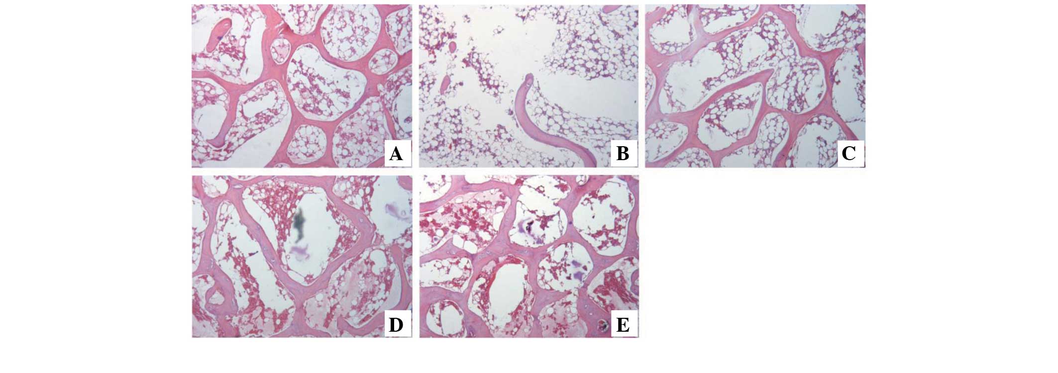
Histological analysis of lumbar
vertebrae
Under the light microscope, following 60 and 120
days of treatment, the histology of the third lumbar vertebra of
the sham group exhibited the normal size, shape, density and
architecture of the trabecular bone (Figs. 1A and 2A), while sections of the OVX group
exhibited sparse, disrupted, spacing-enlarged and area-diminished
trabecular bone tissue (Figs. 1B
and 2B). The OsteoKing-treated
group exhibited partial trabecular restoration following 60 days of
treatment (Fig. 1D and E) and
exhibited almost complete restoration of normal architecture
following 120 days of treatment (Fig.
2D and E); similar effects were also observed in the nilestriol
treatment group (Figs. 1C and
2C).
MicroCT evaluation
At 150 days after ovariectomy, the rabbits of the
OVX group exhibited lower values for BV/TV, Tb.Th and Tb.N, and
higher values for BS/BV and Tb.Sp (P<0.01), when compared with
those in the sham group (Table IV
and Fig 4). Treating OVX rabbits
with OsteoKing (300 or 600 mg/kg) or nilestriol partly abrogated
the OVX-mediated changes in the abovementioned parameters,
resulting in levels similar to those in the sham group (Table IV). Typical three-dimensional
reconstructed MicroCT images of the first lumbar vertebra (Figs. 3, 4 and 5),
including the cortical bone, revealed differences in trabecular
micro-architecture among the various groups. Images of the
representative samples with the BV/TV closest to the mean BV/TV
were reconstructed in each group (32).
| Table IVEffect of OsteoKing or nilestriol on
the micro-architecture parameters of the first lumbar vertebra in
ovariectomized rabbits. |
Table IV
Effect of OsteoKing or nilestriol on
the micro-architecture parameters of the first lumbar vertebra in
ovariectomized rabbits.
| Time-point
(days) | Group | BV/TV (%) | BS/BV (1/mm) | Tb.Th (mm) | Tb.Sp (mm) | Tb.N (1/mm) |
|---|
| 0 | Sham | 0.385±0.052 | 6.161±0.312 | 0.325±0.017 | 0.891±0.053 | 1.178±0.102 |
| Ovariectomized | 0.193±0.048b | 7.489±0.454b | 0.268±0.016b | 1.278±0.075b | 0.713±0.130b |
| 60 | Sham | 0.386±0.047 | 6.269±0.298 | 0.320±0.015 | 0.866±0.080 | 1.203±0.093 |
| Model | 0.200±0.043b | 7.405±0.334b | 0.271±0.012b | 1.244±0.093b | 0.735±0.129b |
| Nilestriol | 0.291±0.049a,c | 6.784±0.457 | 0.296±0.020 | 1.018±0.099d | 0.977±0.102a,c |
| OsteoKing 300 | 0.268±0.046b | 6.928±0.411 | 0.290±0.017 | 1.070±0.123a | 0.922±0.118b |
| OsteoKing 600 | 0.295±0.046b | 6.668±0.411c | 0.301±0.019c | 1.038±0.096c | 0.975±0.099a,d |
| 120 | Sham | 0.387±0.042 | 6.173±0.265 | 0.324±0.014 | 0.864±0.081 | 1.190±0.098 |
| Model | 0.198±0.041b | 7.512±0.368b | 0.267±0.013b | 1.254±0.089b | 0.737±1.122b |
| Nilestriol | 0.348±0.051d | 6.319±0.283d | 0.317±0.014d | 0.862±0.111d | 1.096±0.124d |
| OsteoKing 300 | 0.314±0.053d | 6.623±0.440d | 0.303±0.020d | 0.906±0.106d | 1.034±0.126d |
| OsteoKing 600 | 0.345±0.060d | 6.383±0.356d | 0.314±0.018d | 0.879±0.098d | 1.094±0.132d |
Discussion
OsteoKing is a Traditional Chinese Medicine, which
is widely used in the treatment of bone disease, particularly for
femoral head necrosis, prolapse of the lumbar intervertebral disc
and osteoarthritis. Previous studies have demonstrated that
OsteoKing has an effect on the prevention of fracture and the
improvement of the micro-architecture in the necrotic femoral head
of rabbits, which indicates that OsteoKing may have
anti-osteoporotic effects (17,18,21,23).
The present study was the first, to the best of our knowledge, to
demonstrate the beneficial effects of OsteoKing against the
reduction of bone mass and bone strength, and the deterioration of
the micro-architecture of the bone induced by ovariectomy in
rabbits.
Experimental animal models are important in
improving knowledge of the aetiology, pathophysiology and diagnosis
of osteoporosis, as well as in the prevention of the condition and
development of therapeutics (2).
It is well known that estrogen deficiency is an important risk
factor in the pathogenesis of osteoporosis and estrogenic
deprivation has been the most commonly used experimental model of
osteoporosis in animals (33,37).
Although the ovariectomy rat model is the most frequently used
animal model of osteoporosis, rats do not experience a natural
menopause, fail to achieve true skeletal maturity, lack the
Haversian system and remodeling differs from that in humans
(38–41). By contrast, rabbits have a short
developmental period and fast bone turnover, achieve skeletal
maturity shortly after reaching complete sexual development at ~six
months of age, and exhibit an active Haversian remodeling;
therefore, rabbits are often selected for the investigation of
osteoporosis (25,37,41).
Osteoporotic rabbit models induced by OVX or glucocorticoid (GC)
alone and OVX combined with GC have been used to investigate the
effects of loss of bone mass and have exhibited promising results
of reductions of BMD (24,25,37,42,43).
In the present study, six-month-old rabbits underwent bilateral
ovariectomy alone (without GC administration), and after 150 days,
the BMD of the vertebrae in the OVX group decreased by 14.0%, which
was significantly lower compared with that in the sham group
(P<0.01). Furthermore, a significant reduction of mechanical
strength in the vertebral load compression assessments and
micro-architectural deterioration of the lumbar vertebra were
observed in rabbits of the OVX group. The osteoporotic rabbit model
induced by OVX was successfully established after a longer period
than that reported in previous studies (24,25,28,37,38).
Bone turnover markers reflect the rates of bone
resorption and bone formation in the whole body, and they provide a
representative index of the overall skeletal bone loss (44–47).
Estrogen deficiency following a natural or artificial menopause
results in an increase in the levels of markers of bone remod-eling
and bone turnover (48). Previous
studies on menopausal women demonstrated that the increase in the
markers of bone resorption (of 50–150%) is rapid and precedes the
increase (of 50–100%) in markers of bone formation by several
months. This imbalance and disproportionately high rate of bone
resorption compared with formation exists within several years
following ovariectomy and remains in late postmenopausal women
(48–50). Similar changes were observed in the
present study: 150 days after ovariectomy, the expression of OC and
PINP, which are the biomarkers of bone formation, increased by 37.6
and 56.9% respectively, and TRAP5b and NTX, which are biomarkers of
bone resorption, increased by 45.2 and 40.0%, respectively, as
compared with those in the sham group. Following treatment with
OsteoKing, the levels of all the above bone turnover biomarkers
returned to a normal level and the rate of decrease in bone
resorption markers was slightly faster, which indicated that
OsteoKing may prevent bone loss by suppressing bone resorption.
However, long-term suppression of bone turnover may eventually lead
to an accumulation of fatigue-induced damage, as was observed
following bisphosphonate treatment (51,52).
Therefore, although the levels of bone turnover biomarkers returned
to normal levels, it was important to measure the mechanical
properties and micro-architecture of bone.
Compression testing of the lumbar vertebrae is
recommended for the assessment of the mechanical properties of
cancellous bone (28). The
biomechanical definition of bone fragility includes at least three
components: Strength (maximum load), brittleness (reciprocal of the
displacement) and work to failure (energy absorption) (25,52).
A fourth biomechanical measure, stiffness, is also used to assess
the mechanical integrity of bone, but is not a direct measure of
fragility (52). In the present
study, the strength, brittleness, stiffness and energy absorption
of the lumbar vertebrae decreased significantly in the OVX group
compared with that in the sham group 150 days after ovariectomy.
Following OsteoKing treatment, the strength, stiffness and energy
absorption increased gradually and almost recovered to normal
levels, but no effect on the brittleness was observed. This was
consistent with a previous study, which demonstrated that it was
rare for a treatment to improve strength and decrease brittleness
simultaneously (53).
Previous studies have demonstrated that BMD and the
aspects of the trabecular micro-architecture affect trabecular bone
strength (53). In the present
study, the BMD of the vertebrae decreased significantly and
deteriorated micro-architecture was observed in the MicroCT
analysis of the lumbar vertebrae 150 days after OVX. Increased
vertebral BMD and trabecular restoration of the lumbar vertebrae
were observed following OsteoKing treatment by the DXA and
histopathological evaluation, respectively. Furthermore, MicroCT
analysis demonstrated an increased travbecular BV/TV, Tb.Th, Tb.N
and decreased BS/BV, Tb.Sp following treatment with OsteoKing
compared with those in the model group. These results were
consistent with the consequences of the mechanical assessment and
previous studies, which indicated that the suppression of bone
turnover did not lead to bone damage (52).
In conclusion, OsteoKing was able to elevate the BMD
of the vertebrae, reduce the bone turnover rate, restore the
trabecular network and ameliorate the mechanical properties of
lumbar vertebra in a rabbit model of ovariectomy-induced
osteoporosis. These findings suggested that OsteoKing is
potentially useful in the treatment of postmenopausal osteoporosis,
which occurs in women as a result of estrogen deficiency.
Acknowledgments
The present study was supported by the National
Natural Science Foundation of China (no’s. 81060361, 30660212,
81160401 and 30860389), the Natural Science Foundation of Yunnan
Province (no’s. 2008CC009, 2004C0044 M, 2010ZC169, 2012fa002 and
2012HB043), the Department of Education, Yunnan Province (no.
ZD2012006) and the Natural Science Foundation of Kunming City (no.
0S090202. 2012-01-01-A-R-07-0006).
References
|
1
|
Du Z, Chen J, Yan F and Xiao Y: Effects of
simvastatin on bone healing around titanium implants in
osteoporotic rats. Clin Oral Implants Res. 20:145–150. 2009.
View Article : Google Scholar
|
|
2
|
Lasota A and Danowska-Klonowska D:
Experimental osteoporosis - different methods of ovariectomy in
female white rats. Rocz Akad Med Bialymst. 49(Suppl 1): 129–131.
2004.
|
|
3
|
Reginster JY and Burlet N: Osteoporosis: a
still increasing prevalence. Bone. 38(Suppl 1): S4–S9. 2006.
View Article : Google Scholar : PubMed/NCBI
|
|
4
|
Shirke SS, Jadhav SR and Jagtap AG:
Methanolic extract of Cuminum cyminum inhibits ovariectomy-induced
bone loss in rats. Exp Biol Med (Maywood). 233:1403–1410. 2008.
View Article : Google Scholar
|
|
5
|
Canalis E, Giustina A and Bilezikian JP:
Mechanisms of anabolic therapies for osteoporosis. N Engl J Med.
357:905–916. 2007. View Article : Google Scholar : PubMed/NCBI
|
|
6
|
Grey A and Reid IR: Emerging and potential
therapies for osteoporosis. Expert Opin Investig Drugs. 14:265–278.
2005. View Article : Google Scholar : PubMed/NCBI
|
|
7
|
Lane NE and Kelman A: A review of anabolic
therapies for osteoporosis. Arthritis Res Ther. 5:214–222. 2003.
View Article : Google Scholar : PubMed/NCBI
|
|
8
|
Dominguez LJ, Scalisi R and Barbagallo M:
Therapeutic options in osteoporosis. Acta Biomed. 81(Suppl 1):
55–65. 2010.PubMed/NCBI
|
|
9
|
Lacey JV Jr, Mink PJ, Lubin JH, Sherman
ME, Troisi R, Hartge P, Schatzkin A and Schairer C: Menopausal
hormone replacement therapy and risk of ovarian cancer. JAMA.
288:334–341. 2002. View Article : Google Scholar : PubMed/NCBI
|
|
10
|
Reginster JY and Sarlet N: The treatment
of severe postmenopausal osteoporosis: a review of current and
emerging therapeutic options. Treat Endocrinol. 5:15–23. 2006.
View Article : Google Scholar
|
|
11
|
Rossouw JE, Anderson GL, Prentice RL,
LaCroix AZ, Kooperberg C, Stefanick ML, Jackson RD, Beresford SA,
Howard BV, Johnson KC, Kotchen JM and Ockene J; Writing Group for
the Women’s Health Initiative Investigators: Risks and benefits of
estrogen plus progestin in healthy postmenopausal women: principal
results from the Women’s Health Initiative randomized controlled
trial. JAMA. 288:321–333. 2002. View Article : Google Scholar : PubMed/NCBI
|
|
12
|
Salari Sharif P, Abdollahi M and Larijani
B: Current, new and future treatments of osteoporosis. Rheumatol
Int. 31:289–300. 2011. View Article : Google Scholar
|
|
13
|
Putnam SE, Scutt AM, Bicknell K, Priestley
CM and Williamson EM: Natural products as alternative treatments
for metabolic bone disorders and for maintenance of bone health.
Phytother Res. 21:99–112. 2007. View
Article : Google Scholar
|
|
14
|
Cheng M, Wang Q, Fan Y, Liu X, Wang L, Xie
R, Ho CC and Sun W: A traditional Chinese herbal preparation,
Er-Zhi-Wan, prevent ovariectomy-induced osteoporosis in rats. J
Ethnopharmacol. 138:279–285. 2011. View Article : Google Scholar : PubMed/NCBI
|
|
15
|
Chen XQ, Wang QQ, Li JM, Wang YQ, Zhang XZ
and Li K: Comparison of simultaneous distillation and extraction,
static headspace and headspace-solid phase microextraction coupled
with GC/MS to measure the flavour components of Tricholoma
matsutake. Asian J Chem. 25:6059–6063. 2013.
|
|
16
|
Fan L, Ma J, Chen YH and Chen XQ:
Antioxidant and antimicrobial phenolic compounds from Setaria
viridis. Chem Nat Comp. 50:433–437. 2014. View Article : Google Scholar
|
|
17
|
Hu M, Zhao H, Dong X, Luo D and Zhou X:
General and light microscope observation on histological changes of
femoral heads between SANFH rabbit animal models and it were
intervened by Osteoking. Zhongguo Zhong Yao Za Zhi. 35:2912–2916.
2010.In Chinese.
|
|
18
|
Hu M, Zhao HB, Qian CY, Wei W, Xu HH, Tang
W, Luo DJ and Zhang Y: Ultrastructural evaluation of the SANFH
rabbit animal models intervened by Osteoking. Zhong Hua Zhong Yi
Yao Za Zhi. 26:486–489. 2011.
|
|
19
|
Zhao HB, Hu M and Liang HS: Experimental
study on osteoking in promoting gene expression of core binding
factor alpha 1 in necrotic femoral head of rabbits. Zhongguo Zhong
Xi Yi Jie He Za Zhi. 26:1003–1006. 2006.In Chinese. PubMed/NCBI
|
|
20
|
Zhao HB, Hu M, Wang WQ and Li LZ: Effects
of the VEGF gene expressions of Osteoking in the treatment of
femoral head necrosis. Zhongguo Gu Shang. 20:757–759. 2007.
|
|
21
|
Luo DJ, Zhao HB, Dong XL, Li LZ, Wang WZ
and Xiong H: The study on treatment of vertebral ostoporotic
fracture by salmon calcitonin and Heng Gu bong healing reagent.
Chin J Endocr Surg. 5:158–160. 2011.
|
|
22
|
Zhao HB, Wang B, Zhao XL, Li LZ, Li SH, Li
YG, Chen ZY and Luo XR: Osteoking in the treatment of ischemic
necrosis of femoral head: long-term results in 76 patients. Chin J
Pract Chin Modern Med. 4:2906–2907. 2004.
|
|
23
|
Zhao HB, Hu M, Zheng HY, Liang HS and Zhu
XS: Clinical study on effect of Osteoking in preventing
postoperational deep venous thrombosis in patients with
intertrochanteric fracture. Chin J Integr Med. 11:297–299. 2005.
View Article : Google Scholar
|
|
24
|
Castañeda S, Largo R, Calvo E,
Rodríguez-Salvanés F, Marcos ME, Díaz-Curiel M and Herrero-Beaumont
G: Bone mineral measurements of subchondral and trabecular bone in
healthy and osteoporotic rabbits. Skeletal Radiol. 35:34–41. 2006.
View Article : Google Scholar
|
|
25
|
Liu X, Lei W, Wu Z, Cui Y, Han B, Fu S and
Jiang C: Effects of glucocorticoid on BMD, micro-architecture and
biomechanics of cancellous and cortical bone mass in OVX rabbits.
Med Eng Phys. 34:2–8. 2012. View Article : Google Scholar
|
|
26
|
Cao DP, Zheng YN, Qin LP, Han T, Zhang H,
Rahman K and Zhang QY: Curculigo orchioides, a traditional Chinese
medicinal plant, prevents bone loss in ovariectomized rats.
Maturitas. 59:373–380. 2008. View Article : Google Scholar : PubMed/NCBI
|
|
27
|
Nian H, Qin LP, Zhang QY, Zheng HC, Yu Y
and Huang BK: Antiosteoporotic activity of Er-Xian Decoction, a
traditional Chinese herbal formula, in ovariectomized rats. J
Ethnopharmacol. 108:96–102. 2006. View Article : Google Scholar : PubMed/NCBI
|
|
28
|
Baofeng L, Zhi Y, Bei C, Guolin M,
Qingshui Y and Jian L: Characterization of a rabbit osteoporosis
model induced by ovariectomy and glucocorticoid. Acta Orthop.
81:396–401. 2010. View Article : Google Scholar : PubMed/NCBI
|
|
29
|
Schorlemmer S, Ignatius A, Claes L and
Augat P: Inhibition of cortical and cancellous bone formation in
glucocorticoid-treated OVX sheep. Bone. 37:491–496. 2005.
View Article : Google Scholar : PubMed/NCBI
|
|
30
|
Akhter MP, Cullen DM, Gong G and Recker
RR: Bone biome-chanical properties in prostaglandin EP1 and EP2
knockout mice. Bone. 29:121–125. 2001. View Article : Google Scholar : PubMed/NCBI
|
|
31
|
Scholz-Ahrens KE, Delling G, Stampa B,
Helfenstein A, Hahne HJ, Acil Y, Timm W, Barkmann R, Hassenpflug J,
Schrezenmeir J and Glüer CC: Glucocorticosteroid-induced
osteoporosis in adult primiparous Göttingen miniature pigs: effects
on bone mineral and mineral metabolism. Am J Physiol Endocrinol
Metab. 293:E385–395. 2007. View Article : Google Scholar : PubMed/NCBI
|
|
32
|
Arens AM, Barr B, Puchalski SM, Poppenga
R, Kulin RM, Anderson J and Stover SM: Osteoporosis associated with
pulmonary silicosis in an equine bone fragility syndrome. Vet
Pathol. 48:593–615. 2011. View Article : Google Scholar
|
|
33
|
Zhang Y, Yu L, Ao M and Jin W: Effect of
ethanol extract of Lepidium meyenii Walp. on osteoporosis in
ovariectomized rat. J Ethnopharmacol. 105:274–279. 2006. View Article : Google Scholar : PubMed/NCBI
|
|
34
|
Chen H, Wu M and Kubo KY: Combined
treatment with a traditional Chinese medicine, Hachimi-jio-gan
(Ba-Wei-Di-Huang-Wan) and alendronate improves bone micro-structure
in ovariectomized rats. J Ethnopharmacol. 142:80–85. 2012.
View Article : Google Scholar : PubMed/NCBI
|
|
35
|
Hordon LD, Itoda M, Shore PA, Shore RC,
Heald M, Brown M, Kanis JA, Rodan GA and Aaron JE: Preservation of
thoracic spine microarchitecture by alendronate: comparison of
histology and microCT. Bone. 38:444–449. 2006. View Article : Google Scholar
|
|
36
|
Zhao FD, Pollintine P, Hole BD, Adams MA
and Dolan P: Vertebral fractures usually affect the cranial
endplate because it is thinner and supported by less-dense
trabecular bone. Bone. 44:372–379. 2009. View Article : Google Scholar
|
|
37
|
Castañeda S, Calvo E, Largo R,
González-González R, de la Piedra C, Díaz-Curiel M and
Herrero-Beaumont G: Characterization of a new experimental model of
osteoporosis in rabbits. J Bone Miner Metab. 26:53–59. 2008.
View Article : Google Scholar
|
|
38
|
Bellido M, Lugo L, Castañeda S, Roman-Blas
JA, Rufián-Henares JA, Navarro-Alarcón M, Largo R and
Herrero-Beaumont G: PTH increases jaw mineral density in a rabbit
model of osteoporosis. J Dent Res. 89:360–365. 2010. View Article : Google Scholar : PubMed/NCBI
|
|
39
|
Mosekilde L: Assessing bone quality-animal
models in preclinical osteoporosis research. Bone. 17(Suppl):
343S–352S. 1995. View Article : Google Scholar
|
|
40
|
Rodgers JB, Monier-Faugere MC and Malluche
H: Animal models for the study of bone loss after cessation of
ovarian function. Bone. 14:369–377. 1993. View Article : Google Scholar : PubMed/NCBI
|
|
41
|
Turner AS: Animal models of osteoporosis -
necessity and limitations. Eur Cells Mater. 1:66–81. 2001.
|
|
42
|
Cao T, Shirota T, Ohno K and Michi KI:
Mineralized bone loss in partially edentulous trabeculae of
ovariectomized rabbit mandibles. J Periodontal Res. 39:37–41. 2004.
View Article : Google Scholar
|
|
43
|
Cao T, Shirota T, Yamazaki M, Ohno K and
Michi KI: Bone mineral density in mandibles of ovariectomized
rabbits. Clin Oral Implants Res. 12:604–608. 2001. View Article : Google Scholar : PubMed/NCBI
|
|
44
|
Chaki O, Yoshikata I, Kikuchi R, Nakayama
M, Uchiyama Y, Hirahara F and Gorai I: The predictive value of
biochemical markers of bone turnover for bone mineral density in
postmenopausal Japanese women. J Bone Miner Res. 15:1537–1544.
2000. View Article : Google Scholar : PubMed/NCBI
|
|
45
|
Eastell R, Colwell A, Hampton L and Reeve
J: Biochemical markers of bone resorption compared with estimates
of bone resorption from radiotracer kinetic studies in
osteoporosis. J Bone Miner Res. 12:59–65. 1997. View Article : Google Scholar : PubMed/NCBI
|
|
46
|
Garnero P, Sornay-Rendu E, Duboeuf F and
Delmas PD: Markers of bone turnover predict postmenopausal forearm
bone loss over 4 years: the OFELY study. J Bone Miner Res.
14:1614–1621. 1999. View Article : Google Scholar : PubMed/NCBI
|
|
47
|
Whitaker M, Guo J, Kehoe T and Benson G:
Bisphosphonates for osteoporosis - where do we go from here? N Engl
J Med. 366:2048–2051. 2012. View Article : Google Scholar : PubMed/NCBI
|
|
48
|
Delmas PD, Eastell R, Garnero P, Seibel MJ
and Stepan J; Committee of Scientific Advisors of the International
Osteoporosis Foundation: The use of biochemical markers of bone
turnover in osteoporosis. Committee of Scientific Advisors of the
International Osteoporosis Foundation. Osteoporosis Int. 11(Suppl
6): S2–S17. 2000. View Article : Google Scholar
|
|
49
|
Garnero P, Sornay-Rendu E, Chapuy MC and
Delmas PD: Increased bone turnover in late postmenopausal women is
a major determinant of osteoporosis. J Bone Miner Res. 11:337–349.
1996. View Article : Google Scholar : PubMed/NCBI
|
|
50
|
Szulc P and Delmas PD: Biochemical markers
of bone turnover: potential use in the investigation and management
of postmenopausal osteoporosis. Osteoporos Int. 19:1683–1704. 2008.
View Article : Google Scholar : PubMed/NCBI
|
|
51
|
Armamento-Villareal R, Napoli N, Panwar V
and Novack D: Suppressed bone turnover during alendronate therapy
for high-turnover osteoporosis. N Engl J Med. 355:2048–2050. 2006.
View Article : Google Scholar : PubMed/NCBI
|
|
52
|
Turner CH: Biomechanics of bone:
determinants of skeletal fragility and bone quality. Osteoporos
Int. 13:97–104. 2002. View Article : Google Scholar : PubMed/NCBI
|
|
53
|
Bouxsein Ml: Determinants of skeletal
fragility. Best Pract Res Clin Rheumatol. 19:897–911. 2005.
View Article : Google Scholar : PubMed/NCBI
|