Article
Evaluation of the effects of Angelicae dahuricae radix on the morphology and viability of mesenchymal stem cells
- Authors:
- Su‑Hyeon Jeong
- Bo‑Bae Kim
- Ji‑Eun Lee
- Youngkyung Ko
- Jun‑Beom Park
-
View Affiliations / Copyright
Affiliations:
Department of Rehabilitation Medicine of Korean Medicine, Chungju Hospital of Korean Medicine, College of Korean Medicine, Semyung University, Jecheon-si, Chungcheongbuk-do 390-711, Republic of Korea, Department of Periodontics, College of Medicine, The Catholic University of Korea, Seoul 137‑701, Republic of Korea
-
Pages:
1556-1560
|
Published online on:
March 9, 2015
https://doi.org/10.3892/mmr.2015.3456
- Expand metrics +
Metrics:
Total
Views: 0
(Spandidos Publications: | PMC Statistics:
)
Metrics:
Total PDF Downloads: 0
(Spandidos Publications: | PMC Statistics:
)
This article is mentioned in:
Abstract
Angelicae dahuricae radix is a traditional herbal medicine used to treat various diseases in China and Korea, such as colds, headaches, rhinitis and psoriasis. Angelicae dahuricae radix has been used as an anti‑inflammatory, analgesic, antipyretic and antioxidant remedy. This study was performed in order to evaluate the effects of the extracts of Angelicae dahuricae radix on the morphology and viability of mesenchymal stem cells derived from the gingiva. Mesenchymal stem cells derived from the gingiva were grown in the presence of Angelicae dahuricae radix at final concentrations that ranged from 0.001 to 100 µg/ml. The morphology of the cells was viewed under an inverted microscope, and the analysis of cell proliferation was performed with cell counting kit‑8 (CCK‑8) on days 1, 3 and 7. The cells in the control group had spindle‑shaped, fibroblast‑like morphology at days 1, 3 and 7 under optical microscopy. The shapes of the cells in 0.001, 0.01, 0.1, 1, 10 and 100 µg/ml Angelicae dahuricae radix were similar to the shapes of the cells in the control group. The relative values of the CCK‑8 assays of 0.001, 0.01, 0.1, 1, 10, and 100 µg/ml Angelicae dahuricae radix were 102.5±0.6, 133.3±9.6, 148.4±20.5, 147.7±12.6, 132.3±27.7 and 101.1±4.6%, respectively, when the CCK‑8 result of the control group on day 1 was considered to be 100%. There was a marginal increase in cell proliferation at 0.1 and 1 µg/ml groups at day 1; however, this did not achieve statistical significance (P=0.052). The relative values of the CCK‑8 assays of 0.001, 0.01, 0.1, 1, 10 and 100 µg/ml Angelicae dahuricae radix were 96.5±1.3, 89.3±0.9, 90.3±3.0, 84.8±12.2, 92.3±4.5 and 86.8±11.7%, respectively, when the CCK‑8 result of the control group on day 3 was considered to be 100% (P>0.05). The relative values of the CCK‑8 assays of 0.001, 0.01, 0.1, 1, 10 and 100 µg/ml Angelicae dahuricae radix day 7 were 94.9±22.3, 102.8±22.1, 127.4±7.4, 130.4±1.3, 129.2±10.8 and 124.8±9.1%, respectively, when the CCK‑8 result of the control group on day 7 was considered to be 100%, but there were no statistically significant differences among the groups (P>0.05). Within the limits of this study, Angelicae dahuricae radix at the tested concentrations did not produce statistically significant differences in the viability of stem cells derived from the gingiva.
View Figures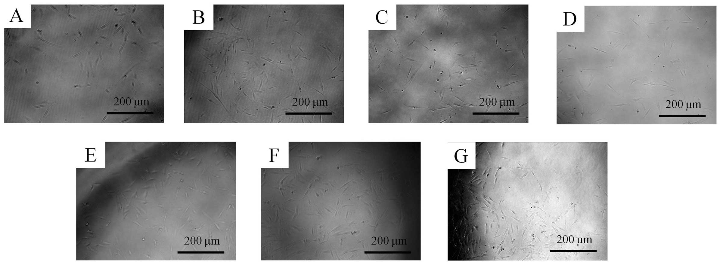 |
Figure 1
|
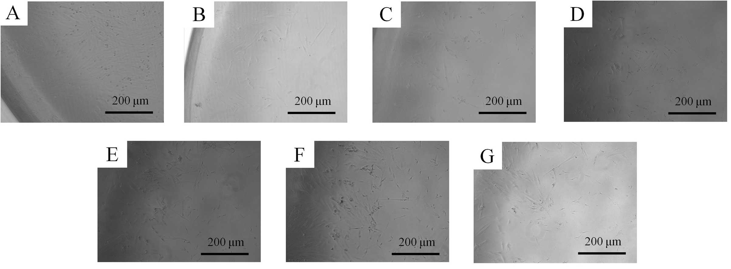 |
Figure 2
|
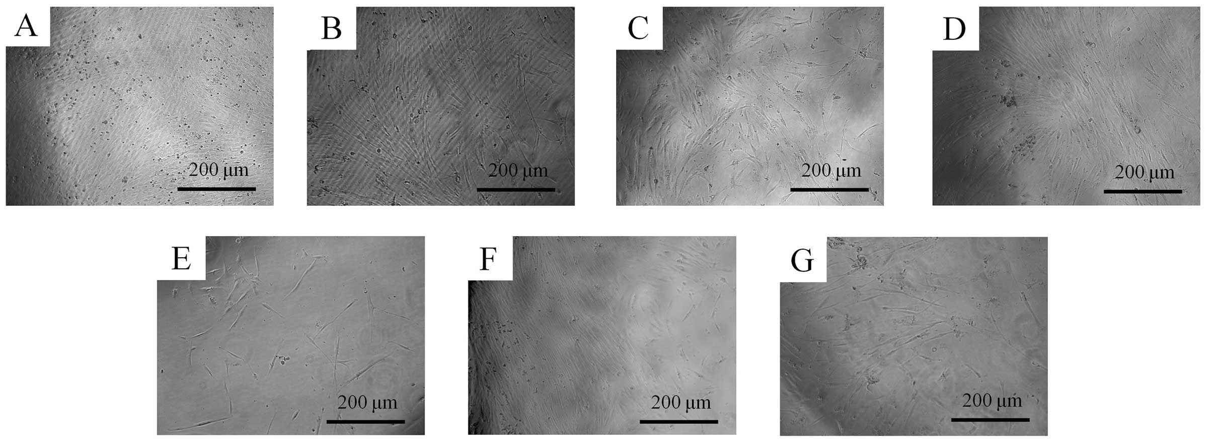 |
Figure 3
|
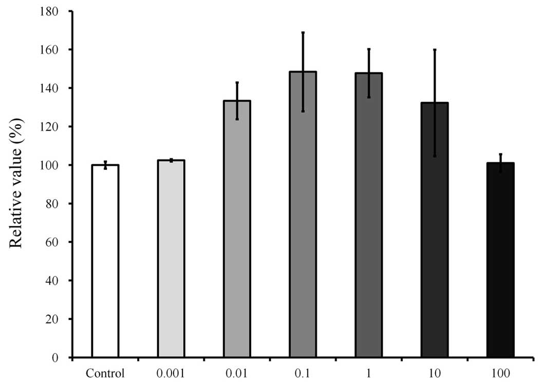 |
Figure 4
|
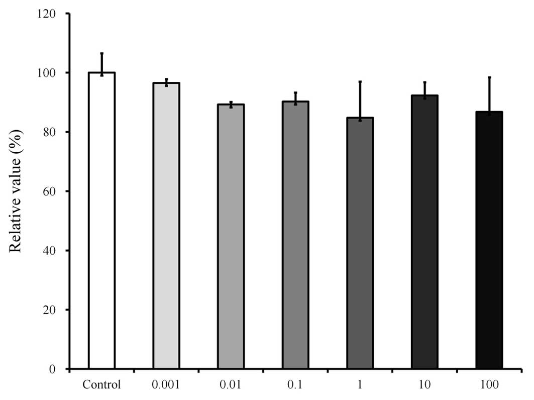 |
Figure 5
|
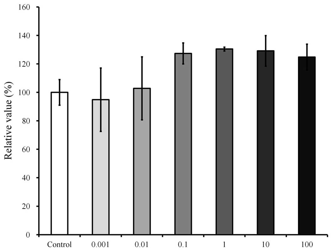 |
Figure 6
|
View References
|
1
|
Wang Y, Cao SE, Tian J, Liu G, Zhang X and
Li P: Auraptenol attenuates vincristine-induced mechanical
hyperalgesia through serotonin 5-HT1A receptors. Sci Rep.
3:33772013.PubMed/NCBI
|
|
2
|
Zhou RH: Resource science of Chinese
medicinal materials. China Medical & Pharmaceutical Sciences
Press; Beijing: pp. 19–32. 1993
|
|
3
|
Lee H, Lee JK, Ha H, Lee MY, Seo CS and
Shin HK: Angelicae dahuricae radix inhibits dust mite
extract-induced atopic dermatitis-like skin lesions in NC/Nga mice.
Evid Based Complement Alternat Med. 2012:7430752012.PubMed/NCBI
|
|
4
|
Li H, Dai Y, Zhang H and Xie C:
Pharmacological studies on the Chinese drug radix Angelicae
dahuricae. Zhongguo Zhong Yao Za Zhi. 16:560–562. 5761991.In
Chinese.
|
|
5
|
Kang OH, Chae HS, Oh YC, et al:
Anti-nociceptive and anti-inflammatory effects of Angelicae
dahuricae radix through inhibition of the expression of inducible
nitric oxide synthase and NO production. Am J Chin Med. 36:913–928.
2008. View Article : Google Scholar : PubMed/NCBI
|
|
6
|
Yi S, Cho JY, Lim KS, et al: Effects of
Angelicae tenuissima radix, Angelicae dahuricae radix and
Scutellariae radix extracts on cytochrome P450 activities in
healthy volunteers. Basic Clin Pharmacol Toxicol. 105:249–256.
2009. View Article : Google Scholar : PubMed/NCBI
|
|
7
|
Zhao G, Peng C, Du W and Wang S:
Pharmacokinetic study of eight coumarins of Radix Angelicae
dahuricae in rats by gas chromatography-mass spectrometry.
Fitoterapia. 89:250–256. 2013. View Article : Google Scholar : PubMed/NCBI
|
|
8
|
Tang W and Eisenbrand G: Angelica spp.
Chinese drugs of plant origin, chemistry, pharmacology and use in
traditional and modern medicine. Springer; Berlin: pp. 113–125.
1992, View Article : Google Scholar
|
|
9
|
Szucs V and Freitag R: Comparison of a
three-peptide separation by capillary electrochromatography,
voltage-assisted liquid chromatography and nano-high-performance
liquid chromatography. J Chromatogr A. 1044:201–210. 2004.
View Article : Google Scholar : PubMed/NCBI
|
|
10
|
Xie Y, Chen Y, Lin M, Wen J, Fan G and Wu
Y: High-performance liquid chromatographic method for the
determination and pharmacokinetic study of oxypeucedanin hydrate
and byak-angelicin after oral administration of Angelica dahurica
extracts in mongrel dog plasma. J Pharm Biomed Anal. 44:166–172.
2007. View Article : Google Scholar : PubMed/NCBI
|
|
11
|
Zheng X, Zhang X, Sheng X, et al:
Simultaneous characterization and quantitation of 11 coumarins in
Radix Angelicae dahuricae by high performance liquid chromatography
with electrospray tandem mass spectrometry. J Pharm Biomed Anal.
51:599–605. 2010. View Article : Google Scholar
|
|
12
|
Liu R, Li A and Sun A: Preparative
isolation and purification of coumarins from Angelica dahurica
(Fisch ex Hoffn) Benth, et Hook f (Chinese traditional medicinal
herb) by high-speed counter-current chromatography. J Chromatogr A.
1052:223–227. 2004. View Article : Google Scholar : PubMed/NCBI
|
|
13
|
Zhu M, Liang XL, Zhao LJ, et al:
Elucidation of the transport mechanism of baicalin and the
influence of a Radix Angelicae dahuricae extract on the absorption
of baicalin in a Caco-2 cell monolayer model. J Ethnopharmacol.
150:553–559. 2013. View Article : Google Scholar : PubMed/NCBI
|
|
14
|
Thanh PN, Jin W, Song G, Bae K and Kang
SS: Cytotoxic coumarins from the root of Angelica dahurica. Arch
Pharm Res. 27:1211–1215. 2004. View Article : Google Scholar
|
|
15
|
Kang OH, Lee GH, Choi HJ, et al: Ethyl
acetate extract from Angelica dahuricae Radix inhibits
lipopolysaccharide-induced production of nitric oxide,
prostaglandin E2 and tumor necrosis factor-alpha via
mitogen-activated protein kinases and nuclear factor-kappaB in
macrophages. Pharmacol Res. 55:263–270. 2007. View Article : Google Scholar : PubMed/NCBI
|
|
16
|
Zhao W, Cao Y and Liu J: Research on
chronic toxicology of Compound Radix Angelicae dahuricae capsule. J
Shaxi Med Univ. 37:160–165. 2006.
|
|
17
|
Moshaverinia A, Chen C, Xu X, et al: Bone
regeneration potential of stem cells derived from periodontal
ligament or gingival tissue sources encapsulated in RGD-modified
alginate scaffold. Tissue Eng Part A. 20:611–621. 2014.
|
|
18
|
Jeong SH, Lee JE, Jin SH, Ko Y and Park
JB: Effects of Asiasari radix on the morphology and viability of
mesenchymal stem cells derived from the gingiva. Mol Med Rep.
10:3315–3319. 2014.PubMed/NCBI
|
|
19
|
Tomar GB, Srivastava RK, Gupta N, et al:
Human gingiva-derived mesenchymal stem cells are superior to bone
marrow-derived mesenchymal stem cells for cell therapy in
regenerative medicine. Biochem Biophys Res Commun. 393:377–383.
2010. View Article : Google Scholar : PubMed/NCBI
|
|
20
|
Park JB, Kim YS, Lee G, Yun BG and Kim CH:
The effect of surface treatment of titanium with
sand-blasting/acid-etching or hydroxyapaptite-coating and
application of bone morphogenetic protein-2 on attachment,
proliferation and differentiation of stem cells derived from buccal
fat pad. Tissue Eng Regen Med. 10:115–121. 2013. View Article : Google Scholar
|
|
21
|
Fournier BP, Larjava H and Häkkinen L:
Gingiva as a source of stem cells with therapeutic potential. Stem
Cells Dev. 22:3157–3177. 2013. View Article : Google Scholar : PubMed/NCBI
|
|
22
|
Yang H, Gao LN, An Y, et al: Comparison of
mesenchymal stem cells derived from gingival tissue and periodontal
ligament in different incubation conditions. Biomaterials.
34:7033–7047. 2013. View Article : Google Scholar : PubMed/NCBI
|


