Introduction
Intestinal fibrosis is a common complication of
inflammatory bowel disease (IBD) and occurs in ulcerative colitis
(UC) and Crohn’s disease (CD) (1).
Intestinal fibrosis is characterized by abnormal deposition of
extracellular matrix (ECM) proteins produced by activated
myofibroblasts, and is hypothesized to develop as a result of
abnormal wound repair following local chronic inflammatory
responses (2). Until now, few
therapies have been shown to reliably treat intestinal fibrosis in
IBD (3,4).
Subepithelial myofibroblast are important in these
processes by regulating inflammatory responses and ECM metabolism
(5,6). These generate a plausible link
between mucosal inflammation and destruction of the subepithelial
matrix. Inhibition of these processes represents a lucrative target
for IBD anti-fibrosis therapies.
Triptolide, referred to as PG490, is the major
active component of Tripterygium wilfordii Hook F (TWHF)
extracts, has anti-inflammatory and immunomodulatory activities.
Extracts of TWHF have been used in the treatment of
glomerulonephritis and autoimmune diseases, such as rheumatoid
arthritis and systemic lupus erythematosus (7,8). It
has also been investigated as an immunosuppressant for kidney
transplantation (9). A previous
study showed that triptolide exerted antifibrotic effects in renal
fibrosis (10), hepatic fibrosis
(11) and lung fibrosis (12). However, the data of its effects on
intestinal fibrosis caused by IBD remain to be elucidated. Our
previous study showed that increased activation of chemokines
interleukin-8 and monocyte chemoattractant protein-1 and matrix
metalloproteinase (MMP)-3 expressed by human subepithelial
myofibroblasts stimulated with pro-inflammatory cytokines could be
inhibited by triptolide (13). In
a cohort clinical trial, it was reported that triptolide could
prevent the postoperative recurrence of CD (14). Recently, the therapeutic efficacy
of triptolide in CD was also confirmed and it was shown that
microscopic intestinal inflammation was attenuated with the
modulation of in situ levels of inflammatory cytokines
through the upregulation of Foxp3+ Tregs (regulatory T
cells) (15). Based on these
results, it was hypothesized that triptolide may have antifibrotic
efficacy in vivo in chronic colitis with intestinal fibrosis
through the therapeutic action against chronic inflammation.
Therefore the present study aimed to evaluate the antifibrotic role
of triptolide in rats with colonic fibrosis.
Materials and methods
Induction of colonic fibrosis
According to the described protocol (16), colonic fibrosis was established in
male Sprague-Dawley rats weighing 1550200 g (Shanghai SLAC
Laboratory Animal Co., Shanghai, China) by 6-weekly intrarectal
instillation of increasing doses of TNBS (Sigma Chemical Co., St.
Louis, MO, USA): 60, 60, 67.5, 67.5, 75, 75 mg/kg per week in 45%
EtOH (Sigma). The rats were also administered 45 mg/kg per day of
triptolide (PG490, molecular weight 360, purity 99%)
intraperitoneally or phosphate buffered saline (PBS) starting with
the initial TNBS treatment. Crystalline triptolide was obtained
from the Institute of Dermatology, Chinese Academy of Medical
Sciences (Nanjing, China).
At the time of tissue collection, the rats were
sacrificed by carbon dioxide and the colons were removed intact
from the anus to the ileocecal junction. Sections were taken from
these regions for the following experiments: (i) Serial paraffin
sections of the colon were stained with hematoxylin and eosin and
Masson’s Trichrome to detect connective tissue. A pathologist
examined each slide in a blinded manner. (ii) Isolation of
subepithelial myofibroblasts. The present study was approved by the
Institutional Animal Care and Use Committee for Southeast
University Medical School and performed according to the
institutional ethical guidelines stipulated by the Review Board for
Southeast University Medical School.
Image analysis of ECM content
The paraffin embedded blocks representing the
similar positions of colon were sectioned and stained with Masson’s
Trichrome. Quantitative digital morphometric analysis of ECM was
performed according to a previously described method (17). In brief, 6–12 randomly selected
fields for each section were photographed using a Spot digital
camera (KY-F55MD; Olympus, Tokyo, Japan) and transformed into
digital readings using Olympus Image Analysis software (Olympus
Stream Ver.1.9.1; Olympus), which allowed for quantification of the
various color wavelengths with pixels as the unit of measurement.
The percentage of ECM was then calculated by dividing the pixel
area of the ECM by the pixel area corresponding to the total tissue
in the field of view.
Sircol collagen assay
Total collagen content in the colon was detected
with Sirius red collagen detection kit (Chondrex, Redmond, WA,
USA). Colonic tissue was homogenized in T-PER buffer (Thermal
Science, Amarillo, TX, USA) using a TissueLyser (Qiagen,
Germantown, MD, USA), incubated on ice for 15 min, and centrifuged
for 5 min at 10,600 × g at 4°C (Heraeus™ Primo™/Primo R centrifuge;
Thermo Scientific, Waltham, MA, USA). Each protein sample was
diluted in 0.5 M acetic acid to a final concentration of 100
µg/ml. Optical density was read at 530 nm against the
reagent blank using a DU-530 spectrophotometer (Beckman Coulter,
Inc., Fullerton, CA, USA). Results were calculated based on
collagen per 100 µg/ml protein. Cultured SEMFs were
characterized by immunohistochemistry. Mouse monoclonal antibodies
against α-SMA, vimentin and desmin were used (Sigma). The cells
were grown on glass coverslips and fixed using acetone, prior to
immunoperoxidase staining with the Vectastain ABC peroxidase kit
(Vecta Laboratories, Burlingame, CA, USA). Following incubation
with the primary antibody, biotinylated goat anti-mouse
immunoglobulin(Ig)-G (Sigma) was applied and subsequently
avidin-biotinylated horseradish peroxidase complex. Peroxidase
activity was developed with diaminobenzidine, followed by nuclear
staining using hematoxylin (Sigma).
Isolation and characterization of the
colonic subepithelial myofibroblasts in the rats
We have previously established the isolation and
primary culture of the colonic subepithelial myofibroblasts
(13) according to the described
protocol (18). In the present
study, the method was slightly modified. Briefly, fresh mucosal
samples were obtained from the colons at similar positions. After
washing with calcium- and magnesium-free Hanks’ balanced salt
solution (Gibco-BRL, Gaithersburg, MD, USA), the mucosa samples
were completely denuded of epithelial cells by three 30-min
incubations at 37°C in 1 mM EDTA (Sigma-Aldrich, St. Louis, MO,
USA). The de-epithelialized mucosal samples were cultured in
Dulbecco’s modified Eagle’s medium (Gibco BRL, Gaithersburg, MD,
USA) containing 10% fetal bovine serum, 50 U/ml penicillin and 50
µg/ml streptomycin (Gibco-BRL), and incubated at 37°C in a
5% CO2 atmosphere. The cells in suspension were removed
after every 24- to 72-h culture period, and the denuded mucosal
tissue was maintained in culture for up to 2 weeks. Established
colonies of myofibroblasts possess the physiologic characteristics
with immunostaining for α-smooth muscle actin (SMA) and vimentin.
α-SMA is a contractile protein present in smooth muscle (19) and myofibroblasts (20). Vimentin is commonly used to stain
myofibroblasts and fibroblasts. Desmin is an intermediate
contractile filament that is a muscle specific protein (21). Intestinal myofibroblasts are
immunoreactive for α-SMA as well as for vimentin, but completely
negative for desmin. Myofibroblasts and fibroblasts or muscle cells
also differ morphologically. Myofibroblasts tend to be spreading
with numerous long processes that gave the cultures a complex
overlapping appearance (Fig. 4).
By contrast, muscle cells or fibroblasts appeared as elongated
bipolar cells.
Reverse transcription-quantitative
polymerase chain reaction (RT-qPCR)
The mRNA expression of Collagen Iα1 (COL1A1) was
determined by real-time polymerase chain reaction, as previously
described (13). The cells were
harvested with 0.25% trypsin (Sigma) and 0.02% EDTA, and total RNA
was isolated using RNeasy reagents (Qiagen, Chatsworth, CA, USA),
according to the manufacturer’s instructions. The mRNA
concentration was quantitated by spectrophotometry (Beckman). For
synthesization of cDNA, 1 µg total RNA was treated with
reverse transcriptase (Promega, Madison, WI, US) and oligo (dT)
were used for reverse transcription. The reactions were performed
using the Reverse Transcription system (Promega) under the
following conditions: 42°C for 15 min, 95°C for 5 min and 4°C for 5
min. Samples were stored at −20°C until use.
qPCR analysis was performed using an ABI PRISM 7700
(Perkin-Elmer, Applied Biosystems, Foster City, CA, USA). Specific
primers and dual-labelled fluorescent probes were designed using
the Primer Express primer design program v1.01 (Perkin-Elmer). The
constitutively expressed GAPDH was used as an internal control.
Probes were labeled with the fluorescent reporter dye
5-carboxyfluorescein (FAM) at the 5′ end and the quencher
N,N,N,N′-tetramethyl-6-carboxyrhodamine (TAMRA) or Minor Groove
Binder (MGB) at the 3′ end. The primer and probe sequences were as
follows: Collagen Iα1, forward 5′-AATCAGCCGCTCCCATTCTCCTA-3′,
reverse 5′-GGAGGGCGAGGGAGGAGAGAA-3′ and probe
5′-(FAM)-TCATCCCGCCCCCATTCCCTG-(MGB)-3 and GAPDH, forward
5′-GGCAAATTCAACGGCACAGT-3′, reverse 5′-AGATGGTGATGGGCTTCCC-3′ and
probe 5′-(FAM)-AAGGCCGAGAATGGGAAGCTTGTCATC -(MGB)-3′. The samples
were amplified in a final volume of 25 µl. The primers were
used at a concentration of 900 nM, and probes at 250 nM. GAPDH was
amplified in separate reactions. The cycling conditions were as
follows: 50°C for 2 min, 95°C for 10 min, 45 cycles of 95°C for 30
sec and 60°C for 30 sec. The data were normalized to GAPDH gene
expression and are expressed as fold increase in expression
Western blot analysis
The activity of Collagen I was determined by western
blotting. The cells were harvested and sonicated in solubilization
buffer, containing 20 mM Tris-HCl, (pH 8.0), 150 mM NaCl, 1 mM
EDTA, 1 mM EGTA, 1% Triton X-100, 2.5 mM sodium pyrophosphate, 1 mM
sodium vanadate, 10 µg/ml aprotinin, 10 µg/ml
leupeptin, 1 mM phenylmethylsulfonyl fluoride. The cell debris was
removed by centrifugation at 10,000 × g for 15 min and the
supernatants were boiled in Laemmli sample buffer (Bio-Rad) for 5
min. An equal quantity of protein was subjected to sodium dodecyl
sulfate-10% polyacrylamide gel electrophoresis, and the proteins
were blotted onto a PVDF membrane (Amersham Pharmacia Biotech,
Piscataway, NJ, USA). The membranes were blocked with 5% skimmed
milk in Tris-buffered saline, containing 0.1% Tween 20 (pH 7.6),
overnight at 4°C and were probed with primary rabbit antibodies
(Abcam, Cambridge, MA, US) for 1 h at room temperature. Following
washing, the membranes were incubated with secondary goat
anti-rabbit antibody (Abcam) coupled to horseradish peroxidase for
1 h at room temperature. Antibody-antigen complexes were then
detected using an ECL chemiluminescent detection system (Amersham
Pharmacia Biotech). Quantification was performed by
densitometry.
Statistical analysis
Statistical significance was determined by a t-test
or analysis of variance followed by Fisher’s least significant
difference post hoc test, as appropriate, using SPSS V20 (IBM,
Armonk, NY, USA). Data are expressed as the mean ± standard error.
P<0.05 was considered to indicate a statistically significant
difference.
Results
Triptolide decreases ECM deposition in
vivo in the colon
At the endpoint of the study (42 days post
induction), in the TNBS-treated rats without triptolide treatment,
severe intestinal fibrosis and stricture were found particularly in
the distal 5 cm of the colon. The lamina propria showed pockets of
inflammation alternating with areas of minimal or moderate
inflammation as well as persistent edematous swelling of the
colonic wall (Fig. 1A).
Furthermore, the colon exhibited marked collagen deposition in the
subepithelial and serosal areas determined with Masson Trichrome
staining (Fig. 1B). By contrast,
the rats in the TNBS/triptolide-treated group exhibited less severe
inflammatory changes and only developed slight colonic fibrosis. As
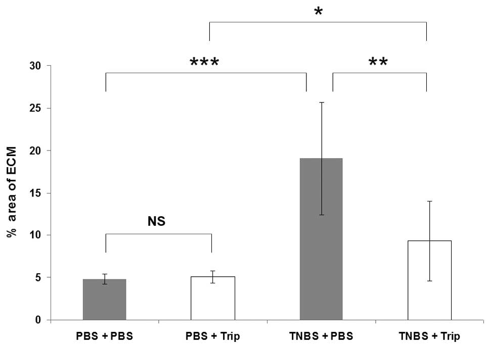
shown in Fig. 2, the area of ECM
deposition quantified from the ‘blue area’ stained with Masson
Trichrome was similar: 4.8±0.6 and 5.1±0.7% in the PBS/PBS and
PBS/triptolide-treated rats, respectively. The area of ECM
deposition was 19±6.6% in the TNBS/PBS-treated rats (P<0.001,
vs. the PBS/PBS-treated rats). It decreased to 9.3±4.7% in the
TNBS/triptolide-treated group, and was significantly lower than the
TNBS/PBS-treated group (P<0.01). No intestinal inflammation or
fibrosis was found in the PBS-treated mice with or without
triptolide treatment as expected.
Triptolide decreases total collagen
production in vivo in the colon
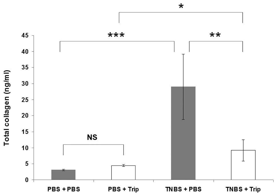
As shown in Fig. 3,
total collagen production in the colon of the PBS/PBS-treated rats
measured with the Sircol assay was 3.1±0.23 ng/ml, and it increased
to 29±10.1 ng/ml in the TNBS/PBS-treated rats (P<0.001). In the
PBS/triptolide-treated rats, total collagen expression was 4.5±0.34
ng/ml, and it increased to only 9.2±3.3 ng/ml in the
TNBS/triptolide-treated rats; however, there was a significant
difference between the two groups (P<0.05). Compared with the
TNBS/PBS-treated rats, total collagen expression in the colon of
the TNBS/triptolide-treated rats significantly decreased
(P<0.01).
Characterization of the colonic
subepithelial myofibroblasts
Intestinal myofibroblasts are immunoreactive for
α-SMA and vimentin, however, completely negative for desmin.
Myofibroblasts and fibroblasts or muscle cells also differ
morphologically. Myofibroblasts tended to be spreading with
numerous long processes, which gave the cultures a complex
overlapping appearance (Fig.
4).
Triptolide suppresses collagen Iα1 mRNA
and collagen I protein expression in the colonic subepithelial
myofibroblasts isolated from the rats with chronic colitis and
colonic fibrosis
Collagen Iα1 mRNA expression was measured ex
vivo in the subepithelial myofibroblasts (Fig. 4) isolated from the rat colon.
Collagen Iα1 mRNA expression increased to 3.4±0.54 fold in the
TNBS/PBS-treated rats compared with the PBS/PBS-treated rats
(P<0.001, Fig. 5A). By
contrast, collagen Iα1 mRNA expression in the
TNBS/triptolide-treated rats decreased to only 1.57±0.30 fold that
of the PBS/PBS-treated rats (P<0.05), and only 1.63±0.45 fold
that of the PBS/triptolide-treated rats (P<0.05). Compared with
the TNBS/PBS-treated rats (3.4±0.54), the expression of collagen
Iα1 in the TNBS/triptolide-treated rats (1.57±0.30) was
significantly lower (P<0.01). As shown in Fig. 5B, similar results were obtained,
when collagen I protein was measured by immunoblot analysis.
Discussion
In the present study, the antifibrotic effect of
triptolide in rats with colonic fibrosis was examined. It was found
that repeated treatment with low-doses of TNBS induced colonic
fibrosis. Triptolide reduced ECM deposition and collagen production
in vivo in the colon, and decreased ex vivo collagen
Iα1 mRNA and collagen I protein production in the colonic
subepithelial myofibroblasts.
After a 6-week induction of low-dose TNBS, there
were no obvious acute colitis signs, such as ruffled coats, hunched
posture or restricted movement as the model used in these studies
is not a model of acute inflammation, but chronic inflammation and
colonic fibrosis. This experimental model of colonic fibrosis
mimics the process of chronic stricture formation and progression
which occurs in IBD. Once this process of fibrosis is initiated in
the susceptible patient, it is self-perpetuating and chronic
inflammation, instead of acute inflammation was induced in this
experimental model of colonic fibrosis (22,23).
Excess extracellular matrix production is central to
the aberrant wound healing process leading to fibrosis in a number
of organs, including the intestine (24). IBD is a chronic, progressive
disease of the gastrointestinal tract with an unknown etiology and
CD is characterized by transmural inflammation of all layers of the
bowel wall (25). The inflammatory
changes in intestinal physiology result in the majority of the
symptomatology associated with CD (26) and significant morbidity results
from the irreversible tissue injury and fibrosis that frequently
occur in chronically inflamed bowel segments (25,27).
For reasons unknown, the reparative process associated with CD can
progress uncontrollably, leading to enhanced proliferation along
with defective programmed cell death of mesenchymal cells, and the
unrestrained deposition of ECM (28,29).
The recurrence of fibrosis is the predominant reason for
obstruction, however, few therapies have reliable effect on the
inhibition of fibrosis (30–33).
To the best of our knowledge the present study is the first to
report that triptolide diminishes ECM deposition in rats with
colonic fibrosis. This may be a potential mechanism by which
triptolide protects the intestine under inflammatory conditions and
act as a therapeutic agent for treatment of IBD (14).
Triptolide has been demonstrated to possess
anti-inflammatory and immunosuppressive effects (34,35).
Triptolide inhibits lymphocyte proliferation, synthesis and
secretion of proinflammatory cytokines (36,37),
and also induces apoptosis in T cells and lymphoma cell lines
(38,39). In addition, triptolide inhibits
dendritic cell-mediated chemoattraction of neutrophils and T cells
by inhibiting Stat3 phosphorylation and nuclear factor-κB
activation (39). We previously
reported that mRNA and protein expression of chemokines, including
interleukin (IL)-8 and monocyte chemotactic protien (MCP)-1, and
stromelysin-1 in colonic subepithelial myofibroblasts were
inhibited in vitro by triptolide in a similar manner
(13). Healthy intestinal
mesenchymal cells produce limited amounts of collagen and
fibronectin, which serve to maintain tissue integrity and
facilitate healing. On exposure to chronic inflammation,
mesenchymal cells transform into activated myofibroblasts, a
pro-repair and pro-fibrogenic cell phenotype, and markedly increase
the production of ECM (40,41).
In addition, the finely tuned balance between tissue matrix
metalloproteinases, which degrade ECM, and tissue inhibitors of
metalloproteinases, which inhibit ECM degradation, is lost,
promoting further structural changes of the colonic bowel wall
(42,43). In the present study, decrease in
the extent of intestinal fibrosis following treatment with
triptolide may be due to downregulation of collagen expression in
the colonic subepithelial myofibroblasts.
Triptolide has several characteristics of particular
interest in relation to IBD (44).
Our previous study demonstrated that increased activation of
chemokines, including IL-8 and MCP-1, and matrix metallo
proteinases-3 expressed by human subepithelial myofibroblasts
stimulated with pro-inflammatory cytokines could be inhibited by
triptolide (45). In a cohort
clinical trial, we reported that triptolide prevented the
postoperative recurrence of CD (46). Recently, we also confirmed the
therapeutic efficacy of triptolide in CD and demonstrated that
microscopic intestinal inflammation was attenuated with the
modulation of in situ levels of inflammatory cytokines
through the upregulation of regulatory T cells (47). It inhibits several proinflammatory
cytokines and adhesion molecules, which are all important mediators
of IBD (48,49). Triptolide has been shown to be safe
and clinically beneficial in rheumatoid arthritis (49). It has also been shown to be
effective in the treatment of several autoimmune diseases, such as
lung fibrosis (12) and
uveoretinitis (50) in animal
models. Yan et al (51)
showed triptolide could inhibit IFN-γ-induced activation of
fibroblasts derived from patients with Graves’ ophthalmopathy. We
previously reported that triptolide could prevent the postoperative
recurrence of CD in a clinical trial (14). Steroids have been administered
widely for their anti-inflammatory activity in IBD, but they are
not free of adverse effects. Such adverse reactions may be avoided
if triptolide proves effective for the treatment of IBD,
particularly for CD. The present study indicated that triptolide
may be a potential therapeutic agent for IBD due to its
extracellular matrix protective and anti-inflammatory properties.
Whether triptolide is a viable adjunctive for treatment of IBD and
is devoid of adverse effects remain to be clarified. Further
studies are required to understand the underlying mechanisms and
potential limitations of treatment.
In conclusion, inhibition of colonic fibrosis by
treatment with triptolide in the experimental rat model may suggest
triptolide as a promising compound for IBD treatment.
Acknowledgments
This study was supported by grants from National
Foundation for Natural Scientific Research of China (grant no.
81000153, PI: Qingsong Tao) and Foundation for Natural Scientific
Research of Jiangsu Province (grant no. BK2010415, PI: Qingsong
Tao).
References
|
1
|
Rieder F and Fiocchi C: Intestinal
fibrosis in inflammatory bowel disease-current knowledge and future
perspectives. J Crohns Colitis. 2:279–290. 2008. View Article : Google Scholar : PubMed/NCBI
|
|
2
|
Rieder F and Fiocchi C: Mechanisms of
tissue remodeling in inflammatory bowel disease. Dig Dis.
31:186–193. 2013. View Article : Google Scholar : PubMed/NCBI
|
|
3
|
Limketkai BN and Bayless TM: Editorial:
can stenosis in ileal Crohn’s disease be prevented by current
therapy? Am J Gastroenterol. 108:1755–1756. 2013. View Article : Google Scholar : PubMed/NCBI
|
|
4
|
Latella G, Sferra R, Speca S, Vetuschi A
and Gaudio E: Can we prevent, reduce or reverse intestinal fibrosis
in IBD? Eur Rev Med Pharmacol Sci. 17:1283–1304. 2013.PubMed/NCBI
|
|
5
|
McKaig BC, McWilliams D, Watson SA and
Mahida YR: Expression and regulation of tissue inhibitor of
metal-loproteinase-1 and matrix metalloproteinases by intestinal
myofibroblasts in inflammatory bowel disease. Am J Pathol.
162:1355–1360. 2003. View Article : Google Scholar : PubMed/NCBI
|
|
6
|
Kruidenier L, MacDonald TT, Collins JE,
Pender SL and Sanderson IR: Myofibroblast matrix metalloproteinases
activate the neutrophil chemoattractant CXCL7 from intestinal
epithelial cells. Gastroenterology. 130:127–136. 2006. View Article : Google Scholar : PubMed/NCBI
|
|
7
|
Tao X and Lipsky PE: The Chinese
anti-inflammatory and immunosuppressive herbal remedy Tripterygium
wilfordii Hook F. Rheum Dis Clin North Am. 26:29–50. viii2000.
View Article : Google Scholar : PubMed/NCBI
|
|
8
|
Ma J, Dey M, Yang H, et al:
Anti-inflammatory and immu-nosuppressive compounds from
Tripterygium wilfordii. Phytochemistry. 68:1172–1178. 2007.
View Article : Google Scholar : PubMed/NCBI
|
|
9
|
Ji SM, Wang QW, Chen JS, Sha GZ, Liu ZH
and Li LS: Clinical trial of Tripterygium Wilfordii Hook F. in
human kidney transplantation in China. Transplant Proc.
38:1274–1279. 2006. View Article : Google Scholar : PubMed/NCBI
|
|
10
|
Yuan XP, He XS, Wang CX, Liu LS and Fu Q:
Triptolide attenuates renal interstitial fibrosis in rats with
unilateral ureteral obstruction. Nephrology (Carlton). 16:200–210.
2011. View Article : Google Scholar
|
|
11
|
Chong LW, Hsu YC, Chiu YT, Yang KC and
Huang YT: Antifibrotic effects of triptolide on hepatic stellate
cells and dimethylnitrosamine-intoxicated rats. Phytother Res.
25:990–999. 2011. View
Article : Google Scholar : PubMed/NCBI
|
|
12
|
Krishna G, Liu K, Shigemitsu H, Gao M,
Raffin TA and Rosen GD: PG490-88, a derivative of triptolide,
blocks bleomycin-induced lung fibrosis. Am J Pathol. 158:997–1004.
2001. View Article : Google Scholar : PubMed/NCBI
|
|
13
|
Tao QS, Ren JA and Li JS: Triptolide
suppresses IL-1beta-induced chemokine and stromelysin-1 gene
expression in human colonic subepithelial myofibroblasts. Acta
Pharmacol Sin. 28:81–88. 2007.
|
|
14
|
Ren J, Tao Q, Wang X, Wang Z and Li J:
Efficacy of T2 in active Crohn’s disease: a prospective study
report. Dig Dis Sci. 52:1790–1797. 2007. View Article : Google Scholar : PubMed/NCBI
|
|
15
|
Li G, Ren J, Wang G, et al: T2 enhances in
situ level of Foxp3 regulatory cells and modulates inflammatory
cytokines in Crohn’s disease. Int Immunopharmacol. 18:244–248.
2014. View Article : Google Scholar
|
|
16
|
Yamada Y, Marshall S, Specian RD and
Grisham MB: A comparative analysis of two models of colitis in
rats. Gastroenterology. 102:1524–1534. 1992.PubMed/NCBI
|
|
17
|
Hogaboam CM, Chensue SW, Steinhauser ML,
et al: Alteration of the cytokine phenotype in an experimental lung
granuloma model by inhibiting nitric oxide. J Immunol.
159:5585–5593. 1997.
|
|
18
|
Mahida YR, Beltinger J, Makh S, et al:
Adult human colonic subepithelial myofibroblasts express
extracellular matrix proteins and cyclooxygenase-1 and -2. Am J
Physiol. 273:G1341–1348. 1997.
|
|
19
|
Roholl PJ, Elbers HR, Prinsen I, Claessens
JA and van Unnik JA: Distribution of actin isoforms in sarcomas: an
immunohistochemical study. Hum Pathol. 21:1269–1274. 1990.
View Article : Google Scholar : PubMed/NCBI
|
|
20
|
Iwanaga K, Murata T, Hori M and Ozaki H:
Isolation and characterization of bovine intestinal subepithelial
myofibroblasts. J Pharmacol Sci. 112:98–104. 2010. View Article : Google Scholar : PubMed/NCBI
|
|
21
|
Paulin D and Li Z: Desmin: a major
intermediate filament protein essential for the structural
integrity and function of muscle. Exp Cell Res. 301:1–7. 2004.
View Article : Google Scholar : PubMed/NCBI
|
|
22
|
Zhu MY, Lu YM, Ou YX, Zhang HZ and Chen
WX: Dynamic progress of 2,4,6-trinitrobenzene sulfonic acid induced
chronic colitis and fibrosis in rat model. J Dig Dis. 13:421–429.
2012. View Article : Google Scholar : PubMed/NCBI
|
|
23
|
Lawrance IC, Wu F, Leite AZ, et al: A
murine model of chronic inflammation-induced intestinal fibrosis
down-regulated by antisense NF-kappaB. Gastroenterology.
125:1750–1761. 2003. View Article : Google Scholar
|
|
24
|
Rieder F, Brenmoehl J, Leeb S, Schölmerich
J and Rogler G: Wound healing and fibrosis in intestinal disease.
Gut. 56:130–139. 2007. View Article : Google Scholar
|
|
25
|
Graham MF: Pathogenesis of intestinal
strictures in Crohn’s disease-an update. Inflamm Bowel Dis.
1:220–227. 1995.PubMed/NCBI
|
|
26
|
Fiocchi C: Intestinal inflammation: a
complex interplay of immune and nonimmune cell interactions. Am J
Physiol. 273:G769–775. 1997.PubMed/NCBI
|
|
27
|
Pucilowska JB, Williams KL and Lund PK:
Fibrogenesis. IV. Fibrosis and inflammatory bowel disease: cellular
mediators and animal models. Am J Physiol Gastrointest Liver
Physiol. 279:G653–659. 2000.PubMed/NCBI
|
|
28
|
Burke JP, Mulsow JJ, O’Keane C, Docherty
NG, Watson RW and O’Connell PR: Fibrogenesis in Crohn’s disease. Am
J Gastroenterol. 102:439–448. 2007. View Article : Google Scholar
|
|
29
|
Luna J, Masamunt MC, Lawrance IC and Sans
M: Mesenchymal cell proliferation and programmed cell death: key
players in fibrogenesis and new targets for therapeutic
intervention. Am J Physiol Gastrointest Liver Physiol.
300:G703–708. 2011. View Article : Google Scholar : PubMed/NCBI
|
|
30
|
Pelletier AL, Kalisazan B, Wienckiewicz J,
Bouarioua N and Soulé JC: Infliximab treatment for symptomatic
Crohn’s disease strictures. Aliment Pharmacol Ther. 29:279–285.
2009. View Article : Google Scholar
|
|
31
|
Froehlich F, Juillerat P, Mottet C, et al:
Fibrostenotic Crohn’s disease. Digestion. 76:113–115. 2007.
View Article : Google Scholar
|
|
32
|
Dietz DW, Laureti S, Strong SA, et al:
Safety and longterm efficacy of strictureplasty in 314 patients
with obstructing small bowel Crohn’s disease. J Am Coll Surg.
192:330–338. 2001. View Article : Google Scholar
|
|
33
|
Fearnhead NS, Chowdhury R, Box B, George
BD, Jewell DP and Mortensen NJ: Long-term follow-up of
strictureplasty for Crohn’s disease. Br J Surg. 93:475–482. 2006.
View Article : Google Scholar : PubMed/NCBI
|
|
34
|
Chen M, et al: Triptolide inhibits TGF-β1
induced proliferation and migration of rat airway smooth muscle
cells by suppressing NF-κB but not ERK1/2. Immunology. 2014.
View Article : Google Scholar
|
|
35
|
Hoyle GW, et al: Identification of
triptolide, a natural diterpenoid compound, as an inhibitor of lung
inflammation. Am J Physiol Lung Mol Physiol. 298:L830–L836. 2010.
View Article : Google Scholar
|
|
36
|
Qiu D, Zhao G, Aoki Y, et al:
Immunosuppressant PG490 (triptolide) inhibits T-cell interleukin-2
expression at the level of purine-box/nuclear factor of activated
T-cells and NF-kappaB transcriptional activation. J Biol Chem.
274:13443–13450. 1999. View Article : Google Scholar : PubMed/NCBI
|
|
37
|
Choi YJ, Kim TG, Kim YH, et al:
Immunosuppressant PG490 (triptolide) induces apoptosis through the
activation of caspase-3 and down-regulation of XIAP in U937 cells.
Biochem Pharmacol. 66:273–280. 2003. View Article : Google Scholar : PubMed/NCBI
|
|
38
|
Yang Y, Liu Z, Tolosa E, Yang J and Li L:
Triptolide induces apoptotic death of T lymphocyte.
Immunopharmacology. 40:139–149. 1998. View Article : Google Scholar : PubMed/NCBI
|
|
39
|
Liu Q, Chen T, Chen G, et al:
Immunosuppressant triptolide inhibits dendritic cell-mediated
chemoattraction of neutrophils and T cells through inhibiting Stat3
phosphorylation and NF-kappaB activation. Biochem Biophys Res
Commun. 345:1122–1130. 2006. View Article : Google Scholar : PubMed/NCBI
|
|
40
|
Rieder F, Zimmermann EM, Remzi FH and
Sandborn WJ: Crohn’s disease complicated by strictures: a
systematic review. Gut. 62:1072–1084. 2013. View Article : Google Scholar : PubMed/NCBI
|
|
41
|
Matsuno K, Adachi Y, Yamamoto H, et al:
The expression of matrix metalloproteinase matrilysin indicates the
degree of inflammation in ulcerative colitis. J Gastroenterol.
38:348–354. 2003. View Article : Google Scholar : PubMed/NCBI
|
|
42
|
Vaalamo M, Karjalainen-Lindsberg ML,
Puolakkainen P, Kere J and Saarialho-Kere U: Distinct expression
profiles of stromelysin-2 (MMP-10), collagenase-3 (MMP-13),
macrophage metalloelastase (MMP-12) and tissue inhibitor of
metalloproteinases-3 (TIMP-3) in intestinal ulcerations. Am J
Pathol. 152:1005–1014. 1998.PubMed/NCBI
|
|
43
|
Zhou ZL, Yang YX, Ding J, Li YC and Miao
ZH: Triptolide: structural modifications, structure-activity
relationships, bioactivities, clinical development and mechanisms.
Nat Prod Rep. 29:457–475. 2012. View Article : Google Scholar : PubMed/NCBI
|
|
44
|
Han R, Rostami-Yazdi M, Gerdes S and
Mrowietz U: Triptolide in the treatment of psoriasis and other
immune-mediated inflammatory diseases. Br J Clin Pharmacol.
74:424–436. 2012. View Article : Google Scholar : PubMed/NCBI
|
|
45
|
Tao QS, Ren JA and Li JS: Triptolide
suppresses IL-1beta-induced chemokine and stromelysin-1 gene
expression in human colonic subepithelial myofibroblasts. Acta
Pharmacol Sin. 28:81–88. 2007.
|
|
46
|
Ren J, Tao Q, Wang X, Wang Z and Li J:
Efficacy of T2 in active Crohn’s disease: A prospective study
report. Dig Dis Sci. 52:1790–1797. 2007. View Article : Google Scholar : PubMed/NCBI
|
|
47
|
Li G, et al: T2 enhances in situ level of
Foxp3 regulatory cells and modulates inflammatory cytokines in
Crohn’s disease. Int Immunopharmacol. 18:244–248. 2013. View Article : Google Scholar
|
|
48
|
Li XJ, Jiang ZZ and Zhang LY: Triptolide
Progress on research in pharmacodynamics and toxicology. J
Ethnopharmacol. 155:67–79. 2014. View Article : Google Scholar : PubMed/NCBI
|
|
49
|
Tao X, Younger J, Fan FZ, Wang B and
Lipsky PE: Benefit of an extract of Tripterygium wilfordii Hook F
in patients with rheumatoid arthritis: a double-blind,
placebo-controlled study. Arthritis Rheum. 46:1735–1743. 2002.
View Article : Google Scholar : PubMed/NCBI
|
|
50
|
Wu Y, Wang Y, Zhong C, Li Y, Li X and Sun
B: The suppressive effect of triptolide on experimental autoimmune
uveoretinitis by down-regulating Th1-type response. Int
Immunopharmacol. 3:1457–1465. 2003. View Article : Google Scholar : PubMed/NCBI
|
|
51
|
Yan SX and Wang Y: Inhibitory effects of
Triptolide on interferon-gamma-induced human leucocyte antigen-DR,
inter-cellular adhesion molecule-1, CD40 expression on retro-ocular
fibroblasts derived from patients with Graves’ ophthalmopathy. Clin
Experiment Ophthalmol. 34:265–271. 2006. View Article : Google Scholar : PubMed/NCBI
|