Introduction
Chronic allograft nephropathy (CAN) is a major cause
of graft loss following kidney transplantation (1). CAN is characterized by renal
interstitial fibrosis and tubular atrophy and may result from the
interactions of various immune and non-immune factors (2). The glomerular mesangium has an
important role in the development of inflammatory diseases, and it
has previously been demonstrated that strategies that suppress the
adverse proliferation of mesangial cells are able to attenuate
inflammatory kidney diseases (3).
Glomerular mesangial cells represent a specialized type of vascular
pericyte and are the most active intrinsic renal cells in glomeruli
(4,5). Mesangial cell proliferation or
dysfunction results in the excessive deposition of mesangial
matrix, which subsequently leads to glomerular mesangial sclerosis
and renal interstitial fibrosis (6–8).
Indoleamine 2,3-dioxygenase (IDO) is a 45 kD iron-containing heme
enzyme monomer, which is the rate-limiting enzyme for the
metabolism of tryptophan, particularly in dendrites. In addition,
IDO is an effective immunosuppressive enzyme (9–11).
Tryptophan is required for T cell proliferation and is an essential
amino acid. IDO can be catalyzed by tryptophan degradation of
serotonin anthranilic acid and 3-alanine, and can also inhibit T
cell proliferation, accelerate the apoptosis of T cells and cause
cell cycle arrest. In addition, IDO can enhance the ability of
local antioxidant defenses, reducing damage caused by allograft
hypoxia, or ischemia caused by rejection, thereby promoting
long-term allograft survival (12,13).
Heme oxygenase (HO) is an antioxidant enzyme, which is the
rate-limiting enzyme of heme metabolism. HO degrades heme to
biliverdin and carbon monoxide (14–20).
HO includes three isoenzymes: HO-1, HO-2 and HO-3. HO-1 is also
termed heat shock protein 32, which is expressed at low levels in
the majority of tissues, with high-level expression being induced
by stimulation injury (21–23).
Expression of HO-1 has been shown to be induced in various organ
transplantation models, where it has a role in protection,
including the prevention of ischemia-reperfusion injury, in order
to reduce the damage caused by acute, chronic and xenograft
rejection (20). Due to the
continued retention of memory CD8+ T cells, chronic
rejection remains a major obstacle to achieving tolerance, and
long-term memory T cells may pose a threat to allograft survival
(10). Interleukin-7 (IL-7) is a T
cell survival promoting cytokine, which maintains essential
cellular homeostasis and regulates the expression of anti-apoptotic
B-cell lymphoma-2 (21). Studies
have demonstrated that IL-7 is important for the survival of memory
T cells, especially for CD8+ memory T cell generation
(22). Therefore, inhibition of
the production of IL-7 may help suppress the generation of memory T
cells, and prevent or delay the process of CAN, thus contributing
to the long-term survival of allografts. The aim of the present
study was to establish an in vitro model of glomerular
mesangial cell injury in order to examine the gene expression
levels of IDO, HO-1 and IL-7 in mesangial cells during the healing
process. The present study also aimed to investigate the effects of
various immunosuppressants on the expression of these genes. This
study may contribute to the understanding of the pathogenesis of
CAN and provide novel strategies for the prevention and treatment
of CAN.
Materials and methods
Cells and reagents
The HBZY-1 glomerular mesangial cell line was
obtained from the Laboratory of Transplant Engineering and
Immunology, West China Hospital, Sichuan University (Chengdu,
China). The Total RNA Isolation kit was purchased from Invitrogen
Life Technologies (Carlsbad, CA, USA). The cDNA synthesis and
polymerase chain reaction (PCR) kits were purchased from Thermo
Fisher Scientific (Waltham, MA, USA). The primary and secondary
antibodies used for western blot analysis for detecting the protein
expression levels of IDO, HO-1 and IL-7 were purchased from Santa
Cruz Biotechnology, Inc. (Austin, TX, USA) and Beijing Zhongshan
Golden Bridge Biological Technology Co., Ltd. (Beijing, China),
respectively. The immunohistochemistry detection kits used for
analyzing the expression of IDO, HO-1 and IL-7 were purchased from
Dako (Glostrup, Denmark) and Beijing Zhongshan Golden Bridge
Biological Technology Co., Ltd. The same primary antibodies were
used for western blotting and immunohistochemical analysis.
Primer design and synthesis
The primers specific for each target gene were
designed based on exon distribution and mRNA sequence, using the
Primer Premier version 5.0 software (Premier Biosoft, Palo Alto,
CA, USA). Each primer spanned >two exons and yielded products of
100–250 bp in length. The primers and TaqMan® probes for
IDO, HO-1, IL-7 and GAPDH were synthesized by Shenggong Biotech
Co., Ltd. (Shanghai, China), and are presented in Table I. The housekeeping gene GAPDH was
used as an internal reference.
 | Table IPolymerase chain reaction primer and
TaqMan® probe sequences of each gene of interest. |
Table I
Polymerase chain reaction primer and
TaqMan® probe sequences of each gene of interest.
| Gene | Primer | Primer
sequence | Amplification
segment (bp) |
|---|
| IDO | F
R
TM |
5′-TGGCAAACTGGAAGAAAAAG-3′
5′-ATTGCTTTGGATTGCAGGAGAA-3′
5′-TTTCCTGGTGGGGACTGCGA-3′ | 151 |
| HO-1 | F
R
TM |
5′-GACAGCATGTCCCAGGATTT-3′
5′-CATCACCAGCTTAAAGCCTT-3′
5′-CACCTCCTTGGTGGCCTCCTTC-3′ | 135 |
| IL-7 | F
R
TM |
5′-CATCAATCAACTGGACAAAATG-3′
5′-GTCATTGAATTCCTCACTGAT-3′
5′-CCTCAACTTGCGAGCAGCAC-3′ | 172 |
| GAPDH | F
R
TM |
5′-CCTCAAGATTGTCAGCAAT-3′
5′-CCATCCACAGTCTTCTGAGT-3′
5′-FAM-ACCACAGTCCATGCCATCAC-TAMRA-3′ | 141 |
Cell culture and resuscitation
The HBZY-1 glomerular mesangial cell line was
quickly thawed (<1 min) at 37°C. The cell suspension was then
cultured overnight with fresh Dulbecco’s modified Eagle’s medium
(DMEM; Hali Biological Engineering Co., Ltd., Chengdu, China)
supplemented with 10% newborn calf serum (Sino-American
Biotechnology Company, Luoyang, China) and penicillin/streptomycin
(North China Pharmaceutical Corporation, Shijiazhuang, China), at
37°C in an incubator containing 5% CO2. The next day,
the cells were washed twice with phosphate-buffered saline (PBS)
(0.01 mol/l) and cultured in fresh medium. The medium was then
discarded and the cells were washed twice with PBS (0.01 mol/l) and
incubated with 0.25% trypsin solution (Sino-American Biotechnology
Company) at 37°C for 2 min. Once the cells had become rounded, the
trypsin digestion was terminated using fresh DMEM supplemented with
20% newborn calf serum. Cell suspensions were prepared and
centrifuged at 1,300 x g for 5 min. Following removal of the
supernatant, the cells were resuspended in fresh DMEM supplemented
with 10% newborn calf serum and were cultured at 37°C in a
humidified incubator containing 5% CO2.
Experimental protocol
Cytochalasin B (Sigma West, San Francisco, CA, USA)
was administered in vitro to introduce reversible damage to
the glomerular mesangial cells. The effects of various
immunosuppressants on the mRNA and protein expression of IDO, HO-1
and IL-7 in the mesangial cells during cellular repair were then
determined. Briefly, the HBZY-1 proliferating mesangial cells were
cultured in vitro and incubated with cytochalasin B (5
µg/ml) for 2 h. Following pretreatment with cytochalasin B,
the HBZY-1 cells were divided into the following five groups: Blank
control group, in which the cells were treated with media only;
cyclosporine A (CsA) group, in which the cells were incubated with
3 µg/ml CsA (Sino-us East China Pharmaceutical Co., Ltd,
Hangzhou, China); tacrolimus (Tac) group, in which the cells were
incubated with 1 µg/ml Tac (Fujisawa Ireland Ltd.,
Killorglin, Ireland); mycophenolate mofetil (MMF) group, in which
the cells were treated with 0.3 µg/ml MMF (Roche
Pharmaceutical Co., Ltd, Shanghai, China); and rapamycin (RAPA)
group, in which the cells were incubated with 10 ng/ml RAPA (Wyeth
Pharmaceutical Co., Ltd., Philadelphia, PA, USA). The mRNA
expression levels of IDO, HO-1 and IL-7 were analyzed at 6, 12 and
24 h after the administration of the drugs, using reverse
transcription quantitative PCR (RT-qPCR). In addition, the protein
expression levels of the three target proteins were examined by
western blot analysis and immunohistochemistry.
RT-qPCR analysis
Total RNA was extracted from the cultured cells
(5×10⁵) using chloroform and further purified using the
RNA Isolation kit. A portion of the total RNA was subjected to
electrophoresis on a 1% agarose gel (5 µl per lane) (Biowest
Co., Ltd., Nuaillé, France). The remaining isolated total RNA was
stored at −80°C until further use. cDNA was synthesized using the
cDNA synthesis kit on a PCR machine and was then stored at −20°C.
The primers for the amplification of IDO, HO-1, IL-7 and GAPDH, and
the corresponding TaqMan® probes, are listed in Table I. Quantitative analysis of the mRNA
expression levels was conducted using a real-time PCR kit (Takara
Biotechnology Co., Ltd., Dalian, China) on a Real-Time Quantitative
Cycler (FTC2000; Funglyn Biotech, Inc., Ontario, Canada), according
to the manufacturer’s instructions. The PCR cycling conditions were
set as follows: Initial denaturation at 94°C for 2 min; then 45
cycles of denaturation at 94°C for 20 sec, refolding at 50/54°C for
20 sec and extension at 60°C for 30 sec then a final extension for
7 min. Amplification curves were created by plotting the
fluorescence intensity of each sample against the cycle number, and
were used to determine the amplification cycle number at which the
fluorescence intensity of a sample exceeded a specific threshold,
also known as the cycle threshold (Ct) value. GAPDH was used as an
internal reference. PCR results were expressed as the ΔCt values
(ΔCt=Ctsample − CtGADPH). The ΔCt values
represent the relative abundance of the target gene. Larger ΔCt
values indicate lower starting copy numbers of a gene and lower
gene product abundance.
Western blot analysis
The protein expression levels of IDO, HO-1 and IL-7
were determined in the mesangial cells 24 h after the
administration of the drugs by western blot analysis. Preparation
of cell lysates, protein fractionation and transfer were conducted
according to product specifications. Following transfer to
polyvinylidene difluoride membranes as described previously
(24), 5% non-fat milk was used to
block the membranes overnight. The membrane was then incubated with
the following primary antibodies were used: Affinity purified
rabbit polyclonal anti-IDO antibody (cat. no. sc-25809; 1:500
dilution), goat polyclonal anti-HO-1 antibody (cat. no. sc-1796;
1:500 dilution) and goat polyclonal anti-IL-7 antibody (cat. no.
sc-1268; 1:500 dilution) at 4°C overnight or 37°C for 2 h.
Subsequently, the membranes were incubated with
peroxidase-conjugated goat anti-rabbit immunoglobulin (Ig)G and
rabbit anti-goat IgG secondary antibodies (cat. no. BA-5000
1:20,000 dilution) at 37°C for 1 h. The protein-antibody complexes
were visualized using an Enhanced Chemiluminescence (ECL) Detection
system (Pierce ECL Western Blotting Substrate; Thermo Fisher
Scientific). The integrated optical density (IOD) values for the
target proteins and β-actin (internal reference; cat. no. bs-0061R;
1:1,500; Bioss Inc., Woburn, MA, USA) were obtained using the
Quantity One (4.4.0) software (Bio-Rad, Hercules, CA, USA). The
IODsample/IODβ-actin ratio reflects the
relative content of each target protein. Larger
IODsample/IODβ-actin ratios indicate higher
relative target protein content.
Cell immunocytochemistry
The HBZY-1 cells were incubated with various
immunosuppressants for 6, 12 or 24 h. Following treatment with the
various immunosuppressants, the cells were fixed with 4%
paraformaldehyde (Wuhan Boster Biological Technology Co., Ltd.,
Wuhan, China) at room temperature for 15 min and then stored at
4°C. The immunocytochemistry kits used for detection of IDO, HO-1
and IL-7 included Envision™ (anti-rabbit IgG stock solution; cat.
no. K500711; Dako) and PV-9003 (anti-goat IgG stock solution;
Beijing Zhongshan Golden Bridge Biological Technology Co., Ltd.).
Endogenous peroxidase activity was blocked with 3% hydrogen
peroxide at room temperature for 10 min, then the samples were
washed in PBS 3 times, each for 2 min. They were then incubated
with the primary antibodies at 4°C overnight. The primary
antibodies used in the immunocytochemical assay, for the detection
of IDO, HO-1 and IL-7 were the same as those used in the western
blot analysis, albeit at a 1:50 dilution. The samples were then
incubated with the Envision rabbit and rat universal secondary
antibody at 37°C for 45 min, then PV-9003 kit reagent 1 (100
µl) was added at 37°C for 20 min, then the samples were
subjected to 3 washes with PBS. The PV-9003 kit reagent 2 (100
µl) was then added at 37°C for 20 min, then the samples were
washed in PBS. The samples were then dehydrated with gradient
alcohol, then permeabilized in xylene, prior to mounting with
neutral gum. Samples treated with PBS instead of the primary
antibody were used as a negative control. The target proteins were
stained with 3,3′-diaminobenzidine chromogenic reagent (Thermo
Fisher Scientific). The cells positive for target gene expression
exhibited clear brown-yellow staining. Images of the cells were
captured using an Olympus microscope (BX51; Olympus Corporation,
Tokyo, Japan) and were processed using Adobe Photoshop version 7.0
software (Adobe Systems, San Jose, CA, USA).
Statistical analysis
Values are presented as the mean ± standard
deviation. Significant differences between the groups were analyzed
using univariate analysis of variance. P<0.05 was considered to
indicate a statistically significant difference. Statistical
analyses were performed using SPSS version 14.0 software (SPSS
Inc., Chicago, IL, USA).
Results
Effects of immunosuppressive drugs on the
mRNA expression levels of IDO, HO-1 and IL-7 in mesangial cells, as
detected by RT-qPCR
As shown in Fig.
1A, at 6 h, the mRNA expression levels of IDO were markedly
elevated in the CsA and Tac groups as compared with those in the
control group. Furthermore, the mRNA expression levels of HO-1 were
significantly lower in the control group as compared with those in
the other experimental groups (P<0.05). The mRNA expression
levels of HO-1 were higher in the CsA group as compared with those
in the RAPA group. No significant differences were detected in the
HO-1 mRNA expression levels between the CsA, Tac and MMF groups.
The mRNA expression levels of IL-7 were significantly higher in the
CsA group as compared with those in the RAPA, MMF and control
groups (P<0.05).
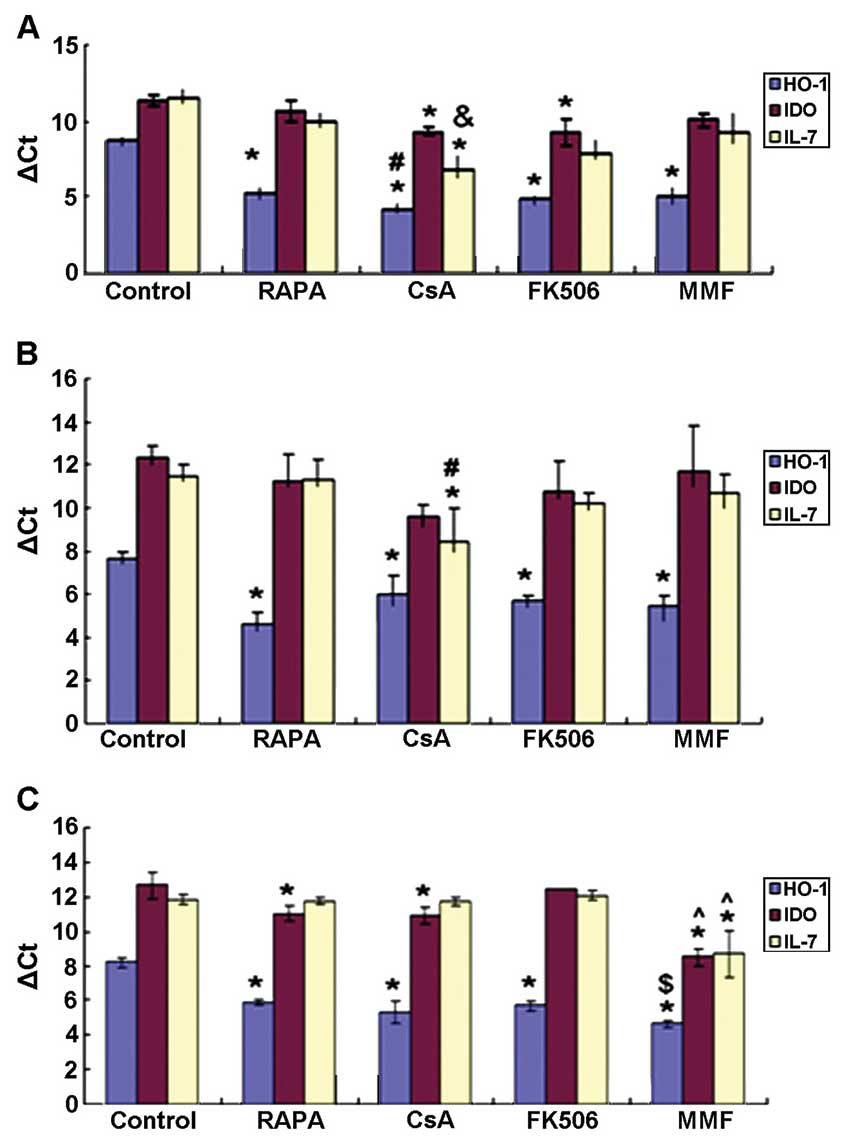 | Figure 1mRNA expression levels of HO-1, IDO
and IL-7 in the HBZY-1 mesangial cells (A) 6 h; (B) 12 h; and (C)
24 h after administration of the immunosuppressive agents.
*P<0.05 vs. control group, #P<0.05 vs.
RAPA group, &P<0.05 vs. RAPA and MMF groups,
^P<0.05 vs. other experimental groups,
$P<0.05 vs. RAPA and Tac groups. IDO, indoleamine
2,3-dioxygenase; HO-1, heme oxygenase-1; IL-7, interleukin-7; RAPA,
rapamycin; CsA, cyclosporine A; FK506, tacrolimus; MMF,
mycophenolate mofetil; ΔCt, Δ cycle threshold. |
As shown in Fig.
1B, after 12 h, no significant differences were observed in the
mRNA expression levels of IDO between the various experimental
groups. The mRNA expression levels of HO-1 were significantly lower
in the control group as compared with those in the drug treatment
groups (P<0.05). However, no significant differences were
detected in the HO-1 mRNA expression levels among the various drug
treatment groups. The mRNA expression levels of IL-7 were
significantly increased in the CsA group as compared with those in
the RAPA and control groups (P<0.05).
As shown in Fig.
1C, after 24 h, the mRNA expression levels of IDO were
significantly higher in the MMF group as compared with those in the
other experimental groups (P<0.05). In addition, the mRNA
expression levels of IDO were markedly increased in the CsA and
RAPA groups as compared with those in the control group
(P<0.05). The mRNA expression levels of HO-1 were significantly
lower in the control group as compared with those in the other
groups (P<0.05), and were markedly higher in the MMF group as
compared with those in the RAPA and Tac groups (P<0.05).
Furthermore, no differences were observed in the mRNA expression
levels of HO-1 between the CsA and Tac groups. The mRNA expression
levels of IL-7 were significantly elevated in the MMF group as
compared with those in the other groups (P<0.05).
Expression of IDO, HO-1 and IL-7, as
detected by cell immunohistochemistry
As shown in Fig. 2,
IDO was markedly expressed in the caryon and cytoplasm of the
mesangial cells treated with MMF, CsA and Tac, particularly in
those treated with MMF. As shown in Fig. 3, HO-1 was markedly expressed in the
caryon and cytoplasm of all of the treatment groups at 24 h;
however, there were no significant differences between them. As
shown in Fig. 4, IL-7 was
predominantly expressed in the cytoplasm of the mesangial cells,
and was most highly expressed in the cells treated with MMF at 24
h.
Protein expression levels of IDO, HO-1
and IL-7
At 24 h, the protein expression levels of HO-1 were
markedly elevated in the CsA, Tac, RAPA and MMF groups, as compared
with those in the control group (P<0.05). Furthermore, no
significant differences were detected in the protein expression
levels of HO-1 between the drug treatment groups. The protein
expression levels of IDO were significantly increased in the CsA,
Tac and MMF groups as compared with those in the control group
(P<0.05). The protein expression levels of IDO and IL-7 were
significantly higher in the MMF group as compared with those in the
CsA, Tac and RAPA groups (P<0.05). There were no significant
differences observed between the Tac and CsA groups. In addition,
RAPA had no significant effect on the protein expression levels of
IDO. The protein expression levels of IL-7 were markedly decreased
in the CsA, Tac, RAPA and MMF groups as compared with those in the
control group (P<0.05). No significant differences were detected
in the protein expression levels of IL-7 between the RAPA, CsA and
Tac groups (Fig. 5).
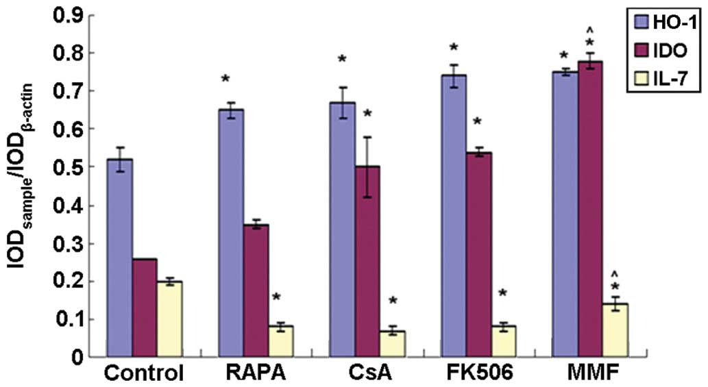 | Figure 5Protein expression levels of HO-1,
IDO, and IL-7 at 24 h after the administration of various
immunosuppressive agents. *P<0.05 vs. control group,
^P<0.05 vs. other experimental groups. IDO,
indoleamine 2,3-dioxygenase; HO-1, heme oxygenase-1; IL-7,
interleukin-7; RAPA, rapamycin; CsA, cyclosporine A; FK506,
tacrolimus; MMF, mycophenolate mofetil; IOD, integrated optical
density. |
Discussion
IDO is a potent immunosuppressive enzyme that has
gained attention in recent years. IDO is a 45-kDa monomeric
heme-containing protein and the rate-limiting enzyme of the
kynurenine pathway, which degrades the essential amino acid
tryptophan (9–11). IDO-induced tryptophan degradation
inhibits the proliferation of T cells, accelerates T-cell apoptosis
and triggers cell cycle arrest (12,13,25).
In addition, the overexpression of IDO has been shown to reduce the
incidence of graft injury or rejection through inhibiting
alloreactive T-cell responses and enhancing the local antioxidative
defense system, leading to peripheral tolerance and long-term graft
survival (12,13). However, inhibition of IDO activity
in graft-tolerant rats has been shown to lead to rapid graft
rejection (26).
HO is the rate-limiting enzyme in the conversion of
ferroheme to billiverdin, free iron ions and carbon monoxide
(14–20). To date, three HO isoforms have been
identified. HO-2 is the constitutively expressed isoform, whereas
HO-1 is the inducible isoform, which is typically expressed at a
rather low level in the majority of cell types. The expression of
HO-1 may be induced by various stimuli, such as ferroheme, heavy
metals, hyperoxia, hypoxia, tissue plasminogen activator, cytokines
and chemokines (21–23,27,28).
The third isoform, HO-3, has yet to be fully defined (21–23,27,28).
HO-1 and its metabolites represent a cytoprotective defense
mechanism in various models of cellular and tissue damage and
exhibit anti-inflammatory, anti-apoptotic, anti-oxidative,
anti-proliferative, anticoagulant and vasodilatory effects through
various pathways (21,29–31).
Previous studies have demonstrated that HO-1 has a protective role
in numerous organ transplantation models (29–33).
HO-1 has also been shown to reduce leukocyte-endothelial
interactions following ischemia/reperfusion (I/R) injury, alleviate
chronic rejection and improve graft survival (29,32,33).
Furthermore, deficient levels of HO-1 may lead to ferroheme
aggregation in the circulation and vascular endothelial damage
(21).
IL-7 is a potent survival factor for T cells, which
has key roles in the maintenance of intracellular homeostasis and
the regulation of the anti- and pro-apoptotic members of the B-cell
lymphoma 2 protein family (34).
IL-7 promotes the transition of CD4+ effector T cells to
persistent memory T cells and is essential for the homeostasis,
proliferation and survival of memory CD8+ T cells. It
has previously been suggested that targeting IL-7 during
transplantation may inhibit the production of allogeneic
antigen-specific memory T cells, thus promoting the induction of
graft tolerance (12).
CAN is a major limiting factor of long-term graft
survival following kidney transplantation (6), which may be induced by various immune
and non-immune factors, including I/R injury and treatment with
immunosuppressants. The immunosuppressive drugs CsA and Tac have
been identified to promote the incidence of CAN (35). The expression levels of IDO, HO-1
and IL-7 in renal grafts, and the effects of various
immunosuppressants on the expression of these genes, are yet to be
fully elucidated. Therefore, the present study aimed to improve the
understanding of the mechanisms of action of immunosuppressants
following transplantation.
In the present study, a model of glomerular
mesangial cell injury was established and investigated. Treatment
with CsA and particularly with MMF significantly upregulated the
mRNA and protein expression levels of IDO. FK-506 (Tac) also
markedly upregulated the protein expression levels of IDO.
Conversely, RAPA exhibited no apparent effect on IDO protein
expression. These results indicated that MMF, and the calcineurin
inhibitors (CNIs) Tac and Csa may have an important role in the
regulation of IDO expression. In addition, RAPA and FK-506 were
shown to regulate IDO expression at the transcriptional and
post-transcriptional level, respectively. This complementary
association between RAPA and FK506 may provide a novel theoretical
basis for the clinical application of a combination therapy of RAPA
and FK-506.
MMF inhibits T-cell proliferation through blocking
DNA synthesis, and CNIs, a class of T-cell directed
immunosuppressive drugs, also suppress the activity of T-cells
(36,37). The results of the present study
suggested that MMF and CNIs may inhibit alloreactive T-cell
responses through the tryptophan catabolic pathway following organ
transplantation. This pathway may therefore account for another
underlying mechanism of the prevention of graft rejection and CAN
by MMF and CNIs.
Various drugs, including statins, RAPA,
erythropoietin and probucol, enhance the expression of HO-1
(22). The results of the present
study indicated that, in addition to RAPA, the immunosuppressants
CsA, FK506 and MMF markedly upregulated the mRNA as well as protein
expression levels of HO-1. No significant differences were observed
in the HO-1 protein expression levels in the cells following
treatment with CsA, FK506, RAPA or MMF, suggesting that CsA, FK506,
RAPA and MMF exert similar inductive effects on HO-1 expression at
the post-transcriptional level. The administration of CsA, FK506,
RAPA and MMF following kidney transplantation is conducive to the
prevention of I/R injury and the reduction of chronic rejection
(33). In addition, these drugs
have been shown to regulate fibrosis and angiogenesis (22). Therefore, the induction of HO-1 by
CsA, FK506, RAPA and MMF is beneficial for the prevention and
treatment of CAN.
Previous studies have demonstrated that
IL-7-mediated homeostatic proliferation of T cells is regulated by
MMF, but not by CNIs or sirolimus (38). The results of the present study
demonstrated that although MMF significantly increased the mRNA
expression levels of IL-7, protein expression levels of IL-7 were
significantly inhibited by MMF and particularly by CNIs and RAPA.
It is evident that all four types of immunosuppressants are
involved in the regulation of IL-7 protein expression in
vitro, suggesting that the four types of immunosuppressants
regulate the proliferation of T cells. The present study also
observed that MMF exerted bidirectional regulatory effects on the
mRNA and protein expression levels of IL-7. Conversely, treatment
with CNIs and RAPA exhibited no significant effect on the
expression levels of IL-7 mRNA, but they did markedly inhibit the
protein expression levels of IL-7. Inhibition of IL-7 protein
expression by immunosuppressants may have an important role in the
prevention of graft rejection. Clinical studies have demonstrated
that prior to recovery, the plasma levels of IL-7 and
CD8+ T cells are increased, suggesting that IL-7 may
result in generation of transplant recipients’ CD8+
memory T cells (39,40); therefore, inhibition of the
production of IL-7 may contribute to suppression of memory T cell
production, and thus serve to prevent graft rejection and promote
long-term allograft survival.
In conclusion, the present study investigated for
the first time, to the best of our knowledge, the gene expression
levels of IDO, HO-1 and IL-7 in glomerular mesangial cells in
vitro, as well as the regulatory effects of various
immunosuppressants on the expression of these genes. The expression
levels of HO-1 were significantly upregulated in response to
treatment with CsA, FK506, RAPA and MMF, whereas the expression
levels of IL-7 were markedly downregulated by treatment with the
above immunosuppressants. CsA, FK506 and MMF significantly enhanced
the expression levels of IDO, whereas RAPA exhibited no apparent
effect on IDO. To understand the effects of IDO, HO-1, and IL-7
expression on chronic graft rejection and CAN, further in-depth
studies are required.
Acknowledgments
The authors of the present study would like to
express their most sincere gratitude to all staff members at the
Laboratory of Transplant Engineering and Immunology, and the
Laboratory of Molecular Genetics at West China Medical School of
Sichuan University (Chengdu, China).
References
|
1
|
Ma X, Lu YP, Yang L, et al: Rapamycin and
cyclosporine have different effects on expression of Ang-1 and
Ang-2 and Tie2 in rat renal allograft with chronic allograft
nephropathy. Transplant Proc. 40:2804–2807. 2008. View Article : Google Scholar : PubMed/NCBI
|
|
2
|
Najafian B and Kasiske BL: Chronic
allograft nephropathy. Curr Opin Nephrol Hypertens. 17:149–155.
2008. View Article : Google Scholar : PubMed/NCBI
|
|
3
|
Weissgarten J, Berman S, Efrati S, et al:
Apoptosis and proliferation of cultured mesangial cells isolated
from kidneys of rosiglitazone-treated pregnant diabetic rats.
Nephrol Dial Transplant. 21:1198–1204. 2006. View Article : Google Scholar : PubMed/NCBI
|
|
4
|
Shultz PJ and Raij L: The glomerular
mesangium: Role in initiation and progression of renal injury. Am J
Kidney Dis. 17(5 Suppl 1): 8–14. 1991.PubMed/NCBI
|
|
5
|
Chen JK, Zou WZ and You JF: Mesangial
cells endothelin receptors. Chinese J Nephrol. 1:52–53. 1997.In
Chinese.
|
|
6
|
Joosten SA, van Kooten C and Paul LC:
Pathogenesis of chronic allograft rejection. Transpl Int.
16:137–145. 2003. View Article : Google Scholar : PubMed/NCBI
|
|
7
|
Libby P and Pober JS: Chronic rejection.
Immunity. 14:387–397. 2001. View Article : Google Scholar : PubMed/NCBI
|
|
8
|
Joosten SA, van Ham V, Borrias MC, et al:
Antibodies against mesangial cells in a rat model of chronic renal
allograft rejection. Nephrol Dial Transplant. 20:692–698. 2005.
View Article : Google Scholar : PubMed/NCBI
|
|
9
|
Hill M, Zagani R, Voisine C, et al: Nitric
oxide and indoleamine 2,3-dioxygenase mediate CTLA4Ig-induced
survival in heart allografts in rats. Transplantation.
84:1060–1063. 2007. View Article : Google Scholar : PubMed/NCBI
|
|
10
|
Brandacher G, Margreiter R and Fuchs D:
Clinical relevance of indoleamine 2,3-dioxygenase for alloimmunity
and transplantation. Curr Opin Organ Transplant. 13:10–15. 2008.
View Article : Google Scholar : PubMed/NCBI
|
|
11
|
Vogel CF, Goth SR, Dong B, et al: Aryl
hydrocarbon receptor signaling mediates expression of indoleamine
2,3-dioxygenase. Biochem Biophys Res Commun. 375:331–335. 2008.
View Article : Google Scholar : PubMed/NCBI
|
|
12
|
Dai H and Dai Z: The role of tryptophan
catabolism in acquisition and effector function of memory T cells.
Curr Opin Organ Transplant. 13:31–35. 2008. View Article : Google Scholar : PubMed/NCBI
|
|
13
|
Arefayene M, Philips S, Cao D, et al:
Identification of genetic variants in the human indoleamine
2,3-dioxygenase (IDO1) gene, which have altered enzyme activity.
Pharmacogenet Genomics. 19:464–476. 2009. View Article : Google Scholar : PubMed/NCBI
|
|
14
|
Lai IR, Chang KJ, Tsai HW and Chen CF:
Pharmacological preconditioning with simvastatin protects liver
from ischemia-reperfusion injury by heme oxygenase-1 induction.
Transplantation. 85:732–738. 2008. View Article : Google Scholar : PubMed/NCBI
|
|
15
|
Chen C, Wang Y, Zhang Z, et al: Toll-like
receptor 4 regulates heme oxygenase-1 expression after hemorrhagic
shock induced acute lung injury in mice: Requirement of p38
mitogen-activated protein kinase activation. Shock. 31:486–492.
2009. View Article : Google Scholar
|
|
16
|
Lin CC, Liu XM, Peyton K, et al: Far
infrared therapy inhibits vascular endothelial inflammation via the
induction of heme oxygenase-1. Arterioscler Thromb Vasc Biol.
28:739–745. 2008. View Article : Google Scholar : PubMed/NCBI
|
|
17
|
Saleem S, Zhuang H, Biswal S, et al:
Ginkgo biloba extract neuroprotective action is dependent on heme
oxygenase 1 in ischemic reperfusion brain injury. Stroke.
39:3389–3396. 2008. View Article : Google Scholar : PubMed/NCBI
|
|
18
|
Krönke G, Kadl A, Ikonomu E, et al:
Expression of heme oxygenase-1 in human vascular cells is regulated
by peroxisome proliferator-activated receptors. Arterioscler Thromb
Vasc Biol. 27:1276–1282. 2007. View Article : Google Scholar
|
|
19
|
Tiroch K, Koch W, von Beckerath N, et al:
Heme oxygenase-1 gene promoter polymorphism and restenosis
following coronary stenting. Eur Heart J. 28:968–973. 2007.
View Article : Google Scholar : PubMed/NCBI
|
|
20
|
Lang D, Reuter S, Buzescu T, et al:
Heme-induced heme oxygenase-1 (HO-1) in human monocytes inhibits
apoptosis despite caspase-3 up-regulation. Int Immunol. 17:155–165.
2005. View Article : Google Scholar
|
|
21
|
Dulak J, Loboda A and Jozkowicz A: Effect
of heme oxygenase-1 on vascular function and disease. Curr Opin
Lipidol. 19:505–512. 2008. View Article : Google Scholar : PubMed/NCBI
|
|
22
|
Dulak J, Deshane J, Jozkowicz A and
Agarwal A: Heme oxygenase-1 and carbon monoxide in vascular
pathobiology: Focus on angiogenesis. Circulation. 117:231–241.
2008. View Article : Google Scholar : PubMed/NCBI
|
|
23
|
Ryter SW, Alam J and Choi AM: Heme
oxygenase-1/carbon monoxide: From basic science to therapeutic
applications. Physiol Rev. 86:583–650. 2006. View Article : Google Scholar : PubMed/NCBI
|
|
24
|
Nginamau ES, Maehle BO and Jonsson R: An
experimental protocol for the fractionation and 2DE separation of
HeLa and A-253 cell lysates suitable for the identification of the
individual antigenic proteome in Sjögren’s syndrome. Autoimmunity.
44:652–663. 2011. View Article : Google Scholar : PubMed/NCBI
|
|
25
|
Radu CA, Bosch N, Bauer TM, et al:
Immunosuppressive effect of tryptophan metabolites in composite
tissue allotransplantation. Plast Reconstr Surg. 119:2023–2028.
2007. View Article : Google Scholar : PubMed/NCBI
|
|
26
|
Niederkorn JY: Emerging concepts in CD8(+)
T regulatory cells. Curr Opin Immunol. 20:327–331. 2008. View Article : Google Scholar : PubMed/NCBI
|
|
27
|
Nath KA: Heme oxygenase-1: A provenance
for cytoprotective pathways in the kidney and other tissues. Kidney
Int. 70:432–443. 2006.PubMed/NCBI
|
|
28
|
Kassovska-Bratinova S, Yang G, Igarashi K
and Dennery PA: Bach1 modulates heme oxygenase-1 expression in the
neonatal mouse lung. Pediatr Res. 65:145–149. 2009. View Article : Google Scholar :
|
|
29
|
von Dobschuetz E, Schmidt R, Scholtes M,
et al: Protective role of heme oxygenase-1 in pancreatic
microcirculatory dysfunction after ischemia/reperfusion in rats.
Pancreas. 36:377–384. 2008. View Article : Google Scholar : PubMed/NCBI
|
|
30
|
Di Francesco L, Totani L, Dovizio M, et
al: Induction of prostacyclin by steady laminar shear stress
suppresses tumor necrosis factor-alpha biosynthesis via heme
oxygenase-1 in human endothelial cells. Circ Res. 104:506–513.
2009. View Article : Google Scholar : PubMed/NCBI
|
|
31
|
Abraham NG and Kappas A: Pharmacological
and clinical aspects of heme oxygenase. Pharmacol Rev. 60:79–127.
2008. View Article : Google Scholar : PubMed/NCBI
|
|
32
|
Roach JP, Moore EE, Partrick DA, et al:
Heme oxygenase-1 induction in macrophages by a hemoglobin-based
oxygen carrier reduces endotoxin-stimulated cytokine secretion.
Shock. 31:251–257. 2009. View Article : Google Scholar
|
|
33
|
Kaczorowski DJ, Nakao A, Mollen KP, et al:
Toll-like receptor 4 mediates the early inflammatory response after
cold ischemia/reperfusion. Transplantation. 84:1279–1287. 2007.
View Article : Google Scholar : PubMed/NCBI
|
|
34
|
Rethi B, Vivar N, Sammicheli S and Chiodi
F: Limited efficiency of endogenous interleukin-7 levels in T cell
reconstitution during HIV-1 infection: Will exogenous interleukin-7
therapy work-AIDS. 23:745–755. 2009.
|
|
35
|
Song J, Lu YP, Luo GH, et al: Effects of
mycophenolate mofetil on chronic allograft nephropathy by affecting
RHO/ROCK signal pathways. Transplant Proc. 40:2790–2794. 2008.
View Article : Google Scholar : PubMed/NCBI
|
|
36
|
Siemionow M and Klimczak A: Basics of
immune responses in transplantation in preparation for application
of composite tissue allografts in plastic and reconstructive
surgery: Part 1. Plast Reconstr Surg. 121:4e–12e. 2008. View Article : Google Scholar
|
|
37
|
Tanabe K: Japanese experience of
ABO-incompatible living kidney transplantation. Transplantation.
84(12 Suppl): S4–S7. 2007. View Article : Google Scholar
|
|
38
|
Monti P, Scirpoli M, Maffi P, et al: Islet
transplantation in patients with autoimmune diabetes induces
homeostatic cytokines that expand autoreactive memory T cells. J
Clin Invest. 118:1806–1814. 2008.PubMed/NCBI
|
|
39
|
Donckier V, Craciun L, Lucidi V, et al:
Acute liver transplant rejection upon immunosuppression withdrawal
in a tolerance induction trial: Potential role of
IFN-gamma-secreting CD8+ T cells. Transplantation. 87(9 Suppl):
S91–S95. 2009. View Article : Google Scholar : PubMed/NCBI
|
|
40
|
Daudt L, Maccario R, Locatelli F, et al:
Interleukin-15 favors the expansion of central memory CD8+ T cells
in ex vivogenerated, antileukemia human cytotoxic T lymphocyte
lines. J Immunother. 31:385–393. 2008. View Article : Google Scholar : PubMed/NCBI
|


