Introduction
Massive blood transfusion is generally defined as
the administration of ≥10 units of packed red blood cells (pRBC) to
a patient (1,2) or the transfusion of more than one
blood volume in 24 h (1,3–5).
Acute clinical situations that warrant the administration of
massive transfusions include a 50% blood volume loss within 3 h or
a blood loss rate of 150 ml/min (3). Massive transfusion is generally
necessary in severely injured military personnel or patients with
multiple injuries. Such patients often require multiple, complex
surgical procedures. A rational blood transfusion protocol can
improve the outcome of surgery, whereas unreasonably excessive
transfusion can lead to mortality, predominantly due to coagulation
disorders, acidosis and hypothermia. The majority of studies
published hitherto have been conducted in western countries and on
trauma patients (5–10). To the best of our knowledge, no
multicenter data are currently available on the effect of massive
transfusion on coagulation during the perioperative period in
Chinese patients.
In the present study, a retrospective investigation
of 1,601 cases of surgical inpatients from 20 large-scale,
comprehensive hospitals in different regions of China was
undertaken and variations in the coagulation indices of patients
were analyzed. The actual measurements of the platelet (PLT) counts
in patients receiving massive transfusion against those determined
by theoretical calculation were also verified.
Materials and methods
Retrospective survey study
Study protocol
In the present study, massive blood transfusion was
defined as the administration of ≥10 units of pRBC in 24 h. Data
was collected from the medical records of surgical inpatients who
received massive transfusion at 20 large-scale hospitals in the
northwest, southwest, central south, north and northeast regions of
China between January 2009 and December 2010. A total of 2,000
copies of the Massive Transfusion Survey Table (hereafter referred
to as Survey Table) were distributed to 20 participants in the
hospital. Members of the National Massive Transfusion Current
Status Investigation Coordination Group (hereafter referred to as
the Coordination Group) were responsible for collecting the data
from these hospitals using the Survey Table. The data analysis was
conducted at Shaanxi Provincial People's Hospital, the Third
Affiliated Hospital of the Medical College of Xi'an Jiaotong
University (Xi'an, China). The present study was approved by the
Ethics Committee of Shaanxi Provincial People's Hospital.
Study population
In the present study, the research group included
patients who received transfusion of ≥10 units of pRBC over a
period of ≤24 h for trauma, cardiac surgery, obstetric conditions
or other common surgeries, including orthopedic, thoracic, general,
urinary, hepatobiliary and neurological surgery. Patients who
received transfusions of <10 U for ≤24 h were assigned to the
control group. By contrast, patients with coagulation disorders
and/or hepatic failure due to medical causes were excluded from the
analysis. Informed consent was obtained from each of the
participants.
Survey table
The directors of the transfusion departments of the
20 participating hospitals discussed the topic, consulted experts
and designed the Survey Table with reference to several
international and domestic sources, in accordance with the
principle of voluntary participation in this study. A meeting of
the Coordination Group was then held, where 35 experts of clinical
transfusion, surgery, anesthesia, gynecology and obstetrics,
hematology and medical statistics discussed the study protocol and
mode of data collection and also perfected and added supplements to
the Survey Table. Suitable training was then offered to the
investigating staff.
Components of the survey table
The survey table comprised the following four
sections: i) Clinical and demographic characteristics of the
patient, including name, gender, age, body weight, blood type,
ethnicity, admission number, admission department, primary
diagnosis, secondary diagnosis, pathological diagnosis, nature of
surgery and vital signs on admission; ii) details regarding
perioperative complications, the clinical condition within 24 h and
after 24 h of the transfusion, and the total quantity of blood
transfused; iii) the results of the following blood tests performed
prior to, within 24 h and after 24 h of transfusion: Routine blood
test, coagulation tests, liver function test, kidney function test
and arterial blood gas analysis; iv) adverse events due to massive
transfusion.
Quality control
The Survey Table was initially subjected to a
small-scale preliminary test at Shaanxi Provincial People's
Hospital so that revisions could be made on the basis of the
results and comments of experts to further improve the table. As
per the Chinese standards, the protocol for massive transfusion was
as follows: One unit of pRBC derived from 200 ml of whole blood
with a volume of 140–172 ml; one unit of fresh frozen plasma (FFP)
derived from 200 ml of whole blood and a volume of 100 ml; one bag
of apheresis PLT of 10 U and a volume of 150–250 ml; and one unit
of PLT concentrate derived from 200 ml of whole blood and with a
volume of 20–30 ml. One bag of apheresis PLT is 10 units of PLT
concentrate. The pRBC were stored at 2–6°C. FFP was stored at
≤−18°C and thawed in a 37°C water bath, for ~10 to 15 min. PLTs
were stored at 20–24°C in a platelet shaker.
The main test devices and reagents used were as
follows: Sysmex XE-2100/XT-1800i hematology analyzer (Sysmex Corp.,
Kobe, Japan), Beckman Coulter LH780 Coulter Hematology Analyzer
(Beckman Coulter, Inc., Brea, CA, USA); Hitachi 7170A/7180
Biochemical Analyzer (Hitachi, Tokyo, Japan); Roche Modular DP
Automatic Biochemical Analyzer (Roche Diagnostics, Indianapolis,
IN, USA); Olympus AU640 Biochemical Analyzer (Olympus, Tokyo,
Japan); Radiometer ABL-77 Blood Gas Analyzer (Radiometer,
Copenhagen, Denmark); Roche Cobas-B123 Blood Gas Analyzer (Roche
Diagnostics); Sysmex CA1500/CA7000 Automatic Blood Coagulation
Analyzer (Sysmex Corp.). All test reagents used were
device-supporting reagents.
Data on the blood tests performed were collected
from the laboratory records: Blood routine, coagulation tests,
liver function test, kidney function and blood gas analysis. The
data were collected for the blood tests performed prior to
transfusion and at 16 different time points during the 24-h
transfusion (2, 4, 6, 8, 10, 12, 14, 16, 18, 20, 22, 24, 26, 28, 30
and 40 U) and subjected to statistical analysis. The tests were
conducted at the laboratory of each participating hospital, which
undergoes internal quality control and an external quality
assessment conducted by the National Center for Clinical
Laboratories (Beijing, China).
In vitro test: The effect of the
addition of pRBC in vitro following hemodilution on PLT count
Since a large quantity of debris of platelet-like
cells was found after performing the PLT count on samples of stored
pRBC units, the present study therefore aimed to determine whether
the PLT count was affected by the transfusion of pRBC. Thus, the
test was performed in vitro.
Participants
Following approval from the ethics committee and
obtaining informed consent, 16 healthy staff members at Shaanxi
Provincial People's Hospital, including 10 females and 6 males were
included in the present study ranging between 19 and 50 years old.
Those with a history of anemia, coagulation disorders, hemorrhage
or kidney disease as well as individuals taking anticoagulant drugs
within 1 week and females undergoing menstruation were
excluded.
Preparation of red blood cells
The pRBCs were provided by the Blood Center of
Shaanxi Province (Xi'an, China). The blood sample of pRBCs was
prepared from the package of the pRBCs stored for 3–5 days.
Experimental procedure
Blood (26 ml) from 16 donors was collected in 3.8%
sodium citrate. The first 2 ml blood was discarded and the
remaining 24 ml blood was used in the experiment. Then, 14 ml was
used as a control, following dilution with saline at ratios of
10:0, 9:1, 8:2, 7:3, 6:4, 5:5, 4:6, 3:7, 8:2 and 1:9 (blood versus
saline). The other 10 ml was used for the experimental group at
dilutions of 7:3, 6:4, 5:5, 4:6, 3:7, 8:2 and 1:9 with saline
(blood versus saline). A total of 1 ml of each diluted blood sample
was allocated to seven tubes and a different quantity of pRBCs was
added to each tube to assess blood routine testing, with reference
to the current international guidelines for massive transfusion and
surgical blood transfusion (3,5,10,11–15).
The critical range of RBC concentration was maintained by ensuring
that the hemoglobin level remained at 60–80 g/l. The quantity of
pRBCs added was determined according to the results of the
experiments regarding the addition of pRBCs under different
dilutions (i.e., 100 μl of pRBCs was found to be required
for a hemodilution of 30%; 200 μl for 40%; 300 μl for
50%; 400 μl for 60%; 500 μl for 70%; 600 μl
for 80% and 700 μl for 90%), which will be published
separately.
Statistical analysis
Statistical analysis was conducted using SPSS
software (version 18.0; SPSS, Inc., Chicago, IL, USA). EpiData
(version 3.01; EpiData Association, Odense, Denmark) was used for
double data entry verification and database construction. The data
on the demographic characteristics and clinical features are
expressed as the mean ± standard deviation or as absolute numbers.
Categorical variables were analyzed by χ2 test, while
continuous variables with normal distribution were analyzed by the
Shapiro-Wilk test, analysis of variance or the Kruskal-Wallis test,
as appropriate. The Bonferroni method was applied for post-hoc
tests to determine the significance of the differences between the
group that received massive transfusion and the control group that
did not. Linear regression was used to describe the association
between units of pRBC transfused and PLT count. A two-sided P-value
of <0.05 was considered to indicate a statistically significant
difference.
Results
Patient characteristics
A total of 1,753 of the 2,000 copies of the Survey
Table were able to be retrieved from 20 hospitals, at a recovery
rate of 87.65%. Following excluding tables with missing
information, 1,601 copies (91.33%; 889 male patients; 702 female
patients) were used for the analysis. The age of the enrolled
patients was 16–91 years (median: 46 years) and weight was 46–105
kg (median: 60 kg). The data regarding age and weight were assessed
by the Shapiro-Wilk test (P<0.01) and demonstrated an abnormal
distribution; therefore, they were presented as median values.
Among the 1,601 patients who received blood transfusion, 1,048
received ≥10 units of pRBC within 24 h (108 died, 940 survived;
mortality rate: 10.31%), whereas 553 patients received <10 units
of pRBC within 24 h (24 died, 529 survived; mortality rate: 4.34%).
The reasons for transfusion in the 1,601 enrolled cases were as
follows: Trauma in 268 patients (34 died, 234 survived; mortality
rate: 12.69%), cardiac surgery in 383 patients (53 died, 330
survived; mortality rate: 13.84%), general surgery in 876 patients
(42 died, 834 survived; mortality rate: 4.79%) and obstetric
complications in 74 patients (3 died, 71 survived; mortality rate:
4.05%). The cases of mortality in the present study refer to
fatalities occurring during the period of hospitalization. The
details of the patient characteristics are provided in Table I.
 | Table IBaseline data of 1,601 patients
receiving massive transfusion. |
Table I
Baseline data of 1,601 patients
receiving massive transfusion.
| Demographics and
clinical data | <10 units of
pRBC | ≥10 units of
pRBC | P-value |
|---|
| Demographics |
| Number of patients,
n (%) | 553 (34.5) | 1,048 (65.5) | |
| Age, years (mean ±
SD) | 46.5±18.2 | 44.9±16.7 | |
| Males, n (%) | 300 (300/553) | 402 (402/1048) | |
| Weight, kg (mean ±
SD) | 56.6±13.9 | 58.5±11.4 | |
| Patients suffering
from trauma, n (%) | 81 (30.2) | 187 (69.8) | |
| Patients who
underwent cardiac surgery, n (%) | 116 (30.3) | 267 (69.7) | |
| Patients who
underwent general surgery, n (%) | 335 (38.2) | 541 (61.8) | |
| Patients with
obstetric complications, n (%) | 21 (28.4) | 53 (71.6) | |
| Clinical data
(prior to transfusion) |
| Respiration, n/min
(mean ± SD) | 20.3±3.5 | 20.5±3.6 | 0.043a |
| Pulse, n/min (mean
± SD) | 94.1±69.8 | 92.5±54.3 | 0.452a |
| SBP, mmHg (mean ±
SD) | 113.5±24.7 | 112.8±30.2 | 0.020a |
| Temperature, °C
(mean ± SD) | 36.6±1.0 | 36.5±0.7 | 0.319a |
| RBC,
×1012/l (mean±SD) | 3.8±1.0 | 3.8±1.1 | 0.323a |
| Hb, g/l (mean ±
SD) | 114.3±30.2 | 117.4±43.2 | 0.213a |
| Hct as % (mean ±
SD) | 21.2±17.7 | 16.6±17.6 | 0.834a |
| PLT,
×109/l (mean±SD) | 179.5±91.5 | 175.6±98.9 | 0.324a |
| PT, sec (mean ±
SD) | 13.7±6.0 | 14.1±5.8 | 0.173a |
| APTT, sec (mean ±
SD) | 33.6±11.7 | 36.3±24.2 | 0.006a |
| TT, sec (mean ±
SD) | 17.1±12.8 | 17.5±7.1 | 0.529a |
| INR (mean ±
SD) | 1.3±2.1 | 1.2±1.1 | 0.041a |
| FIB, g/l (mean ±
SD) | 11.3±44.4 | 11.0±46.6 | 0.801a |
| Clinical data
(following transfusion) |
| Length of hospital
stay, days (mean ± SD) | 24.9±14.3 | 29.8±23.9 | 0.000a |
| Length of stay in
ICU, days (mean ± SD) | 3.8±3.5 | 8.7±23.4 | 0.006a |
| Surgery time, h
(mean ± SD) | 2.5±3.2 | 3.7±3.9 | 0.000a |
| pRBC in 24 h,
units (median) | 9 | 25 | 0.000b |
| FFP in 24 h, units
(median) | 8 | 20 | 0.000b |
| PLT in 24 h, units
(median) | 10 | 6 | 0.009b |
| pRBC in 72 h, units
(median) | 20 | 18 | 0.202b |
| FFP in 72 h, units
(median) | 14 | 13 | 0.499b |
| PLT in 72 h, units
(median) | 8 | 8 | 0.873b |
PLT variations during massive
transfusion
PLT count in patients receiving
massive blood transfusion
Data for the complete blood routine examination at
all the different time points considered in the present study were
available for 883 of the 1,601 patients enrolled in the present
study. Statistical analysis of the data demonstrated that the PLT
count in the patients receiving blood transfusion decreased with an
increase in the number of pRBC units transfused (Fig. 1A). Following administration of 18
units of pRBC, the PLT count decreased to 71×109/l in
patients receiving massive transfusion; Fig. 1B shows the PLT non-intervention
group (n=776). When pRBCs reached 18 U, the average PLT counts
decreased to 72×109/l; Fig.
1C shows the PLT intervention group (n=107); Fig. 1D shows when patients were
transfused with 0.3 units of pRBC per kilogram of body weight (i.e.
3 U/10 kg). The average PLT count decreased to below
75×109/l. With regard to invasive surgery for
underweight adults, the critical level of PLTs may be calculated
according to the units of pRBC administered and body weight.
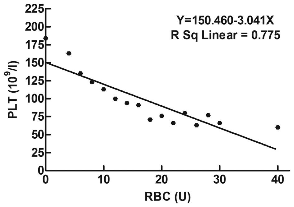
Linear regression analysis of PLT
count measured for different units of pRBC transfused
A linear regression analysis of the number of pRBC
units transfused and PLT count was performed in 776 patients who
received blood transfusion without PLT-intervention, and the
results demonstrated that the two parameters were correlated
negatively. The linear association was defined as R2
linear=0.775, with a regression formula of Y=150.460−3.041X
(Fig. 2).
PLT count prior to and following the
addition of pRBC in the in vitro blood dilution experiment
The data of 16 unrelated healthy volunteers were
analyzed; pRBC were added at different dilutions to maintain the
range of hemoglobin concentration at 60–80 g/l. The results
demonstrated that the PLT count decreased with an increase in the
hemodilution. It was found that administration of pRBC for the
correction of anemia also corrected the PLT count. Further analysis
revealed that the results of the automated device for counting
blood cells were affected by the addition of pRBC (Fig. 3). RBC=(6.29±1.05)
×1012/l, hematocrit=0.5898±0.1 l/l,
hemoglobin=190.4±39.01 g/l and PLT count=(239.8±135.29)
×109/l. These results suggested that RBC in the pRBC
contained PLT-like cell fragments.
Variation in coagulation parameters of
patients with massive transfusion
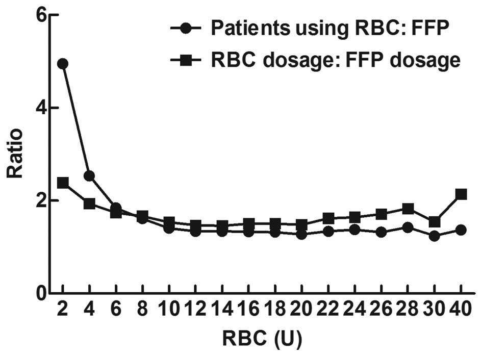
Supplementation of FFP (coagulation
factor supplement)
The data of 1,601 cases enrolled in the present
study demonstrated that all hospitals administered FFP to
supplement pRBC transfusion. When RBC transfusion was 2–8 U, the
ratio of patients transfused with RBC and plasma was 4.88:1–1.61:1
(1601:328–1313:816), and at RBC transfusions of 10–30 U, the ratio
of patients transfused with pRBC and plasma was 1.38:1–1.24:1
(1048:759–62:50). This implies that for transfusion of 10–30 units
of pRBC, the ratio of patients transfused with plasma accounted for
1:1.38–1:1.24 (759:1048–50:62) of those transfused with pRBC, while
the quantity of plasma transfused accounted for 1:1.54–1:1.55
(6939.5:10700–1159.5:1800) that of pRBC transfused (Fig. 4).
Variation in coagulation indices in
patients receiving massive blood transfusion
The results of the present study indicated that the
prothrombin time (PT) varied between 15 and 20 sec when RBC
transfusion was 2–40 U (Fig. 5A).
Activated partial thromboplastin time (APTT) demonstrated a gradual
extension with an increase in the number of RBC units transfused,
reaching 60 sec when 40 units were transfused (Fig. 5B). For pRBC transfusions of 2–40 U,
the international normalized ratio (INR) and fibrinogen (FIB)
concentration varied within the range of 1.0–1.5 (Fig. 5C) and 2–3 g/l (Fig. 5D), respectively.
Discussion
Transfusion is important for treating patients
presenting with emergent, potentially fatal conditions. The timely
administration of blood transfusion in sufficient quantities is
critical to prevent mortality in patients with severe blood loss.
However, the mortality rate in trauma patients receiving massive
transfusion is high, ranging between 19 and 70% (16–18).
In the present study comprising 1,048 cases of massive blood
transfusion (administration of ≥10 units of pRBC within 24 h), the
mortality rate was 10.31%, which is lower than that reported
previously (16–18). This discrepancy may be attributed
to a few characteristic features of the present study. The 20
participating medical institutions in the present study are
large-scale general hospitals, which are well-equipped for
life-saving procedures. Among the cases enrolled, only a few were
of trauma; the majority included patients who had undergone general
surgery with good preoperative preparation of the patient. The
transfusion protocol adopted was immediate administration of FFP at
a high concentration along with pRBC transfusion to correct the
coagulation status at the initial stage. Further studies are
necessary to determine whether the administration of FFP may have
contributed to the reduced mortality rate in the present study.
According to the guidelines for massive transfusion or surgical
blood transfusion (3,5,10,11–15),
the PLT count should be maintained >75×109/l. The
present study demonstrated that the infusion of pRBC has a marked
effect on the PLT count in cases of massive transfusion. On
transfusion of 18, 25 and 0.3 units per kilogram of body weight
(0.3 U/kg) of pRBC, the average PLT count decreased to 71, 60 and
<75×109/l, respectively. In addition, in the present
study 107 patients received PLT therapy with pRBC transfusion.
Thrombocytopenia occurs in patients during the initial stage of RBC
transfusion (thrombocytopenia caused by disease or prior to
transfusion). PLT intervention therapy in the initial transfusion
stage and maintaining the PLT count between
53×109/l–122×109/l ensures smooth operation
of invasive surgery.
A study by Miller et al demonstrated that a
reduction in PLT count due to dilution is the cause of coagulation
disorders during massive transfusion and that the actual measured
value of PLT is higher than the theoretically calculated value,
following blood dilution (19).
The actual measured PLT count was 60×109/l after 25
units of pRBC was transfused, however, the calculated value was
~20×109/l. A linear regression analysis was conducted on
the actual PLT count measured following transfusion of different
units of pRBC and found that the decrease in the PLT count
correlated with the increase in the number of pRBC units transfused
in a linear fashion (Y=150.460−3.041X, R2 linear=0.775).
The decrease in the PLT counts noted in the present study was lower
than that reported by Counts et al
(<100×109/l) following transfusion of 18 units of
pRBC (20). The difference between
the actual PLT count and the value predicted by theoretical
calculation may be attributed to the release of PLTs stored in the
internal organs, including the spleen, lung and liver or early
release of PLTs into the blood circulation by the mobilization of
the marrow, thereby offsetting blood dilution (21). It was hypothesized that this
increase in the actual count measured may be due to the release of
PLT fragments or PLT-like substances into the circulation of the
patient via the transfused pRBC units. This explanation is based on
the results of our in vitro experiments to determine whether
the addition of pRBC affected the PLT count.
Following the addition of pRBC for the correction of
anemia, the PLT counts increased. However, the PLT counts are
theoretically expected to decrease with an increase in the
hemodilution. It was hypothesized that the large number of RBC
transfused may contain PLT-like cellular fragments. Therefore, it
was recommended that close attention should be paid by clinicians
to the PLT levels in patients receiving massive transfusion and
that the actual measurement of the PLT count be relied upon rather
than theoretical calculation.
Previous studies have indicated that immediate
administration of FFP at high concentrations or transfusion with an
appropriate ratio of plasma to RBC (FFP: RBC=1:1−2) can reduce the
mortality rate of patients receiving massive blood transfusion
(6–8,17,22–24).
The abovementioned ratio was maintained in the present study,
implying that for transfusion of 10–30 units of pRBC, the ratio of
patients transfused with plasma accounted for 1:1.38–1:1.24 and the
quantity of FFP transfused accounted for 1:1.54–1:1.55 of RBC
transfused.
Previous studies on massive transfusion (25) have used the criteria of PT >18
sec and APTT >60 sec or INR >1.5 and FIB <1.0 g/l to
define abnormal coagulation. Coagulation is considered to be normal
if FIB is >0.8–1.0 g/l and PT or APTT is <1.5-times the
normal (3,5,10,11–15).
No alterations in the mean values of PT, APTT, INR and FIB were
found during massive blood transfusion. This may be associated with
the infusion of a high concentration of FFP along with pRBC
transfusion in the present study. Several studies have indicated
that when coagulation factors are reduced to 20–30% of the normal
level, the patient can endure invasive surgery and a PT >30–40%
suggests that the levels of coagulation factors are safe (5,10).
If APTT is >1.8-times the normal value or INR>1.5–1.8-times
the normal value, coagulation disorders are highly likely (20,26).
The results demonstrated that in the group with a FFP:RBC ratio of
1:1.54, the differences between the mean value of the coagulation
indices at different time points was not clear. It is possible that
the administration of FFP to supplement pRBC transfusion may have
prevented coagulation disorders. The results of the present study
suggest that supplementation of FFP (coagulation factor) and PLT
during massive transfusion may ensure safety during surgery. In
addition, studies suggest that thromboelastography may be more
useful than the traditional indices of PT/INR and APTT/FIB to
evaluate the coagulation status of patients receiving massive
transfusion (1–3,5,11,13).
Of note, the present study has certain limitations.
For example, this was a retrospective study. Subsequent studies
with prospective design are required to overcome this limitation.
Furthermore, the transfusion protocol of immediate administration
of FFP with pRBC requires further validation.
In conclusion, the present study revealed that in
patients undergoing massive blood transfusion, the PLT count
declined with an increase in the number of pRBC units transfused,
with a linear association between the two parameters
(Y=150.460−3.041X, R2 linear=0.775). The present study
also demonstrated that the actual PLT count was significantly
greater than that estimated theoretically in patients undergoing
massive blood transfusion; therefore, the latter estimation should
only be used as a supplementary reference. Variations in the mean
values of the traditional coagulation indices (PT, APTT, INR and
FIB) assessed in the present study were not apparent, which may be
due to the transfusion of FFP (supplement coagulation factor) along
with pRBC. In vitro experiments also revealed that the
addition of pRBC affected the PLT count.
Acknowledgments
This study was supported by a grant from Johnson
& Johnson (China) Medical Equipment Co., Ltd. (Shanghai,
China). The authors would like to thank the other 19 centers
participating in this study: Professor Shi-Jie Mu, Professor Ai-Jun
Xia and Dr Xian-Qin Zhang from Xijing Hospital, The Fourth Military
Medical University (Xi'an, China); Professor Dai-Yu Li from The
Affiliated Hospital of Luzhou Medical College (Luzhou, China); Dr
Shu-Min Zhao from Xinang Southwest Hospital, The Third Military
Medical University (Chongqing, China); Professor Wei Jiao from the
People's Hospital of Guangxi Zhuang Autonomous Region (Guangxi,
China); Professor Li Tong from The First Affiliated Hospital of
Kunming Medical University (Kunming, China); Professor Qing-Bao
Meng from Shenzhen People's Hospital (Shenzhen, China); Professor
Jie Li from The Fourth Clinical Medical College of Hebei Medical
University (Shijiazhuang, China); Professor Shi-Ming Yang from
Tangdu Hospital, The Fourth Military Medical University (Xi'an,
China); Professor Suo-Liang Yao from Xi'an Hong Hui Hospital
(Xi'an, China); Dr Bi-Juan Li from Xiangya Hospital Center of South
University (Changsha, China); Dr Qiu-Shi Wang from Shengjing
Hospital of China Medical University (Shenyang, China); Professor
Cui-Ying Li from the General Hospital of Chengdu Military Region
(Chengdu, China); Professor Mei-Ning Han from The Second Affiliated
Hospital of Medical College of Xi'an Jiaotong University (Xi'an,
China); Professor Zhi-Xi Hu from Yan'an University Affiliated
Hospital (Yan'an, China); Professor Jin-Shan Jiao from The First
Affiliated Hospital of Shanxi Medical University (Taiyuan, China);
Professor Xian-Ping Lv from The First Affiliated Hospital of
Zhengzhou University (Zhengzhou, China); Professor Yan-Li Bai from
Xi'an Central Hospital (Xi'an, China); Professor Xiao-Xia Shi from
Xianyang 215 Hospital (Xianyang, China); and Professor Fang-Xiang
Chen from Daping Hospital, The Third Military Medical
University.
References
|
1
|
Malone DL, Hess JR and Fingerhut A:
Massive transfusion practices around the globe and a suggestion for
a common massive transfusion protocol. J Trauma. 60(6 Suppl):
S91–S96. 2006. View Article : Google Scholar : PubMed/NCBI
|
|
2
|
Schuster KM, Davis KA, Lui FY, Maerz LL
and Kaplan LJ: The status of massive transfusion protocols in
United States trauma centers: Massive transfusion or massive
confusion? Transfusion. 50:1545–1551. 2010. View Article : Google Scholar : PubMed/NCBI
|
|
3
|
Stainsby D, MacLennan S, Thomas D, Isaac J
and Hamilton PJ: British Committee for Standards in Haematology:
Guidelines on the management of massive blood loss. Br J Haematol.
135:634–641. 2006. View Article : Google Scholar : PubMed/NCBI
|
|
4
|
Hewitt PE and Machin SJ: ABC of
transfusion. Massive blood transfusion. BMJ. 300:107–109. 1990.
View Article : Google Scholar : PubMed/NCBI
|
|
5
|
Kozek-Langenecker S: Management of massive
operative blood loss. Minerva Anestesiol. 73:401–415.
2007.PubMed/NCBI
|
|
6
|
Zink KA, Sambasivan CN, Holcomb JB,
Chisholm G and Schreiber MA: A high ratio of plasma and platelets
to packed red blood cells in the first 6 hours of massive
transfusion improves outcomes in a large multicenter study. Am J
Surg. 197:565–570; discussion 570. 2009. View Article : Google Scholar : PubMed/NCBI
|
|
7
|
Ho AM, Dion PW, Yeung JH, Ng CS, Karmakar
MK, Critchley LA, Rainer TH, Cheung CW and Tay BA: Fresh-frozen
plasma transfusion strategy in trauma with massive and ongoing
bleeding. Common (sense) and sensibility. Resuscitation.
81:1079–1081. 2010. View Article : Google Scholar : PubMed/NCBI
|
|
8
|
Borgman MA, Spinella PC, Perkins JG,
Grathwohl KW, Repine T, Beekley AC, Sebesta J, Jenkins D, Wade CE
and Holcomb JB: The ratio of blood products transfused affects
mortality in patients receiving massive transfusions at a combat
support hospital. J Trauma. 63:805–813. 2007. View Article : Google Scholar : PubMed/NCBI
|
|
9
|
Yuan S, Ferrell C and Chandler WL:
Comparing the prothrombin time INR versus the APTT to evaluate the
coagulopathy of acute trauma. Thromb Res. 120:29–37. 2007.
View Article : Google Scholar
|
|
10
|
Miller RD: Massive blood transfusions: The
impact of Vietnam military data on modern civilian transfusion
medicine. Anesthesiology. 110:1412–1416. 2009. View Article : Google Scholar : PubMed/NCBI
|
|
11
|
Stainsby D, MacLennan S and Hamilton PJ:
Management of massive blood loss: A template guideline. Br J
Anaesth. 85:487–491. 2000. View Article : Google Scholar : PubMed/NCBI
|
|
12
|
Samama CM, Djoudi R, Lecompte T, Nathan N
and Schved JF; French Health Products Safety Agency (AFSSAPS)
Expert Group: Perioperative platelet transfusion. Recommendations
of the French Health Products Safety Agency (AFSSAPS) 2003. Minerva
Anestesiol. 72:447–452. 2006.PubMed/NCBI
|
|
13
|
American Society of Anesthesiologists Task
Force on Perioperative Blood Transfusion and Adjuvant Therapies:
Practice guidelines for perioperative blood transfusion and
adjuvant therapies: An updated report by the American Society of
Anesthesiologists Task Force on Perioperative Blood Transfusion and
Adjuvant Therapies. Anesthesiology. 105:198–208. 2006. View Article : Google Scholar : PubMed/NCBI
|
|
14
|
Liumbruno G, Bennardello F, Lattanzio A,
Piccoli P and Rossetti G; Italian Society of Transfusion Medicine
Immunohaematology (SIMTI) Work Group: Recommendations for the
transfusion of plasma and platelets. Blood Transfus. 7:132–150.
2009.PubMed/NCBI
|
|
15
|
Kor DJ, Stubbs JR and Gajic O:
Perioperative coagulation management - fresh frozen plasma. Best
Pract Res Clin Anaesthesiol. 24:51–64. 2010. View Article : Google Scholar : PubMed/NCBI
|
|
16
|
Cinat ME, Wallace WC, Nastanski F, West J,
Sloan S, Ocariz J and Wilson SE: Improved survival following
massive transfusion in patients who have undergone trauma. Arch
Surg. 134:964–968; discussion 968–970. 1999. View Article : Google Scholar : PubMed/NCBI
|
|
17
|
Riskin DJ, Tsai TC, Riskin L,
Hernandez-Boussard T, Purtill M, Maggio PM, Spain DA and Brundage
SI: Massive transfusion protocols: The role of aggressive
resuscitation versus product ratio in mortality reduction. J Am
Coll Surg. 209:198–205. 2009. View Article : Google Scholar : PubMed/NCBI
|
|
18
|
Como JJ, Dutton RP, Scalea TM, Edelman BB
and Hess JR: Blood transfusion rates in the care of acute trauma.
Transfusion. 44:809–813. 2004. View Article : Google Scholar : PubMed/NCBI
|
|
19
|
Miller RD, Robbins TO, Tong MJ and Barton
SL: Coagulation defects associated with massive blood transfusions.
Ann Surg. 174:794–801. 1971. View Article : Google Scholar : PubMed/NCBI
|
|
20
|
Counts RB, Haisch C, Simon TL, Maxwell NG,
Heimbach DM and Carrico CJ: Hemostasis in massively transfused
trauma patients. Ann Surg. 190:91–99. 1979. View Article : Google Scholar : PubMed/NCBI
|
|
21
|
Hardy JF, De Moerloose P and Samama M;
Groupe d'intérêt en Hémostase Périopératoire: Massive transfusion
and coagulopathy: Pathophysiology and implications for clinical
management. Can J Anaesth. 51:293–310. 2004. View Article : Google Scholar : PubMed/NCBI
|
|
22
|
Stansbury LG, Dutton RP, Stein DM,
Bochicchio GV, Scalea TM and Hess JR: Controversy in trauma
resuscitation: Do ratios of plasma to red blood cells matter?
Transfus Med Rev. 23:255–265. 2009. View Article : Google Scholar : PubMed/NCBI
|
|
23
|
Peiniger S, Nienaber U, Lefering R, Braun
M, Wafaisade A, Wutzler S, Borgmann M, Spinella PC and Maegele M:
Trauma Registry of the Deutsche Gesellschaft für Unfallchirurgie:
Balanced massive transfusion ratios in multiple injury patients
with traumatic brain injury. Crit Care. 15:R682011. View Article : Google Scholar
|
|
24
|
Murad MH, Stubbs JR, Gandhi MJ, Wang AT,
Paul A, Erwin PJ, Montori VM and Roback JD: The effect of plasma
transfusion on morbidity and mortality: A systematic review and
meta-analysis. Transfusion. 50:1370–1383. 2010. View Article : Google Scholar : PubMed/NCBI
|
|
25
|
Brohi K, Singh J, Heron M and Coats T:
Acute traumatic coagulopathy. J Trauma. 54:1127–1130. 2003.
View Article : Google Scholar : PubMed/NCBI
|
|
26
|
Ciavarella D, Reed RL, Counts RB, Baron L,
Pavlin E, Heimbach DM and Carrico CJ: Clotting factor levels and
the risk of diffuse microvascular bleeding in the massively
transfused patient. Br J Haematol. 67:365–368. 1987. View Article : Google Scholar : PubMed/NCBI
|