Introduction
Cartilage tissues are degenerated and are destroyed
in osteoarthritic joints, which are more prevalent in elderly
individuals (1). Although
arthroplasty can efficiently relieve the symptoms of
osteoarthritis, implant loosening is inevitable in the years
following arthroplasty (1). Tissue
engineered cartilage has been suggested as an improved substitution
for conventional arthroplasty. Therefore, it is necessary to
understand the molecular mechanisms underlying cartilage generation
and degeneration.
Subsequent to mesenchymal condensation, mesenchymal
stem cells sense cell-cell and cell-extracellular matrix (ECM)
contact, which is termed focal adhesion (2), followed by differentiation into
chondrocytes and expression of ECM. There are other exogenous
stimuli and intracellular signaling pathways regulating
chondrogenesis and cartilage degeneration, including the
phosphoinositide 3-kinase(PI3K)-Akt, mammalian target of rapamycin
(mTOR) and epidermal growth factor pathways (3–7).
MicroRNAs (miRNAs) are short, non-coding
single-stranded RNAs, which have been identified as important
post-transcriptional regulators. miRNAs specifically bind to the
3′-untranslational region (UTR) of target gene mRNAs by
complementary base pairing in the RNA-induced silencing complex,
and they degrade mRNA or repress the translation of target genes
(8). miRNAs are essential for
multiple biological processes, including cartilage formation and
degeneration (9).
A previous study profiled the miRNA expression
levels in chondrogenic human adipose-derived mesenchymal stem cells
(hADSCs), and observed that the expression levels of miRNA-92a
(miR-92a) were significantly altered (10). The present study hypothesized that
miR-92a is involved in chondrogenesis and cartilage degeneration,
and investigated the presence and biological function of miR-92a in
chondrogenesis and cartilage degeneration.
Materials and methods
The Ethics Committee of Sun Yat-Sen University
(Guangzhou, China) approved the experiments performed in the
present study. Procedures involving human subjects were performed
in accordance with The Code of Ethics of the World Medical
Association (Declaration of Helsinki). Informed consent was
obtained from the patients prior to inclusion in the study. The
experiments involving mice were performed in accordance with the
Laboratory Animal Center of Sun Yat-Sen University and the Guide
for the Care and Use of Laboratory Animals.
Primary chondrocyte isolation
Subsequent to obtaining informed consent, primary
human chondrocytes (PHCs) were isolated from the cartilage of
patients undergoing hip surgery. The patients included two females,
aged 31 and 24 years, who were undergoing surgery for a femoral
neck fracture at the First Affiliated Hospital of Sun Yat-Sen
University. Patients with degraded cartilages, local or systemic
immunological disorders or tumors were excluded from the
investigation. The cartilage was carefully cut into sections and
digested sequentially in pronase (cat. no. 10165921001; Roche
Diagnostics, Basel, Switzerland) for 90 min and collagenase P (cat.
no. 11213865001; Roche Diagnostics) for ~7 h on a 37°C
stirring-plate. The chondrocytes were then collected by
centrifugation (1,000 × g for 3 min) of the digestion solution and
then were rinsed with Ca/Mg-free phosphate-buffered saline (cat.
no. 14190-094; Gibco Life Technologies, Paisley, UK) three times.
The chondrocytes were seeded into flasks containing Dulbecco's
modified Eagle's medium (DMEM/F12; cat. no. SH30023.01B; GE
Healthcare Life Sciences, Logan, UT, USA) with 5% fetal bovine
serum (FBS; cat. no. 12657; Gibco Life Technologies), 2% penicillin
and streptomycin (cat. no. 15140-122; Gibco Life Technologies,) and
ITS+ Premix (cat. no. 354352; BD Biosciences, Franklin Lakes, NJ,
USA).
The isolation of primary mouse chondrocytes (PMCs)
was performed, as previously reported (11). A total of 25 male mice were
purchased from the Laboratory Animal Center of Sun Yat-Sen
University (Guangzhou, China), and were maintained in five isolator
cages in pathogen-free conditions. Mice were fed by the Laboratory
Animal Center, and maintained at 18–22°C under 12 h light/dark
cycles, until the end of the experiments. The mice were washed with
70% ethanol prior to sacrifice. The mice were sacrificed via
cervical dislocation under anesthesia with a sponge of 50 ml
anhydrous diethyl ether (Laboratory Animal Center of Sun Yat Sen
University). Cartilage was carefully collected from the femoral
head, femoral condyle and tibial plateau, and was digested in 3
mg/ml collagenase D (cat. no. 11088866001; Roche Diagnostics) at
37°C with agitation for 90 min, followed by further digestion in
collagenase D overnight. The cells were collected, filtered through
a 48 µm nylon mesh, and were then seeded into a culture
flask at a density of 8,000 cells/cm2 in M199 (Gibco
Life Technologies; 10% FBS, 1% penicillin and streptomycin).
Cell culture and chondrogenic
differentiation
The hADSCs were purchased from Cyagen Bioinformatics
(Suzhou), Inc. (cat. no. HUXMD-01001; Taicang, China). The hADSCs
were maintained to permit expansion in DMEM (Cyagen Bioinformatics
(Suzhou), Inc. 11965–092; Gibco Life Technologies) with 10% FBS,
100 µ/ml penicillin and 100 mg/ml streptomycin at 37°C in 5%
CO2 at saturated humidity. The cells were subcultured at
80% confluence and third generation cells were used for the
subsequent characterization and chondrogenesis experiments.
The third generation hADSCs were harvested and
resuspended in incomplete mesenchymal stem cell chondrogenic
differentiation medium [cat. no. GUXMX-90041; Cyagen Bioinformatics
(Suzhou), Inc.; 194 ml basal medium, 20 µl dexamethasone,
600 µl ascorbic acid, 2 ml ITS, 200 µl sodium
pyruvate, 200 µl proline, 2 ml transforming growth
factor-β3] at 2×10⁷ cells/ml. Droplets (12.5 µl)
were then carefully added to each well of a 24-well plate. The
hADSCs were allowed to adhere at 37°C for 90 min, followed by the
addition of 500 µl chondrogenic differentiation medium
(10,12–14),
which was replaced every 3 days.
ATDC5 mouse cells (Riken Cell Bank, Ibaraki, Japan)
were cultured with DMEM/F12, 5% FBS and 1% penicillin and
streptomycin in a 37°C, 5% CO2 humidified atmosphere.
The culture medium was replaced every 2 days and the cells were
subcultured when cells reached 90–100% confluence during the
expansion culture. All the experiments were completed within 20
passages. The chondrogenic differentiation was induced using ITS+
Premix (15–17). The chondrogenic culture medium was
then replaced daily.
PHCs were cultured in DMEM/F12, 5% FBS, 1%
penicillin and streptomycin and ITS+ at 37°C in a 5% CO2
humidified atmosphere.
The PMCs were cultured for expansion in M199 (cat.
no. 11150-059; Gibco Life Technologies), 10% FBS, 1% penicillin and
streptomycin, basic fibroblast growth factor (cat. no. 450-33;
PeproTech, Oak Park, CA, USA) and epidermal growth factor (cat. no.
315-09; PeproTech), at 37°C in a 5% CO2 humidified
atmosphere.
Interleukin-1β (IL-1β)-treated
chondrocytes
ATDC5 cells were maintained in chondrogenic medium
with 1% ITS+ for 14 days at 37°C to form chondrogenic ATDC5 cells.
Chondrogenic ATDC5 cells, PHCs and PMCs, at the fourth passage,
were treated with recombinant IL-1β (cat. no. 200-01B; PeproTech)
at 1 ng/ml for 4 h (18–20).
Morphological analysis
The stained ATDC5 cells were fixed in formalin for 4
h at room temperature, and were stained with 1 mg/ml alcian blue
8GX for 20 min at room temperature, followed by examination using
microscopy (Axio Imager Z1; Carl Zeiss AG, Oberkochen, Germany).
The micromass was harvested at 0, 7 and 14 days. The
macromorphology was examined by imaging with the M205 FA microscope
[Leica Microsystems AG, Heerbrugg, Switzerland]. The micromass was
fixed in formalin, embedded in paraffin, and stained with alcian
blue (14). Images were then
captured under microscopy.
Reverse transcription-quantitative
polymerase chain reaction (RT-qPCR) assays
Total RNA was extracted from the cells using an
miRNeasy Mini kit (cat. no. 217004; Qiagen, Hilden, Germany),
according to the manufacturer's instructions. The concentration and
purity of the extracted RNA was analyzed using an Epoch
Multi-Volume Spectrophotometer System (BioTek Instruments, Inc.,
Winooski, VT, USA). The cDNA of was obtained from mRNA and miRNAs
using a PrimeScript® miRNA cDNA Synthesis kit (cat. no.
DRR350; Takara Bio, Inc., Otsu, Japan), according to the
manufacturer's instructions.
Semi-qPCR was performed using SYBR®
Premix Ex Taq™ II (cat. no. DRR081; Takara Bio, Inc.) and a Bio-Rad
IQ5 system (Bio-Rad Laboratories, Inc., Hercules, CA, USA). The
concentration of reagents and cycling conditions were according to
the manufacturer's instructions. The cycles began at 95°C for 30
sec, followed by 40 cycles of 95°C for 5 sec and 60°C for 30 sec.
Ten nanograms of cDNA was added into the 25 µl reaction
volume. The primer sequences are presented in Table I. The reverse primer for the miRNAs
was Uni-miR qPCR Primer (cat. no. D352; Takara Bio, Inc.). Quality
control was performed by monitoring the melting curve. Fold
differences in mRNA expression were calculated using the ΔΔCt
method (21). All samples were
measured in triplicate.
 | Table IPrimer sequences for reverse
transcription-quantitative polymerase chain reaction. |
Table I
Primer sequences for reverse
transcription-quantitative polymerase chain reaction.
| Gene | Primer sequence
(5′-3′) |
|---|
| Mmu/hsa-U6 | Forward:
CTCGCTTCGGCAGCACA |
| Reverse:
AACGCTTCACGAATTTGCGT |
| Mmu-GAPDH | Forward:
TGTGTCCGTCGTGGATCTGA |
| Reverse:
TTGCTGTTGAAGTCGCAGGAG |
|
Mmu/hsa-mir-92a |
TATTGCACTTGTCCCGGCCTG |
| Mmu-col2a1 | Forward:
CCCGCCTTCCCATTATTGAC |
| Reverse:
GGGAGGACGGTTGGGTATCA |
| Mmu-Sox9 | Forward:
GGGGGTGAGCTTTGATTAATTC |
| Reverse:
GGGATTTAAGGCTCAAGGTGTTT |
| Mmu-Col10a1 | Forward:
TTCTGCTGCTAATGTTCTTGACC |
| Reverse:
GGGATGAAGTATTGTGTCTTGGG |
| Mmu-Runx2 | Forward:
ATGCTTCATTCGCCTCACAAA |
| Reverse:
GCACTCACTGACTCGGTTGG |
| Mmu-mmp13 | Forward:
ATGCATTCAGCTATCCTGGCCA |
| Reverse:
AAGATTGCATTTCTCGGAGCCTG |
| Mmu-TNF-α | Forward:
GACGTGGAACTGGCAGAAGAG |
| Reverse:
TTGGTGGTTTGTGAGTGTGAG |
| Hsa-GAPDH | Forward:
GGAGCGAGATCCCTCCAAAAT |
| Reverse:
GGCTGTTGTCATACTTCTCATGG |
| Hsa-mmp13 | Forward:
TCCTGATGTGGGTGAATACAATG |
| Reverse:
GCCATCGTGAAGTCTGGTAAAAT |
| Hsa-col2a1 | Forward:
GAGGGCAATAGCAGGTTCACGTA |
| Reverse:
TGGGTGCAATGTCAATGATGG |
| Hsa-col10a1 | Forward:
CACCAGGCATTCCAGGATTCC |
| Reverse:
AGGTTTGTTGGTCTGATAGCTC |
Transfection assays
The condition and efficiency of transfection assays
were verified using a CY3-labelled siR-Ribo™ Transfection Control
(cat. no. siN05815122149-1-1; Guangzhou RiboBio Co., Ltd.,
Guangzhou, China). The ATDC5 cells (4×10⁴) were seeded
into a 6-well plate with DMEM/F12 with 10% FBS, and were allowed to
grow at 37°C until they had reached 50–70% confluence.
Lipofectamine® 2000 transfection reagent (cat. no.
11668; Invitrogen Life Technologies, Carlsbad, CA, USA) was then
used to transfect the micrON™ mmu-miR-92a-3p mimic/inhibitor (cat.
nos. miR10000539-1-2 and miR20000539-1-2; Guangzhou RiboBio Co.,
Ltd.) and micrON™ mimic/inhibitor negative control (cat. no.
miR01101-1-2 and miR02101-1-2; Guangzhou RiboBio Co., Ltd.) into
the cells, according to the manufacturer's instructions. Subsequent
to 6 h transfection, chondrogenic differentiation was induced by
replacing the medium with chondrogenic medium containing 1% ITS+
Premix.
Target prediction
The potential target genes of miRNAs were predicted
using the following online algorithms: miRanda (August 2010
release; http://www.microrna.org/), miRDB
(MirTarget2_v4.0; http://www.mirdb.org/miRDB/), CLIP-Seq (2012-03-28;
http://mirtarclip.mbc.nctu.edu.tw/)
and TargetScan (version 6.2; http://targetscan.org/). Genes predicted by three or
four separate algorithms were considered as potential target
genes.
Based on these predicted target genes, the signaling
pathways potentially regulated by miR-92a were analyzed using the
Kyoto Encyclopedia of Genes and Genomes (KEGG, kobas2.0-20120208;
http://www.genome.jp/kegg/) database and
the possible function of miR-92a was predicted.
Statistical analysis
All experiments were performed in triplicate. The
quantitative data was expressed as the mean ± confidence interval
(mean ± 1/2 CI). Differences between the groups were analyzed using
Student's t-text or analysis of variance with SPSS, version 13.0
(SPSS, Inc., Chicago, IL, USA). The least significant difference
test and Tamhane's T2 test were used in conditions with, and
without, equal variances, respectively. The qualitative data was
analyzed using Fisher's Exact test. P<0.05 was considered to
indicate a statistically significant difference.
Results
Expression of miR-92a is elevated in
chondrogenic differentiation
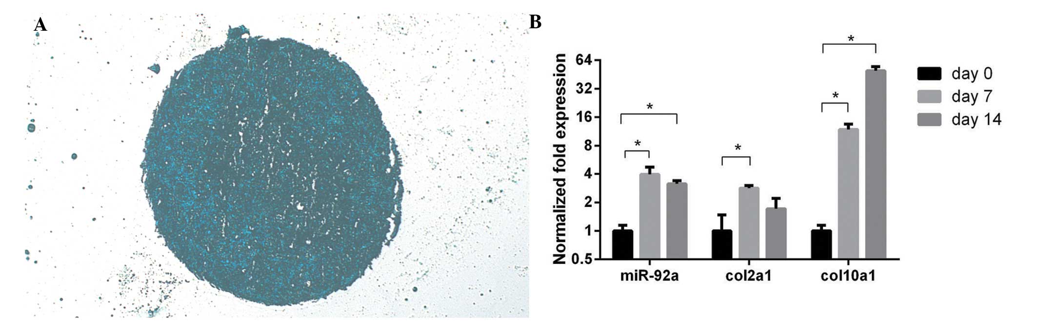
The micromass produced from the hADSCs in
chondrogenic medium was embedded in paraffin, cut into sections and
stained with alcian blue (Fig.
1A). The expression levels of miR-92a, col2a1 and col10a1
increased in the chondrogenic hADSCs cells (Fig. 1B). The expression levels of mir-92a
and the chondrogenic marker of col2a1 peaked at day 7 of
chondrogenic induction, and the hypertrophic marker of col10a1
peaked at day 14.
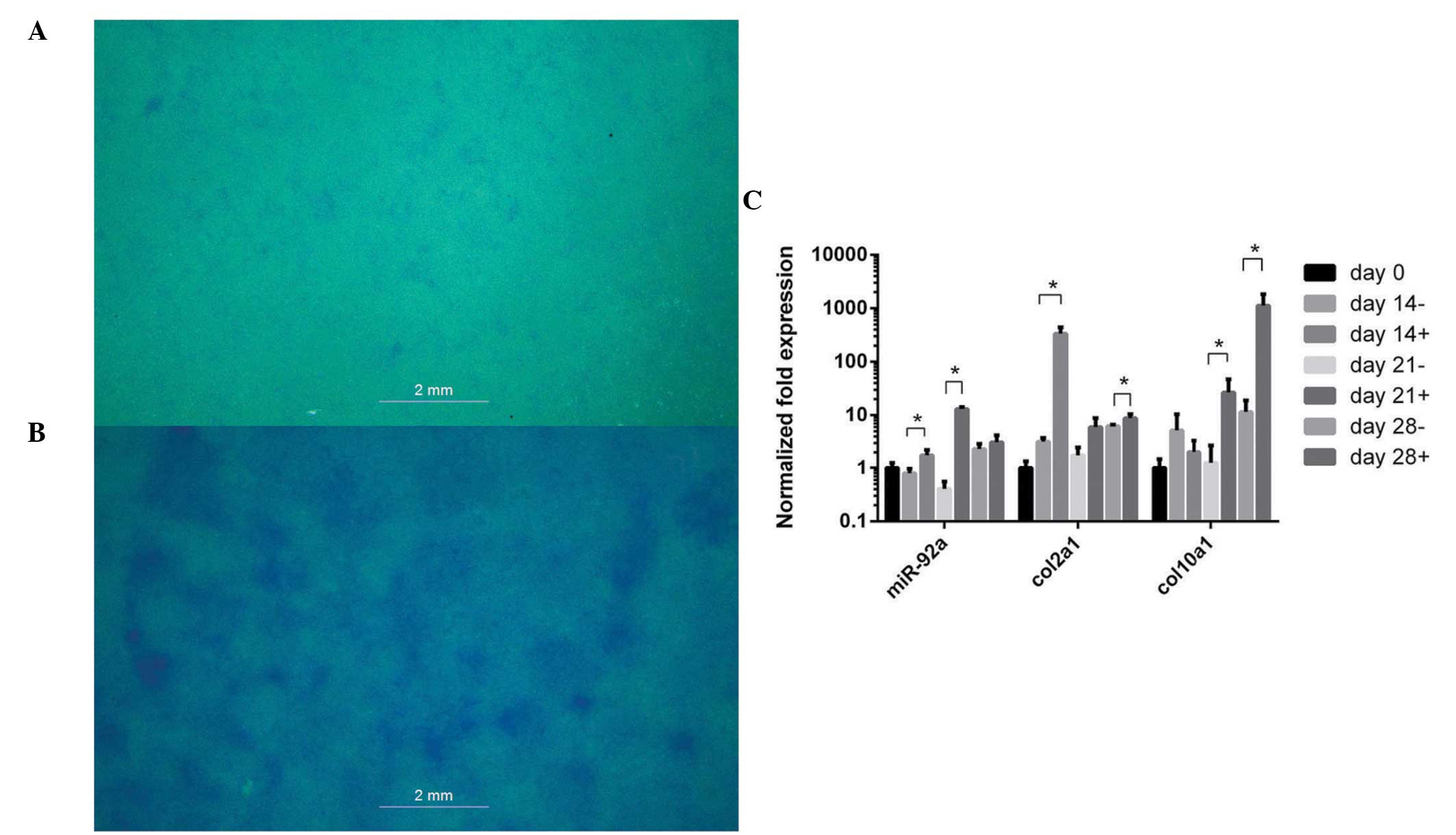
Following 14 days of chondrogenic differentiation
with ITS+ Premix, ATDC5 cells exhibited marked staining with alcian
blue, compared with the cells without ITS+ Premix (Fig. 2A and B). The expression of col2a1
peaked at day 14, col10a1 peaked at day 28 and miR-92a peaked at
day 21 (Fig. 2C).
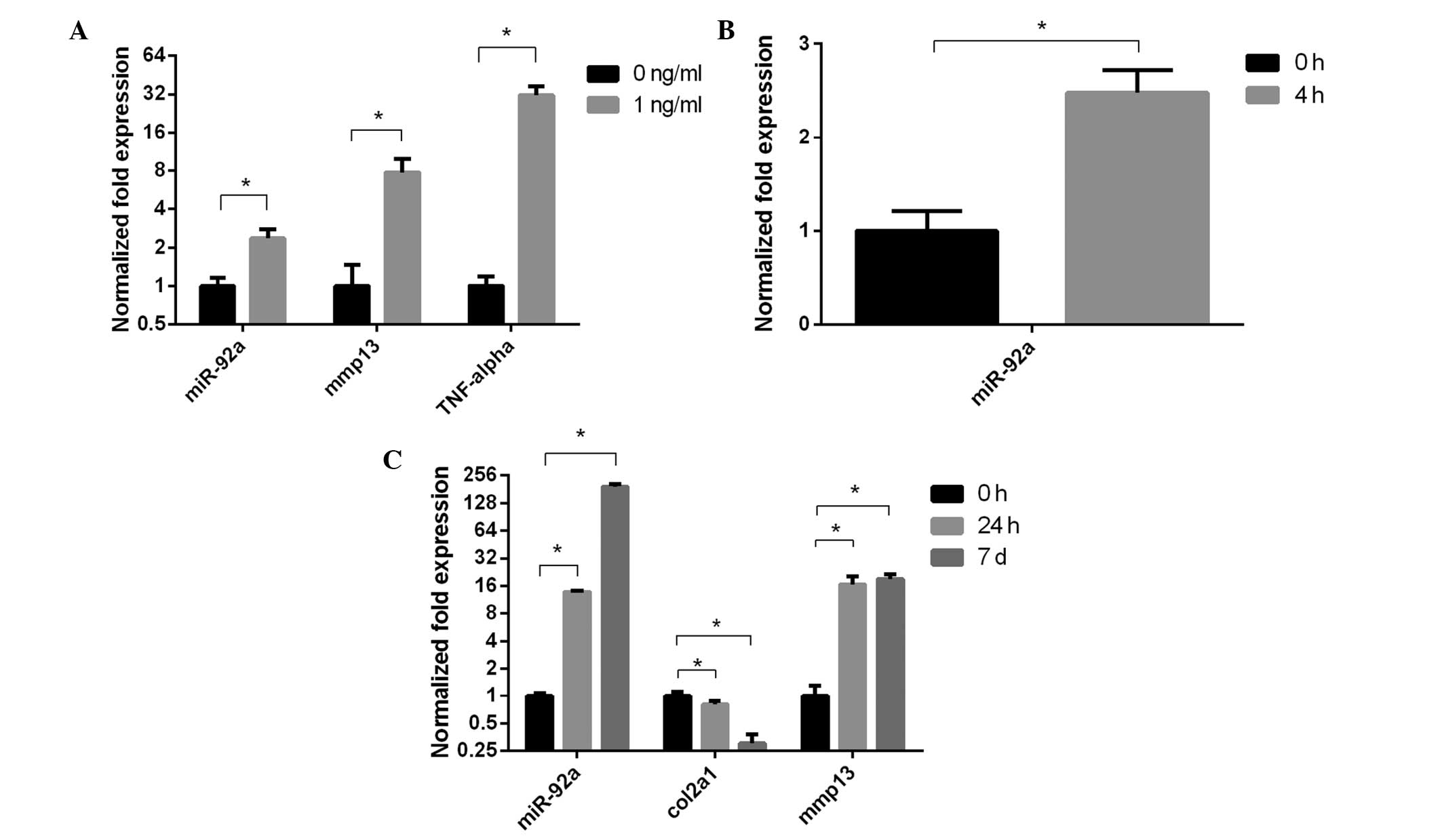
Expression of miR-92a is increased in
IL-1β-treated chondrocytes
Expression levels of miR-92a and mmp13 were
upregulated in the PHCs and PMCs treated with 1 ng/ml IL-1β for 4
h, compared with the control (Fig. 3A
and B). The expression levels of miR-92a and mmp13 were
elevated, and that of col2a1 was suppressed in a time-dependent
manner in the chondrogenic ATDC5 cells treated with 1 ng/ml IL-1β
(Fig. 3C).
Col9a2 and aggrecan may be regulated by
miR-92a
In order to investigate the effects of miR-92a on
chondrogenesis, the expression levels of miR-92a were manipulated
via transfection with a mimic or inhibitor. The altered expression
of miR-92a affected chondrogenic markers, increasing the expression
levels of col9a2 and aggrecan with the miR-92a mimic in a
dose-dependent manner, compared with the untransfected control. In
addition, the miR-193b-3p inhibitor reduced the expression levels
of col9a2 and aggrecan in a dose-dependent manner, compared with
the control (Table II).
| Table IIRelative mRNA expression levels in
ATDC5 cells transfected with miR-92a mimic or inhibitor. |
Table II
Relative mRNA expression levels in
ATDC5 cells transfected with miR-92a mimic or inhibitor.
| Gene | Mimic (50 nM)
| Mimic (100 nM)
| Inhibitor (50 nM)
| Inhibitor (100 nM)
|
|---|
| Fold change | P-value | SD | Fold change | P-value | SD | Fold change | P-value | SD | Fold change | P-value | SD |
|---|
| Col2a1 | 0.89 |
0.24 | 0.14 | 0.74 |
0.02 | 0.10 | 1.11 |
0.14 | 0.10 | 0.40 | <0.001 | 0.10 |
| Col10a1 | 1.10 |
0.17 | 0.06 | 1.11 |
0.12 | 0.01 | 1.36 | <0.001 | 0.12 | 0.75 | 0.001 | 0.05 |
| Comp | 0.61 | <0.001 | 0.07 | 1.24 |
0.001 | 0.07 | 0.91 |
0.014 | 0.06 | 0.24 | <0.001 | 0.04 |
| Agc | 3.52 |
0.001 | 0.62 | 6.89 | <0.001 | 0.76 | 0.56 | <0.001 | 0.01 | 0.26 | <0.001 | 0.01 |
| Mmp-13 | 1.94 |
0.004 | 0.47 | 3.03 | <0.001 | 0.14 | 1.00 |
0.994 | 0.03 | 0.67 | <0.001 | 0.02 |
| Col9a2 | 3.80 |
0.01 | 0.60 | 14.97 | <0.001 | 1.95 | 0.37 | <0.001 | 0.09 | 0.10 | <0.001 | 0.02 |
| Sox9 | 2.45 | <0.001 | 0.26 | 4.15 | <0.001 | 0.50 | 1.18 |
0.15 | 0.16 | 1.10 |
0.40 | 0.23 |
| Runx2 | 1.59 | <0.001 | 0.16 | 2.16 | <0.001 | 0.12 | 0.91 |
0.19 | 0.02 | 1.00 |
0.98 | 0.13 |
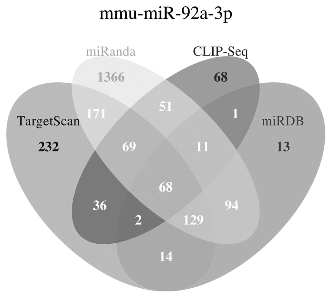
Predicted target genes, signaling
pathways and functions of miR-92a
The four algorithms, miRanda, miRDB, CLIP-Seq and
TargetScan, were used for prediction of the miR-92a target genes.
The general distribution of the predicted potential target genes is
shown in Fig. 4. A total of 279
genes were predicted by three or four algorithms and were
considered as potential target genes of miR-92a.
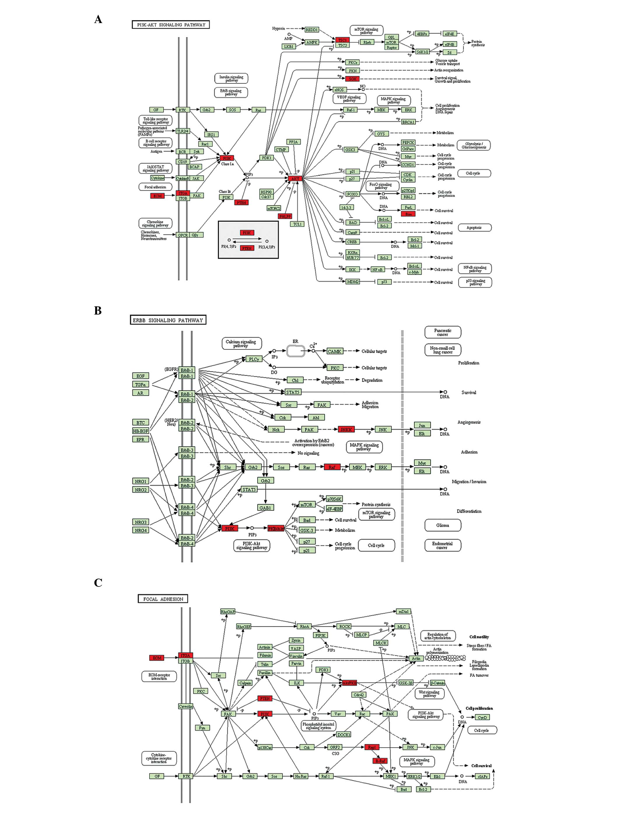
In addition to RT-qPCR, KEGG analysis was used to
investigate the signaling pathways by which miR-92a may regulate
chondrogenesis, based on the predicted potential target genes. The
potential target genes were clustered based on their involvement in
signaling pathways, and signaling pathways, which have been
previously identified to be involved in chondrogenesis or cartilage
degeneration were selected (5,22–25).
The P-value was determined based on the number of potential target
genes and the number of total genes in each signaling pathway. Of
all of the predicted signaling pathways, the PI3K-Akt (P=0.064),
ErbB (P=0.076) and focal adhesion kinase pathways (P=0.014),
ECM-receptor interaction (P=0.024) and the mTOR signaling pathway
(P=0.007) were identified as significant (Table III, Fig. 5).
| Table IIIPredicted signaling pathways, based
on the potential target genes of miR-92a. |
Table III
Predicted signaling pathways, based
on the potential target genes of miR-92a.
| Signaling
pathway | P-value | Predicted target
gene | Function in
chondrogenesis |
|---|
| PI3K-Akt | 0.064 | Sgk3, Phlpp2, Pten,
Pik3r3, Tsc1, Itga5, Itga6, Col1a2, Akt1, Bcl2l11and Itgav | Synergizing with
runx2 to enhance normal hypertrophic differentiation and
endochondral bone growth; promoting matrix synthesis and
chondrocyte survival in adult articular chondrocytes (22). |
| ErbB | 0.076 | Akt1, Pik3r3,
Map2k4 and Braf | Contributing to
expression of aggrecanases and matrix metalloproteinases, delayed
chondrogenesis and inhibition of the PI3K-Akt signaling pathway via
downstream MAPK activation (23). |
| Focal adhesion | 0.014 | Rap1b, Pten,
Pik3r3, Itga5, Itga6, Braf, Col1a2, Akt1 and Itgav | Inhibiting
chondrogenesis via expression of actin and activation of the
RhoA/ROCK pathway (24). |
| ECM-receptor
interaction | 0.024 | Col1a2, Sdc2,
Itga5, Itga6 and Itgav | Inhibiting
chondrogenesis via Itga5-mediated cellular-ECM interaction
(25). |
| mTOR | 0.007 | Pten, Tsc1, Pik3r3,
Braf and Akt1 | Reducing bone
growth and hypertrophy; enhancing insulin-like growth factor I
mediated proteoglycan synthesis in adult articular chondrocytes
(5). |
Discussion
Previous studies have suggested a role for miR-92a
in renal tumorigenesis via the gene expression of VHL (26), and in human acute promyelocytic
leukemia via the expression of p63 (27). An additional study identified the
positive effects of miR-92a on the proliferation, differentiation
and survival of chondrogenic progenitors via the targeting of nog3,
an inhibitor of the bone morphogenetic protein (BMP) signaling
pathway (28). Although miR-92a
was observed to contribute to chondrogenesis by enhancing the
expression of col2a1 in the study by Ning et al (28), no significant trend in the
expression of col2a1 was not observed in the present study
following transfection of either the miR-92a mimic or inhibitor.
This discrepancy may be due to differences in experimental subjects
and signaling pathways. In the study by Ning et al (28), the BMP signaling pathway
(smad1/5/8) and the inhibitor of BMP pathway (nog3) were observed
to mediate the effects of miR-92a on in vivo pharyngeal
chondrogenesis. In the present study, cultured ATDC5 cells were
used for the investigation of miR-92a and chondrogenesis, which are
associated with the autocrine transforming growth factor-β
(smad2/3) signaling pathway (29).
In the study by Ning et al (28), the morphological defects resulting
from the inhibition of nog3, one of the target genes of miR-92a's,
were partially reversed by p53 co-inhibition, suggesting a
contribution of miR-92a-nog3-apoptosis/proliferation to in
vivo morphological regulation of pharyngeal cartilage
formation. During chondrogenesis, high levels of type 9 collagen
and aggrecan are expressed, along with additional matrix proteins
to form the cartilage matrix, with col9a2 and aggrecan considered
as chondrogenic markers (30).
Col9a2 and aggrecan were previously demonstrated to be associated
with a number of diseases, including osteoarthritis (31,32),
degeneration of intervertebral discs (33,34)
and multiple epiphyseal dysplasia, characterized as the deformed
deposition of cartilage at the ends of the bones (32,35,36).
The present study hypothesized that col9a2 may be another mediator
of the degeneration of cartilage, followed by miR-92a knockdown.
For the upstream regulation of col9a2 and aggrecan, Sox9 has been
previously suggested as to be critical in initiating the expression
of col9a2 and aggrecan (37),
although multiple enhancers have been observed to initiate
expression of aggrecan (38).
However, more detailed information is required on the regulation of
the expression levels of col9a2 and aggrecan in order to identify
the cure for these diseases. In the present study, the results
indicated that miR-92a may contribute to the upregulation of col9a2
and aggrecan, without enhancing the expression of sox9. These
results provided novel insight into the upstream regulation of
col9a2 and aggrecan, beyond what is already known about sox9 in
relation to col9a2 and aggrecan. In addition, the results suggested
another possible mechanism of a miR-92a-col9a2-cartilage deformity
axis contributing to cartilage deformity following miR-92a
knockdown. Further investigations are required in order to verify
the effect of miR-92a on the in vivo expression levels of
col9a2, aggrecan, and cartilage degeneration, and to determine the
underlying mechanisms.
Several previous studies investigating miRNAs used
one or two algorithms to predict the target genes, with subsequent
mechanistic experiments, based on the predicted genes (39,40).
However, each of these widely used algorithms has an intrinsic
false positive rate. The false positive rate is 22–31% for
TargetScan, 24–39% for miRanda and ~30% for PicTar (41). In the present study, four
algorithms were used, and an intersection set of predicted genes
from at least three algorithms was identified as a potential target
gene. Based on the potential target genes, KEGG analysis was then
used to predict several signaling pathways that possibly contribute
to the effect of miR-92a on chondrogenesis. KEGG is a database,
which is usually used for the prediction of function and signaling
pathways from large scale molecular information of high-throughput
experiments, including sequencing. This prediction method enables
the minimization of false positive rates and assist in
understanding the possible function of miR-92a in a wider context
(42). Investigations of
underlying mechanisms can be performed using these predictions,
including luciferase reporter assays of miR-92a and 3′-UTR of
Akt1.
In conclusion, the present study demonstrated the
presence of miR-92a in chondrogenesis and the chondrocyte response
induced by IL-1β. The positive contribution of miR-92a in the
expression of col9a2 and aggrecan was observed and the PI3K-Akt,
ErbB and focal adhesion kinase pathways, ECM-receptor interaction,
and mTOR signaling pathway were indicated as potential mediators of
the effects of miR-92a on chondrogenesis and cartilage
degeneration.
Acknowledgments
The authors would like to thank to Dr Xuerong Li, Dr
Shan Li and Dr Shang Mei at the Department of Parasitology,
Zhongshan School of Medicine, Sun Yat-sen University (Guangzhou,
China) for their technical assistance.
The present study was supported by the National
Natural Science Foundation of China (grant nos. 81301558, 81371941
and 81171709), the Doctoral Scientific Fund Project of the Ministry
of Education of China (grant no. 20130171120074) and the Natural
Science Foundation of Guangdong Province, China (grant no.
s2013040016269). The sponsors had no involvement in the study
design; collection, analysis and interpretation of data; the
writing of the manuscript; or in the decision to submit the
manuscript for publication.
References
|
1
|
Li Y, Wei X, Zhou J and Wei L: The
age-related changes in cartilage and osteoarthritis. Biomed Res
Int. 2013:9165302013. View Article : Google Scholar : PubMed/NCBI
|
|
2
|
Mathieu PS and Loboa EG: Cytoskeletal and
focal adhesion influences on mesenchymal stem cell shape,
mechanical properties and differentiation down osteogenic,
adipogenic and chondrogenic pathways. Tissue Eng Part B Rev.
18:436–444. 2012. View Article : Google Scholar : PubMed/NCBI
|
|
3
|
Beier F and Loeser RF: Biology and
pathology of Rho GTPase, PI-3 kinase-Akt and MAP kinase signaling
pathways in chondrocytes. J Cell Biochem. 110:573–580. 2010.
View Article : Google Scholar : PubMed/NCBI
|
|
4
|
Chen J, Crawford R and Xiao Y: Vertical
inhibition of the PI3K/Akt/mTOR pathway for the treatment of
osteoarthritis. J Cell Biochem. 114:245–249. 2013. View Article : Google Scholar
|
|
5
|
Rokutanda S, Fujita T, Kanatani N, Yoshida
CA, Komori H, Liu W, Mizuno A and Komori T: Akt regulates skeletal
development through GSK3, mTOR and FoxOs. Dev Biol. 328:78–93.
2009. View Article : Google Scholar : PubMed/NCBI
|
|
6
|
Malemud CJ: Intracellular signaling
pathways in rheumatoid arthritis. J Clin Cell Immunol. 4:1602013.
View Article : Google Scholar
|
|
7
|
Xian CJ: Roles of epidermal growth factor
family in the regulation of postnatal somatic growth. Endocr Rev.
28:284–296. 2007. View Article : Google Scholar : PubMed/NCBI
|
|
8
|
Carthew RW and Sontheimer EJ: Origins and
Mechanisms of miRNAs and siRNAs. Cell. 136:642–655. 2009.
View Article : Google Scholar : PubMed/NCBI
|
|
9
|
Hong E and Reddi AH: MicroRNAs in
chondrogenesis, articular cartilage and osteoarthritis:
Implications for tissue engineering. Tissue Eng Part B Rev.
18:445–453. 2012. View Article : Google Scholar : PubMed/NCBI
|
|
10
|
Zhang Z, Kang Y, Zhang Z, Zhang H, Duan X,
Liu J, Li X and Liao W: Expression of microRNAs during
chondrogenesis of human adipose-derived stem cells. Osteoarthritis
Cartilage. 20:1638–1646. 2012. View Article : Google Scholar : PubMed/NCBI
|
|
11
|
Thirion S and Berenbaum F: Culture and
phenotyping of chondrocytes in primary culture. Methods Mol Med.
100:1–14. 2004.PubMed/NCBI
|
|
12
|
Zhang ZJ, Zhang H, Kang Y, Sheng PY, Ma
YC, Yang ZB, Zhang ZQ, Fu M, He AS, Liao WM, et al: miRNA
expression profile during osteogenic differentiation of human
adipose-derived stem cells. J Cell Biochem. 113:888–898. 2012.
View Article : Google Scholar : PubMed/NCBI
|
|
13
|
Zhang L, Su P, Xu C, Yang J, Yu W and
Huang D: Chondrogenic differentiation of human mesenchymal stem
cells: A comparison between micromass and pellet culture systems.
Biotechnol Lett. 32:1339–1346. 2010. View Article : Google Scholar : PubMed/NCBI
|
|
14
|
Estes BT, Diekman BO, Gimble JM and Guilak
F: Isolation of adipose-derived stem cells and their induction to a
chondrogenic phenotype. Nat Protoc. 5:1294–1311. 2010. View Article : Google Scholar : PubMed/NCBI
|
|
15
|
Yao Y and Wang Y: ATDC5: An excellent in
vitro model cell line for skeletal development. J Cell Biochem.
114:1223–1229. 2013. View Article : Google Scholar
|
|
16
|
Newton PT, Staines KA, Spevak L, Boskey
AL, Teixeira CC, Macrae VE, Canfield AE and Farquharson C:
Chondrogenic ATDC5 cells: An optimised model for rapid and
physiological matrix mineralisation. Int J Mol Med. 30:1187–1193.
2012.PubMed/NCBI
|
|
17
|
Atsumi T, Miwa Y, Kimata K and Ikawa Y: A
chondrogenic cell line derived from a differentiating culture of
AT805 teratocarcinoma cells. Cell Differ Dev. 30:109–116. 1990.
View Article : Google Scholar : PubMed/NCBI
|
|
18
|
Miyaki S, Nakasa T, Otsuki S, Grogan SP,
Higashiyama R, Inoue A, Kato Y, Sato T, Lotz MK, Asahara H, et al:
MicroRNA-140 is expressed in differentiated human articular
chondrocytes and modulates interleukin-1 responses. Arthritis
Rheum. 60:2723–2730. 2009. View Article : Google Scholar : PubMed/NCBI
|
|
19
|
Simsa-Maziel S and Monsonego-Ornan E:
Interleukin-1β promotes proliferation and inhibits differentiation
of chondrocytes through a mechanism involving down-regulation of
FGFR-3 and p21. Endocrinology. 153:2296–2310. 2012. View Article : Google Scholar : PubMed/NCBI
|
|
20
|
MacRae VE, Farquharson C and Ahmed SF: The
restricted potential for recovery of growth plate chondrogenesis
and longitudinal bone growth following exposure to pro-inflammatory
cytokines. J Endocrinol. 189:319–328. 2006. View Article : Google Scholar : PubMed/NCBI
|
|
21
|
Schmittgen TD and Livak KJ: Analyzing
real-time PCR data by the comparative C(T) method. Nat Protoc.
3:1101–1108. 2008. View Article : Google Scholar : PubMed/NCBI
|
|
22
|
Kita K, Kimura T, Nakamura N, Yoshikawa H
and Nakano T: PI3K/Akt signaling as a key regulatory pathway for
chondrocyte terminal differentiation. Genes Cells. 13:839–850.
2008. View Article : Google Scholar : PubMed/NCBI
|
|
23
|
Fisher MC, Clinton GM, Maihle NJ and Dealy
CN: Requirement for ErbB2/ErbB signaling in developing cartilage
and bone. Dev Growth Differ. 49:503–513. 2007. View Article : Google Scholar : PubMed/NCBI
|
|
24
|
Takahashi I, Onodera K, Sasano Y, et al:
Effect of stretching on gene expression of beta1 integrin and focal
adhesion kinase and on chondrogenesis through cell-extracellular
matrix interactions. Eur J Cell Biol. 82:182–192. 2003. View Article : Google Scholar : PubMed/NCBI
|
|
25
|
Knudson CB: Hyaluronan and CD44: strategic
players for cell-matrix interactions during chondrogenesis and
matrix assembly. Birth Defects Res C Embryo Today. 69:174–196.
2003. View Article : Google Scholar : PubMed/NCBI
|
|
26
|
Valera VA, Walter BA, Linehan WM and
Merino MJ: Regulatory Effects of microRNA-92 (miR-92) on VHL Gene
Expression and the Hypoxic Activation of miR-210 in clear cell
renal cell carcinoma. J Cancer. 2:515–526. 2011. View Article : Google Scholar : PubMed/NCBI
|
|
27
|
Sharifi M, Salehi R, Gheisari Y and Kazemi
M: Inhibition of microRNA miR-92a induces apoptosis and inhibits
cell proliferation in human acute promyelocytic leukemia through
modulation of p63 expression. Mol Biol Rep. 41:2799–2808. 2014.
View Article : Google Scholar : PubMed/NCBI
|
|
28
|
Ning G, Liu X, Dai M, Meng A and Wang Q:
MicroRNA-92a upholds Bmp signaling by targeting noggin3 during
pharyngeal cartilage formation. Dev Cell. 24:283–295. 2013.
View Article : Google Scholar : PubMed/NCBI
|
|
29
|
Kawai J, Akiyama H, Shigeno C, Ito H,
Konishi J and Nakamura T: Effects of transforming growth
factor-beta signaling on chondrogenesis in mouse chondrogenic EC
cells, ATDC5. Eur J Cell Biol. 78:707–714. 1999. View Article : Google Scholar : PubMed/NCBI
|
|
30
|
Okazaki K and Sandell LJ: Extracellular
matrix gene regulation. Clin Orthop Relat Res. 427(Suppl):
S123–S128. 2004. View Article : Google Scholar : PubMed/NCBI
|
|
31
|
Nakki A, Videman T, Kujala UM, Suhonen M,
Männikkö M, Peltonen L, Battié MC, Kaprio J and Saarela J:
Candidate gene association study of magnetic resonance
imaging-based hip osteoarthritis (OA): Evidence for COL9A2 gene as
a common predisposing factor for hip OA and lumbar disc
degeneration. J Rheumatol. 38:747–752. 2011. View Article : Google Scholar
|
|
32
|
Gleghorn L, Ramesar R, Beighton P and
Wallis G: A mutation in the variable repeat region of the aggrecan
gene (AGC1) causes a form of spondyloepiphyseal dysplasia
associated with severe, premature osteoarthritis. Am J Hum Genet.
77:484–490. 2005. View
Article : Google Scholar : PubMed/NCBI
|
|
33
|
Aladin DM, Cheung KM, Chan D, Yee AF, Jim
JJ, Luk KD and Lu WW: Expression of the Trp2 allele of COL9A2 is
associated with alterations in the mechanical properties of human
intervertebral discs. Spine (Phila Pa 1976). 32:2820–2826. 2007.
View Article : Google Scholar
|
|
34
|
Kim NK, Shin DA, Han IB, Yoo EH, Kim SH
and Chung SS: The association of aggrecan gene polymorphism with
the risk of intervertebral disc degeneration. Acta Neurochir
(Wien). 153:129–133. 2011. View Article : Google Scholar
|
|
35
|
Fiedler J, Stöve J, Heber F and Brenner
RE: Clinical phenotype and molecular diagnosis of multiple
epiphyseal dysplasia with relative hip sparing during childhood
(EDM2). Am J Med Genet. 112:144–153. 2002. View Article : Google Scholar : PubMed/NCBI
|
|
36
|
Briggs MD, Choi H, Warman ML, Loughlin JA,
Wordsworth P, Sykes BC, Irven CM, Smith M, Wynne-Davies R, Lipson
MH, et al: Genetic mapping of a locus for multiple epiphyseal
dysplasia (EDM2) to a region of chromosome 1 containing a type IX
collagen gene. Am J Hum Genet. 55:678–684. 1994.PubMed/NCBI
|
|
37
|
Bi W, Deng JM, Zhang Z, Behringer RR and
de Crombrugghe B: Sox9 is required for cartilage formation. Nat
Genet. 22:85–89. 1999. View
Article : Google Scholar : PubMed/NCBI
|
|
38
|
Hu G, Codina M and Fisher S: Multiple
enhancers associated with ACAN suggest highly redundant
transcriptional regulation in cartilage. Matrix Biol. 31:328–337.
2012. View Article : Google Scholar : PubMed/NCBI
|
|
39
|
Ge YZ, Xu LW, Xu Z, et al: Expression
Profiles and Clinical Significance of MicroRNAs in Papillary Renal
Cell Carcinoma: A STROBE-Compliant Observational Study. Medicine.
(Baltimore); 94. pp. e7672015, View Article : Google Scholar
|
|
40
|
Xie J, Tan ZH, Tang X, et al: MiR-374b-5p
suppresses RECK expression and promotes gastric cancer cell
invasion and metastasis. World J Gastroenterol. 20:17439–17447.
2014. View Article : Google Scholar : PubMed/NCBI
|
|
41
|
Bentwich I: Prediction and validation of
microRNAs and their targets. Febs Lett. 579:5904–5910. 2005.
View Article : Google Scholar : PubMed/NCBI
|
|
42
|
ElHefnawi M, Soliman B, Abu-Shahba N and
Amer M: An integrative meta-analysis of microRNAs in hepatocellular
carcinoma. Genomics Proteomics Bioinformatics. 11:354–367. 2013.
View Article : Google Scholar : PubMed/NCBI
|