Introduction
MicroRNAs (miRNAs/miRs) are a class of small RNAs
found in a diverse range of eukaryotes, including animals and
plants, as well as DNA viruses, which range in size between 19 and
24 nucleotides (nt) (1,2). miRNAs regulate mRNA or protein levels
either by promoting mRNA degradation or by attenuating protein
translation (3,4). As with mRNAs, certain miRNAs are
differentially expressed among tissues or developmental stages
(5). miRNAs are important in the
regulation of numerous cellular processes, including cell
proliferation and differentiation, apoptosis, development, tumor
metastasis and other biological processes (6,7).
miR-26b, at a length of 22 nt, is important in the regulation of
cell biology processes. With the development of novel molecular
biology techniques, the investigation of miR-26b has gained
specificity. A number of studies have indicated that miR-26b is
involved in early embryonic development, cell proliferation
regulation, hormone expression regulation in the anterior pituitary
and other physiological activities (8–11).
Therefore, miR-26b is essential for reproductive function. The
present study provides a basis for the further investigation into
the function of miR-26b in embryonic development, pituitary hormone
secretion and other reproductive processes.
Materials and methods
Sample collection
The present study enrolled Yanbian cattle reared
under the same conditions and sacrificed by the Haoyue Company,
three were six months old and three were 24 months old. The bovine
pituitary gland was quickly collected in a liquid nitrogen tank and
stored at −80°C prior to total RNA extraction. The animal slaughter
experiments were conducted in accordance with the guidelines of
Jilin University (Changchun, China) on the Review of Welfare and
Ethics of Laboratory Animals approved by the Jilin Province
Administration Office of Laboratory Animals. All animal procedures
were approved by Institutional Animal Care and Use Committee of
Jilin University (permit no. 20140204). All surgery was performed
under 50 mg/kg sodium pentobarbital (Sigma-Aldrich, St. Louis, MO,
USA) anesthesia, and all efforts were made to minimize
suffering.
Reagents
TRIzol reagent, pMD18-T vector, LA Taq DNA
polymerase, dNTP mixture, DNA marker, T4 DNA ligase, XhoI
and XbaI were purchased from Takara Bio Inc. (Otsu, Japan).
The ReverTra Ace qPCR RT kit and SYBR® Green Realtime
PCR master mix (QPK-201) were purchased from Toyobo Co., Ltd.
(Osaka, Japan) for reverse transcription quantitative polymerase
chain reaction (RT-qPCR). The endotoxin-free plasmid extraction kit
was purchased from Tiangen Biotech Co., Ltd. (Beijing, China). The
pmirGLO Dual-Luciferase miRNA Target Expression vector and
Dual-Luciferase® Reporter Assay system were purchased
from Promega Corporation (Madison, WI, USA). Dulbecco's modified
Eagle's medium: Nutrient mixture F-12 (DMEM/F12) was purchased from
Gibco-BRL (Invitrogen Life Technologies, Carlsbad, CA, USA). Fetal
bovine serum (FBS) was purchased from Hyclone (GE Healthcare,
Logan, UT, USA). miR-26b and the negative controls were synthesized
by Shanghai Jima International Trading Co., Ltd. (Shanghai, China).
The X-tremeGENE small interfering (si)RNA transfection reagent was
purchased from Roche Diagnostics (Basel, Switzerland).
Main instruments
A myECL gel imaging system was purchased from Alpha
Innotech (San Leandro, CA, USA). The Genesys 10 Nanodrop
spectrophotometer, Arktik PCR instrument, 37°C CO2
incubator, −80°C freezer and high-speed desktop centrifuge were
purchased from Thermo Fisher Scientific, Inc. (Waltham, MA, USA). A
PCR 5332 instrument, micropipettes and a Mastercycler Ep Realplex
qPCR instrument were purchased from Eppendorf (Hamburg, Germany).
The multifunctional microplate reader Mithras LB960 was purchased
from Berthold Technologies GmbH & Co. KG (Bad Wildbad,
Germany). A 37XB inverted microscope was purchased from Shanghai
Yiguang Optical Instrument Co., Ltd. (Shanghai, China).
Total RNA extraction of pituitary tissue
from Yanbian cattle
The pituitary tissue was removed from the liquid
nitrogen and placed in a mortar with 5 ml
diethylpyrocarbonate-treated water. TRIzol was added, grinding was
performed and total RNA was extracted. The total RNA was then
dissolved in 20.0 μl RNase-free water and stored at −80°C.
The RNA was separated by 1.0% agarose gel electrophoresis and the
blots were visualized under ultra violet light, following ethidium
bromide staining.
Specific identification of pituitary
tissues
Based on the mRNA sequence of the follicle
stimulating hormone (FSH) β gene in cattle available on GenBank
(http://www.ncbi.nlm.nih.gov/genbank/), the upstream
and the downstream primer sequences, with an annealing temperature
of 60°C, are presented in Table I.
The primers were synthesized by Huada Limited Co. (Shanghai,
China)
 | Table IReverse transcription-quantitative
polymerase chain reaction primer sequences. |
Table I
Reverse transcription-quantitative
polymerase chain reaction primer sequences.
| Name | Primer
sequence | Product length
(bp) |
|---|
| miR-26b | RT:
5′-GTCGTATCCAGTGCGTGTCGTGGAGCGCAATTGCACTGGATACGACAACCTAT-3′ | 67 |
| F:
5′-GGGGTTCAAGTAATTCAGG-3′ | |
| R:
5′-CAGTGCGTGTCGTGGAGT-3′ | |
| U6 | RT:
5′-CGCTTCACGAATTTGCGTGTCAT-3′ | 89 |
| F:
5′-GCTTCGGCAGCACATATACTAAAAT-3′ | |
| R:
5′-CGCTTCACGAATTTGCGTGTCAT-3′ | |
| EphA2 qPCR | F:
5′-CAACGACGACATCAAGAGGAT-3′ | 176 |
| R:
5′-CAGAAGGAAGCCAGTCCATTT-3′ | |
| GADPH | F:
5′-ATTCTGGCAAAGTGGACATCG-3′ | 214 |
| R:
5′-ACATACTCAGCACCAGCATCAC-3′ | |
| FSHβ | F:
5′-CAGCAAGGCCCAATATCCA-3′ | 817 |
| R:
5′-CCAGAGGAGGAGAGGCGAAACA-3′ | |
| EphA2 PCR | F:
5′-TTTCTCGAGGACAGCCCTCGCA-3′ | 725 |
| R:
5′-GCTCTAGAGGAATGCCCGAAGC-3′ | |
| Epha2 3′-UTR
plasmid site-detected primers | F:
5′-ATCTGCTGAGAATACTTTCCAAATGGACTGGCTTC-3′ | 713 |
| R:
5′-GAAGCCAGTCCATTTGGAAAGTATTCTCAGCAGAT-3′ | |
The ReverTra Ace qPCR RT kit was used according to
the manufacturer's instructions and a 10.0-μl RT system was
developed and optimized. cDNA was synthesized and stored at −20°C.
PCR was used for the amplification of FSHβ fragments. The cycling
conditions were as follows: Initial denaturation for 1 min at 95°C,
followed by 35 cycles of denaturation at 95°C for 35 sec, annealing
at 60°C for 35 sec and extension at 72°C for 35 sec. Final
extension was performed at 72°C for 5 min.
miR-26b-specific stem-loop RT primers and
qPCR primer design and cDNA synthesis via RT
Based on the mature sequence of miR-26b provided by
miRBase (http://www.mirbase.org/), miR-26b and
U6-specific stem-loop RT primers were designed and are shown in
Table I.
The ReverTra Ace qPCR RT kit was used according to
the manufacturer's instructions and the miR-26b and U6-specific RT
primers were used. cDNA was synthesized and stored at −20°C.
Based on the stem-loop primer and miR-26b sequences,
the quantitative primers were designed and the primer sequences are
shown in Table I.
miR-26b RT-qPCR detection in pituitary
tissues of cattle at different developmental stages
The Realplex RT quantitative PCR instrument was used
and a three-step method was adopted to perform the miR-26b
expression analysis. The qPCR reaction system was as follows:
Diethylpyrocarbonate-treated water (10.5 μl),
SYBR® Green Realtime PCR master mix (12.5 μl),
upstream primer (0.5 μl), downstream primer (0.5 μl)
and 10 μmol/l cDNA (1 μl) in a total volume of 25.0
μl. A total of three biological replicates were included for
each time period and three replicates were included in each sample.
The reaction conditions were as follows: 95°C denaturation for 1
min; 95°C denaturation for 15 sec, 68°C annealing for 15 sec and
72°C extension for 45 sec for 50 cycles. Subsequently, melting
curve analysis of the PCR products was performed to assess the
specificity of the PCR products.
miR-26b target gene prediction
According to the miRNA sequence provided by miRBase,
the two miRNA target gene databases TargetScan (http://www.targetscan.org/) and RNA22 (https://cm.jefferson.edu/rna22/) were used for
the prediction of the target genes of miR-26b (12). The binding model (complementary
region) was predicted using RNA mfold software (http://mfold.rna.albany.edu/?q=mfold).
Relative expression levels of candidate
target gene mRNA
Based on the mRNA sequence of EphA2 in
cattle, qPCR primers were designed. GAPDH was used as internal
reference and the specific sequences are shown in Table I. The primers were synthesized by
BGI (Shenzhen, China).
The EphA2 qPCR system was the same as that for
miR-26b. Subsequently, the results of the qPCR were analyzed.
Construction of a dual-luciferase
reporter gene vector of miR-26b of the target gene
In order to perform PCR amplification of the target
gene, a target gene 3′-untranslated region (3′-UTR) was designed
and synthesized, which covered a sequence of miRNA loci.
XhoI endonuclease was added in the 5′ region and XbaI
endonuclease was added in the 3′ region. The primers were
synthesized by BGI. The specific sequences are presented in
Table I. Following purification,
the PCR product was ligated into the vector pMD18-T and DH5α
competent cells were added and agitated for vector amplification
for 12 h. The bacterial liquid was then subjected to PCR
amplification. In addition, plasmid DNA was extracted for
XhoI and XbaI double digestion and purified gene
fragments were recovered. XhoI and XbaI double
digestion of pmirGLO Dual-Luciferase miRNA target expression vector
was then performed. The recovered target gene fragments were
ligated into digested vectors, which were transformed into
Escherichia (E.) coli, and monoclonal E.
coli was selected. The positive clones were extracted and sent
to BGI for sequencing. The correct plasmid was termed EphA2
3′-UTR pmirGLO.
Site-directed mutagenesis and
construction of miR-26b target gene dual-luciferase reporter
plasmid
The recombinant EphA2 3′-UTR pmirGLO plasmid
was used and three consecutive bases on the binding sites of the
miR-26b and EphA2 3′-UTR were mutated with a site-directed
mutagenesis kit. The primer sequences used in site-directed
mutagenesis are presented in Table
I.
The PCR amplification reaction system consisted of
34.0 μl nuclease-free water, 5.0 μl 10X reaction
buffer, 5.0 μl mutated template plasmid, 4.0 μl
desoxyribonucleotide triphosphate mix (2.5 mM/l) and 1.0 μl
up- and downstream primer (10 μM) each (total volume, 49.0
μl). 1.0 μl Pyrococcus furiosus DNA polymerase
(10 μmol/l) was added to achieve a total volume of 50.0
μl. The reaction conditions were as follows: Initial
denaturation for 1 min at 95°C, followed by 18 cycles of
denaturation at 95°C for 40 sec, annealing at 60°C for 1 min and
extension at 68°C for 8 min. Final extension was performed at 72°C
for 10 min. 1.0 μl Dpn I was added directly to the PCR
products, and following mixing, they were incubated at 37°C for 1
h.
A total of 10 μl of the above digested
products were added to 100 μl competent cells (E.
coli; DH5α, 100 μl, 5×10⁷/ml), and
transformation and cloning were performed for plasmid extraction.
XhoI and XbaI restriction enzymes were used to
perform double digestion site-directed mutagenesis of the
recombinant plasmid. Following electrophoresis, positive test
plasmids were sent to BGI for sequencing. The correct plasmid was
termed EphA2 3′-UTR pmirGLO-MUT.
Cell culture and luciferase activity
detection
HeLa cells were cultured in DMEM/F12 medium
containing 5% FBS at 37°C and 5% CO2. HeLa cells (Saiqi
Cellbio Co., Ltd., Shanghai, China) in the logarithmic phase were
seeded into a 96-well cell culture plate. After 24 h, when adherent
cells had reached 70% confluence, X-tremeGENE siRNA transfection
reagents were used for transfection. The miR-26b mimics and
EphA2 3′-UTR pmirGLO plasmid containing the 3′-UTR or the
EphA2 3′-UTR pmirGLO-MUT plasmid were co-transfected into
HeLa cells. The negative control group was set to co-transfect the
miR-26b control and the EphA2 3′-UTR pmirGLO plasmid or
EphA2 3′-UTR pmirGLO-MUT plasmid. A total of three
replicates were performed for each combination. After 24 h, the
HeLa cell culture DMEM/F12 medium containing fresh 5% FBS was
replaced. After 48 h, the Dual-Luciferase® reporter
assay system was used to perform the reporter gene assay, according
to the manufacturer's instructions. After 48 h, the cells were
lysed and the multifunctional microplate reader Mithras LB960 was
used to detect the fluorescence intensity. All experiments were
repeated three times.
Statistical analysis
The data were expressed as the mean ± standard
deviation. Differences were assessed by Student's two-tailed t-test
using the SPSS software, version 13.0 (SPSS, Inc., Chicago, IL,
USA). P<0.05 was considered to indicate a statistically
significant difference.
Results
Total RNA extraction
Total RNA in pituitary tissue of Yanbian cattle was
extracted and the electrophoretic analysis is shown in Fig. 1. The optical density at 260 nm
(OD260)/OD280 ratio of the total RNA concentration was 1.8–2.0,
indicating that the integrity of the RNA was good and had not been
contaminated with genetic material, proteins or other impurities.
It was therefore deemed suitable for use in subsequent
experiments.
Identification of pituitary tissue
After the total RNA was reverse transcribed into
cDNA, FSHβ gene primers were used for PCR amplification and the PCR
electrophoresis results are shown in Fig. 2. They were amplified in order to
confirm that specific bands were present, which were consistent in
size with the designed amplified fragments. No apparent dimers or
non-specific bands were observed, suggesting that the pituitary
tissue samples were accurately identified, and were therefore
deemed suitable for use in subsequent experiments.
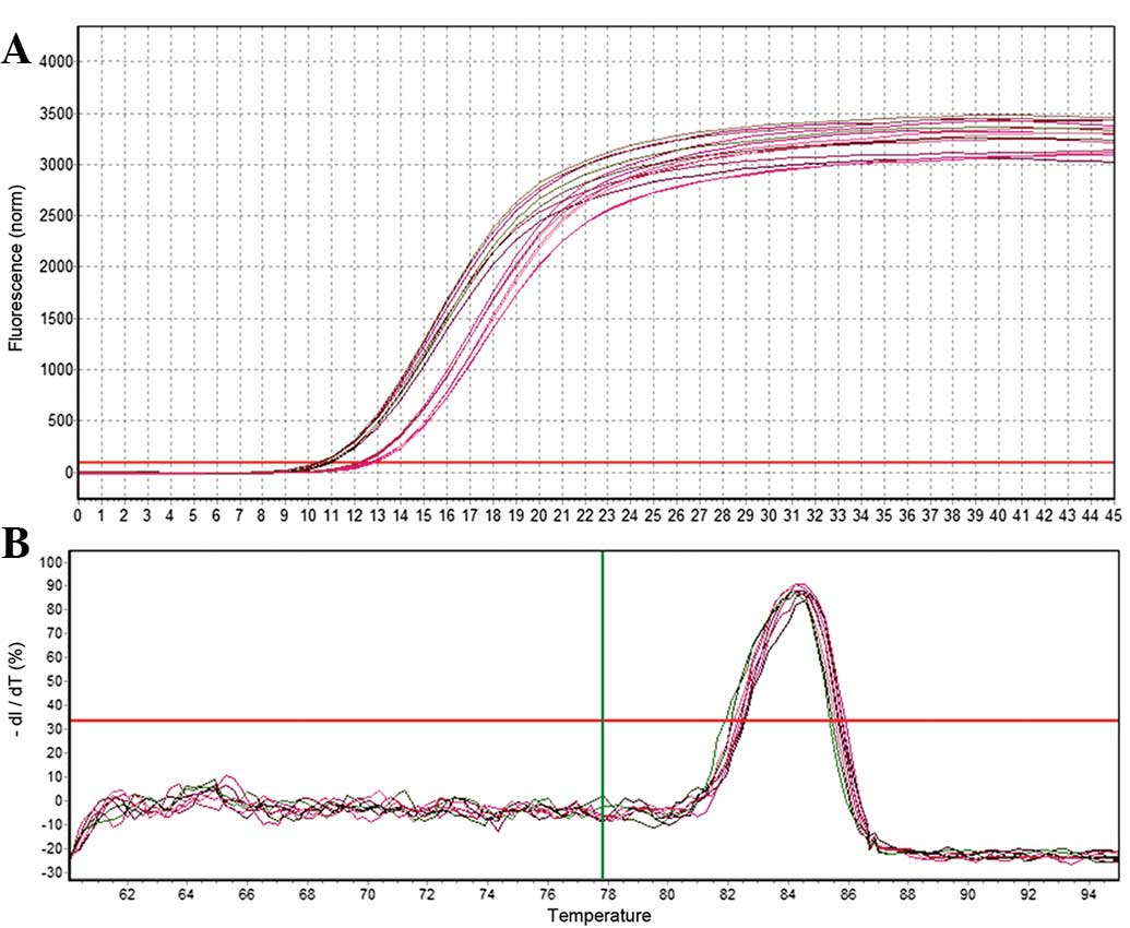
miR-26b RT-qPCR
RT-qPCR was used to analyze the relative expression
of mature miR-26b. A total of three samples were assessed at each
developmental stage and three replicates were performed for each
sample. U6 was selected as a reference gene for normalization of
RT-qPCR reactions and samples. The 2−ΔΔCt method
(13) was used to calculate the
relative expression levels of miR-26b. As shown in Figs. 3 and 4, the U6 and miR-26b samples exhibited a
good amplification curve and the melting curve suggested that the
designed primers had a high specificity.
Relative expression levels of miR-26b in
pituitary tissues of Yanbian cattle at different developmental
stages
RT-qPCR analysis was used to detect the relative
expression of miR-26b in the pituitary tissue of Yanbian cattle at
six and 24 months of age. The results revealed that miR-26b was
expressed in the pituitary tissue of Yanbian cattle at different
developmental stages, revealing significant differences
(P<0.01). The relative expression levels in 24-month-old cattle
were 2.41 times greater than those in the six-month-old cattle, as
shown in Fig. 5.
Predicted target genes of miR-26b
Using TargetScan, 561 predicted target genes were
identified in the prediction database of the miRNA target gene. The
target genes associated with reproductive traits were selected as
the alternative target gene and the RNA22 software was used for
further analysis. The genes predicted by the two software programs
were identified. Following the gene function analysis, EphA2
was selected as the candidate target gene. The 3′-UTR of the
EphA2 gene had a conserved seed sequence at the binding site
of miR-26b. The predicted results of TargetScan and RNA22 are shown
in Fig. 6. The mfold program was
used to predict the seed region complementary of EphA2 mRNA
and miR-26b, and predicted the secondary structure, as shown in
Fig. 7.
RT-qPCR assessment of candidate target
genes of EphA2
RT-qPCR was used to analyze the relative expression
of the candidate target gene of EphA2. A total of three
samples were assessed at each developmental stage and three
replicates were performed for each sample. GAPDH was
selected as a reference gene to allow for differences between
RT-qPCR reactions and samples. The 2−ΔΔCt method was
used to calculate the relative expression of the candidate target
gene of EphA2. As shown in Figs. 8 and 9, GAPDH and the candidate target
gene EphA2 exhibited a good amplification curve and melting
curve, suggesting that the designed primers had a high
specificity.
Relative expression levels of EphA2 in
pituitary tissues of Yanbian cattle at different developmental
stages
RT-qPCR was used to detect the relative expression
of EphA2 in the pituitary tissue of Yanbian cattle at six or
24 months of age. The results revealed that EphA2 expression
was significantly differentially in the pituitary tissue of Yanbian
cattle at different developmental stages (P<0.01). The relative
expression levels in 24-month-old cattle was 3.34 times greater
than that in six-month-old cattle, as shown in Fig. 10.
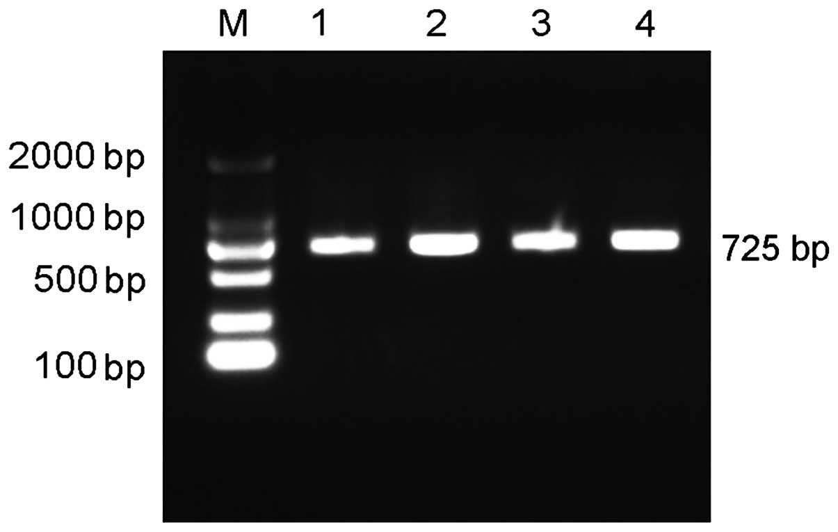
Recombinant plasmid construction
PCR amplification of target fragments of
EphA2 was performed and PCR products were obtained and
detected using agarose gel electrophoresis. As no non-specific
bands and dimers were identified, the fragments were deemed
suitable for the plasmid construction of the dual-luciferase
reporter gene. The PCR results are shown in Fig. 11.
For the construction and identification of miR-26b
candidate target gene dual-luciferase reporter gene vectors and
mutated vectors, the target gene was connected with the vector and
E. coli was transformed to obtain the recombinant plasmid.
PCR and restriction enzyme digestion were used to identify the
positive clones and the bands with a consistent size with the
target fragment are shown in Fig.
12. The recombinant plasmids were sequenced and confirmed by
BGI to be identical with the 3′-UTR amplified sequences available
in the GenBank database.
The XhoI and XbaI restriction enzymes
were used to perform double digestion of the recombinant plasmid
EphA2 3′-UTR pmirGLO-MUT. The recombinant plasmid
EphA2 3′-UTR pmirGLO-MUT cleavage results revealed bands of
the same size as the fragment, as shown in Fig. 13. The recombinant plasmid and
sequencing results matched the expected results.
EphA2 identification of the miR-26b
target gene
miR-26b mimics and negative control, as well as
EphA2 3′-UTR pmirGLO were co-transfected into HeLa cells.
Interaction of miR-26b with the candidate target gene EphA2
was indicated by a decrease in luciferase expression of the cells.
The luciferase expression was significantly decreased in the
miR-26b-transfected group compared with that in the control group
(P<0.01), as shown in Fig. 14.
This suggested that miR-26b may exert its functions through
interacting with the 3′-UTR of EphA2.
Interaction site recognition of the
target gene EphA2 and miR-26b
The dual-luciferase gene reporter assay demonstrated
that the seed region of miR-26b specifically binds with the
mutation in the 3′-UTR of candidate target gene, causing miR-26b
inhibition of the luciferase activity to decrease 60% (100–40%;
Fig. 14) and 26% (100–74%;
Fig. 15) in the wild-type. This
suggested that the predicted binding sites were miR-26b activation
sites. The cell luciferase relative expression results following
transfection are shown in Fig.
15. The results suggested that the miR-26b seed sequence
AUGAACUU exerted a function via binding with the TACTTGAA sequence
of the 3′-UTR in EphA2. Therefore, it was hypothesized that
the TACTTGAA sequence in the 3′-UTR of EphA2 was the target
sequence of miR-26b.
Discussion
miR-26b is important in the regulation of a number
of biological processes in cells (14). It is involved in the regulation of
cell proliferation and the control of cell differentiation
(15). With the advancement of
molecular biology techniques in recent years, investigations into
miR-26b have become increasingly in-depth and specific. A previous
study using a miRNA chip combined with qPCR techniques found that
the placental expression of miR-26b in women with severe
pre-eclampsia was different from that in normal pregnant women
(16). In additional studies, it
was hypothesized that miR-26b was likely to be involved in the
differentiation of embryonic stem cells and the regulation of
nervous system development via inhibiting the expression of
KLF4 and MYCBP (14–17).
Subsequently, a study on pituitary and non-pituitary cell lines
identified that miR-26b is able to target the 3′-UTR of
lef-1, thereby inhibiting the expression of lef-1. In
the GH3 cell line, the expression of lef-1 was inhibited while
simultaneously promoting the expression of Pit-1 and
GH (12). Numerous studies
have revealed that miR-26b is involved in early embryonic
development, cell proliferation regulation and pituitary hormone
secretion, and among other physical activities, it is an miRNA
associated with the function of propagation (18–20).
Therefore, the present study investigated the
differences in the expression of miR-26b in the pituitary tissue of
Yanbian cattle, predicted target genes of miR-26b and selected
reproductive trait-associated candidate target genes for
identification. In the present study, the RT-qPCR method was used
to detect the relative expression levels of miR-26b in the
pituitary tissues of Yanbian cattle at different developmental
stages. The 2−ΔΔCt calculation method was used to
perform the relative quantitative analysis. The results revealed
that miR-26b was expressed in the pituitary tissue of six- and
24-month-old Yanbian cattle, and the relative expression levels of
miR-26b exhibited significant differences between the pituitary
tissues at the different developmental stages. The relative
expression of miR-26b in the pituitary tissues of 24-month-old
Yanbian cattle was 2.41 times that of six-month-old Yanbian cattle.
As studies have revealed that miR-26b is capable of affecting the
secretion of pituitary hormones in mice (18,21–23)
and the relative expression of miR-26b revealed significant
differences in the pituitary tissue of six- and 24-month-old
Yanbian cattle in the present study, it was hypothesized that it
may be associated with the development of the pituitary gland.
The TargetScan and RNA22 prediction databases were
used to analyze and predict the miR-26b sequence provided on
miRBase. It was identified that the TACTTGAA sequence in the 3′-UTR
of EphA2 and miR-26b seed sequence AUGAACUU had conserved
binding sites, suggesting that miR-26b was a target gene.
EphA2 is a member of the receptor tyrosine kinase family.
The Eph receptor and ligand-mediated signal transduction are
involved in numerous physical processes, including nerve signal
transduction, angiogenesis and regulation of the adhesion state
among cells (24–26). The present study found that
EphA2 was widely expressed in human epithelial cells, and
after binding to its ligand, the signaling pathway may be
activated, regulating cell differentiation and adhesion, inhibiting
vascular endothelial growth factor and epithelial growth factor and
inducing cell proliferation (27);
therefore, it may be involved in embryo implantation (28). The RT-qPCR results revealed that in
the pituitary tissue of six-and and 24-month-old Yanbian cattle,
the relative expression of miR-26b exhibited the opposite
expression pattern and the relative expression of miR-26b in the
pituitary tissue of 24 month-old Yanbian cattle was 2.41 times that
of the six-month-old Yanbian cattle. The relative expression of
candidate target gene EphA2 in the pituitary tissue of
six-month-old Yanbian cattle was 3.34 times that of 24-month-old
Yanbian cattle. The results provided a theoretical basis for the
EphA2 gene being a target gene of miR-26b. However, there
are miR-26b binding sites in the EphA2 mRNA 3′-UTR of
bovine, human, murine and other mammalian genomes, suggesting that
the EphA2 gene may be a target gene of miR-26b.
The luciferase reporter system assay revealed that
miR-26b is able to suppress EphA2 expression at the gene
level. The 3′-UTR reporter plasmid of mutant candidate target gene
EphA2 termed EphA2 3′-UTR pmirGLO-MUT and miR-26b
mimics were co-transfected into HeLa cells. A dual-luciferase gene
detection reporter assay revealed that there were three mutations
in the binding sites of the miR-26b seed region and the 3′-UTR of
the candidate target gene, EphA2. It may be the cause of the
decrease in the miR-26b inhibition of luciferase activity from 60%
in the wild-type to 26%, suggesting that miR-26b exerted its
effects via binding with the 3′-UTR in EphA2. It also
revealed that the TACTTGAA sequence in the 3′-UTR of EphA2
was the binding site of miR-26b. miR-26b is able to inhibit
EphA2 mRNA and protein expression in human glioma cells, as
it was previously demonstrated that EphA2 was the direct
target gene of miR-26b in glioma cells (29); these results were consistent with
those of the present study.
In conclusion, the results of the present study have
confirmed in vitro that EphA2 is a target gene of
miR-26b in Yanbian cattle. However, the function, regulatory
mechanism and impact of miR-26b on reproductive traits, including
early embryonic development and pituitary hormone secretion,
remains to be further investigated.
Acknowledgments
The current study was supported by the National Key
Technology R&D Program (grant nos. 2015BAI07B02 and
20150101102JC), the National Beef Cattle Industrial System of the
China Agriculture Research System (grant no. 38) and the National
High-tech R&D Program (grant no. 2013AA102505).
References
|
1
|
Lee RC, Feinbaum RL and Ambros V: The C.
elegans heterochronic gene lin-4 encodes small RNAs with antisense
complementarity to lin-14. Cell. 75:843–854. 1993. View Article : Google Scholar : PubMed/NCBI
|
|
2
|
Pillai RS, Artus CG and Filipowicz W:
Tethering of human Ago proteins to mRNA mimics the miRNA-mediated
repression of protein synthesis. RNA. 10:1518–1525. 2004.
View Article : Google Scholar : PubMed/NCBI
|
|
3
|
Beilharz TH, Humphreys DT and Preiss T:
miRNA Effects on mRNA closed-loop formation during translation
initiation. Prog Mol Subcell Biol. 50:99–112. 2010. View Article : Google Scholar
|
|
4
|
Hausser J, Landthaler M, Jaskiewicz L,
Gaidatzis D and Zavolan M: Relative contribution of sequence and
structure features to the mRNA binding of Argonaute/EIF2C-miRNA
complexes and the degradation of miRNA targets. Genome Res.
19:2009–2020. 2009. View Article : Google Scholar : PubMed/NCBI
|
|
5
|
Trakooljul N, Hicks JA and Liu HC:
Identification of target genes and pathways associated with chicken
microRNA miR-143. Anim Genet. 41:357–364. 2010.PubMed/NCBI
|
|
6
|
Bizuayehu TT, Fernandes JM, Johansen SD
and Babiak I: Characterization of novel precursor miRNAs using next
generation sequencing and prediction of miRNA targets in Atlantic
halibut. PLoS One. 8:e613782013. View Article : Google Scholar : PubMed/NCBI
|
|
7
|
Barros-Carvalho GA, Paschoal AR,
Marcelino-Guimarães FC and Hungria M: Prediction of potential novel
microRNAs in soybean when in symbiosis. Genet Mol Res.
13:8519–8529. 2014. View Article : Google Scholar : PubMed/NCBI
|
|
8
|
Verghese ET, Drury R, Green CA, Holliday
DL, Lu X, Nash C, Speirs V, Thorne JL, Thygesen HH, Zougman A, Hull
MA, et al: MiR-26b is down-regulated in carcinoma-associated
fibroblasts from ER-positive breast cancers leading to enhanced
cell migration and invasion. J Pathol. 231:388–399. 2013.
View Article : Google Scholar : PubMed/NCBI
|
|
9
|
Trompeter HI, Dreesen J, Hermann E,
Iwaniuk KM, Hafner M, Renwick N, Tuschl T and Wernet P: MicroRNAs
miR-26a, miR-26b, and miR-29b accelerate osteogenic differentiation
of unrestricted somatic stem cells from human cord blood. BMC
Genomics. 14:1112013. View Article : Google Scholar : PubMed/NCBI
|
|
10
|
Alijani S, Alizadeh S, Kazemi A, Khatib
ZK, Soleimani M, Rezvani M, Minayi N, Karami F and Tayebi B:
Evaluation of the effect of miR-26b up-regulation on HbF expression
in erythroleukemic K-562 cell line. Avicenna J Med Biotechnol.
6:53–56. 2014.PubMed/NCBI
|
|
11
|
Lin J, Zhang L, Huang H, Huang Y, Huang L,
Wang J, Huang S, He L, Zhou Y and Jia W: MiR-26b/KPNA2 axis
inhibits epithelial ovarian carcinoma proliferation and metastasis
through downregulating OCT4. Oncotarget. 2015. View Article : Google Scholar
|
|
12
|
Miranda KC, Huynh T, Tay Y, Ang YS, Tam
WL, Thomson AM, Lim B and Rigoutsos I: A pattern-based method for
the identification of MicroRNA binding sites and their
corresponding heteroduplexes. Cell. 126:1203–1217. 2006. View Article : Google Scholar : PubMed/NCBI
|
|
13
|
Schmittgen TD, Zakrajsek BA, Mills AG, et
al: Quantitative reverse transcription-polymerase chain reaction to
study mRNA decay: comparison of end-point and real-time methods.
Anal Biochem. 285:194–204. 2000. View Article : Google Scholar : PubMed/NCBI
|
|
14
|
Hu Y, Li P, Hao S, et al: Differential
expression of microRNAs in the placentae of Chinese patients with
severe pre-eclampsia. Clin Chem Lab Med. 47:923–929. 2009.
View Article : Google Scholar : PubMed/NCBI
|
|
15
|
Ji Y, He Y, Liu L and Chong X: MiRNA-26b
regulates the expression of cyclooxygenase-2 in
desferrioxamine-treated CNE cells. FEBS Lett. 584:961–967. 2010.
View Article : Google Scholar : PubMed/NCBI
|
|
16
|
Chen H, Qian K, Tang ZP, et al:
Bioinformatics and microarray analysis of microRNA expression
profiles of murine embryonic stem cells, neural stem cells induced
from ESCs and isolated from E8.5 mouse neural tube. Neurol Res.
32:603–613. 2010. View Article : Google Scholar
|
|
17
|
Zhang Z, Florez S, Gutierrez-Hartmann A,
et al: MicroRNAs regulate pituitary development and microRNA 26b
specifically targets lymphoid enhancer factor 1 (Lef-1), which
modulates pituitary transcription factor 1 (Pit-1) expression. J
Biol Chem. 285:34718–34728. 2010. View Article : Google Scholar : PubMed/NCBI
|
|
18
|
Zhang Z, Kim K, Li X, Moreno M, Sharp T,
Goodheart MJ, Safe S, Dupuy AJ and Amendt BA: MicroRNA-26b
represses colon cancer cell proliferation by inhibiting lymphoid
enhancer factor 1 expression. Mol Cancer Ther. 13:1942–1951. 2014.
View Article : Google Scholar : PubMed/NCBI
|
|
19
|
Palagani A, Op De, Beeck K, Naulaerts S,
Diddens J, Sekhar Chirumamilla C, Van Camp G, Laukens K, Heyninck
K, Gerlo S, Mestdagh P, et al: Ectopic microRNA-150-5p
transcription sensitizes glucocorticoid therapy response in MM1S
multiple myeloma cells but fails to overcome hormone therapy
resistance in MM1R cells. PLoS One. 9:e1138422014. View Article : Google Scholar : PubMed/NCBI
|
|
20
|
Xu G, Shi C, Ji C, Song G, Chen L, Yang L,
Zhao Y and Guo X: Expression of microRNA-26b, an obesity-related
microRNA, is regulated by free fatty acids, glucose, dexamethasone
and growth hormone in human adipocytes. Mol Med Rep. 10:223–228.
2014.PubMed/NCBI
|
|
21
|
Palumbo T, Faucz FR, Azevedo M, Xekouki P,
Iliopoulos D and Stratakis CA: Functional screen analysis reveals
miR-26b and miR-128 as central regulators of pituitary
somatomammotrophic tumor growth through activation of the PTEN-AKT
pathway. Oncogene. 32:1651–1659. 2013. View Article : Google Scholar
|
|
22
|
Song G, Xu G, Ji C, Shi C, Shen Y, Chen L,
Zhu L, Yang L, Zhao Y and Guo X: The role of microRNA-26b in human
adipocyte differentiation and prowliferation. Gene. 533:481–487.
2014. View Article : Google Scholar
|
|
23
|
Xu G, Ji C, Song G, Shi C, Shen Y, Chen L,
Yang L, Zhao Y and Guo X: Obesity associated microRNA26b regulates
the proliferation of human preadipocytes via arrest of the G1/S
transition. Mol Med Rep. 12:3648–3654. 2015.PubMed/NCBI
|
|
24
|
Kinch MS and Carles-Kinch K:
Overexpression and functional alterations of the EphA2 tyrosine
kinase in cancer. Clin Exp Metastasis. 20:59–68. 2003. View Article : Google Scholar : PubMed/NCBI
|
|
25
|
Dodelet VC and Pasquale EB: Eph receptors
and ephrin ligands: embryogenesis to tumorigenesis. Oncogene.
19:5614–5619. 2000. View Article : Google Scholar : PubMed/NCBI
|
|
26
|
Rosenbegr IM, Göke M, Kanai M, et al:
Epithelial cell kinase-B61: an autocrine loop modulating intestinal
epithelial migration and barrier function. Am J Physiol.
273:G824–G832. 1997.
|
|
27
|
Miao H, Wei BR, Peehl DM, et al:
Activation of EphA receptor tyrosine kinase inhibits the Ras/MAPK
pathway. Nat Cell Biol. 3:527–530. 2001. View Article : Google Scholar : PubMed/NCBI
|
|
28
|
Fujii H, Tatsumi K, Kosaka K, et al:
EPh-ephrin A system regulates murine blastocyst attachment and
sreading. Dev Dyn. 235:3250–3258. 2006. View Article : Google Scholar : PubMed/NCBI
|
|
29
|
Wu N, Zhao X, Liu M, et al: Role of
microRNA-26b in glioma development and its mediated regulation on
EphA2. PLoS One. 6:e162642011. View Article : Google Scholar : PubMed/NCBI
|