Introduction
Ovarian cancer is the most life-threatening
gynecologic malignancy, accounting for a mortality rate of ~140,200
in 2008 worldwide (1). Despite
significant advances in surgical techniques and chemotherapy
regimens, the 5-year survival rate for patients with the advanced
disease is ~13%, primarily due to recurrence and drug resistance
(2). In general, the majority of
patients with ovarian cancer initially respond to surgery and
standard chemotherapy; however, the majority ultimately develop
recurrence, and resistance to chemotherapeutic drugs is frequently
observed in recurrent disease (3).
Therefore, understanding the molecular mechanisms involved in
chemotherapy resistance is urgently required to develop novel
therapeutic strategies and improve clinical outcomes of this
life-threatening malignancy.
CLN3 is a recently identified defective gene in
juvenile Batten disease, an inherited neurodegenerative disease of
childhood resulting from accelerated apoptotic death of
photoreceptors and neurons (4).
The CLN3 gene encodes a hydrophobic transmembrane protein, which is
involved in intracellular trafficking and regulation in neuronal
and non-neuronal cells (5–8). Previous studies have indicated that
CLN3 has anti-apoptotic properties in NT2 neuronal precursor cells
and cancer cells (9,10). In particular, Rylova et al
reported that CLN3 mRNA and protein are abundantly expressed in
various cancer cell lines, including glioblastoma, neuroblastoma,
and prostate, ovarian, breast and colon cancer (11,12).
In addition, a series of functional investigations have revealed
that the knockdown of CLN3 by RNA interference (RNAi) inhibits the
proliferation and/or induces apoptosis in several cancer cells
(11,12). CLN3 has, therefore, been indicated
as a potential molecular target for future cancer drug discovery
(11). However, the potential role
of CLN3 in ovarian cancer remains to be fully elucidated.
In the present study, an RNAi-based approach,
specifically targeting CLN3 mRNA, was used to investigate the
effects of CLN3 knockdown on the proliferation, apoptosis and
chemosensitivity of the A2780 human ovarian cancer cell line, and
cells of its cisplatin-resistant (A2780/DDP) and
carboplatin-resistant (A2780/CBP) sublines.
Materials and methods
Cell culture
Human ovarian carcinoma SW626, OVCaR-3 and SK-OV-3
cells were cultured in Dulbecco's modified Eagle's medium (DMEM;
Gibco Life Technologies, Grand Island, NY, USA) supplemented with
10% fetal bovine serum (FBS; GE Healthcare, Logan, UT, USA). Human
ovarian carcinoma HO-8910, COC1, HO-8910PM cells were grown in RPMI
1640 medium (Gibco Life Technologies) containing 10% fetal calf
serum (GE Healthcare). Human ovarian carcinoma ES-2 cells were
maintained in McCoy's 5A medium (Gibco Life Technologies)
containing 10% FBS. All cells were cultured at 37°C in a 5%
CO2 atmosphere. The A2780 human ovarian carcinoma cell
line, and its cisplatin-resistant (A2780/DDP) and
carboplatin-resistant (A2780/CBP) sub-lines were obtained from
KeyGen Biotech, Co., Ltd. (Nanjing, China) and cultured in DMEM,
containing 10% FBS, at 37°C in a 5% CO2 atmosphere. The
resistant sub-lines were established by exposure of the A2780 cells
to stepwise increasing cisplatin or carboplatin (Qilu
Pharmaceutical Co., Ltd., Jinan, China) concentrations,
respectively. To ensure maintenance of the resistant phenotype, the
culture medium of the A2780/DDP and A2780/CBP cells were treated
with 1 mg/l cisplatin and 0.1 µmol/l carboplatin,
respectively. The cisplatin and carboplatin were removed from the
growth medium 2 weeks prior to all subsequent experiments. Normal
ovarian tissues were provided by the Department of Gynaecology and
Obstetrics, The Fourth Affiliated Hospital of Harbin Medical
University (Harbin, China) and the Department of Gynaecology, The
Second Affiliated Hospital of Harbin Medical University (Harbin,
China) following surgery or a laparoscopy. All patients provided
written informed consent for the use of their tissues, and the use
of tissue specimens was approved by the Ethics Committee of Harbin
Medical University. Following primary culture, the cells were grown
in DMEM supplemented with 10% FBS.
Construction and transfection of the CLN3
shRNA lentiviral vector
Oligonucleotides corresponding to CLN3 mRNA or
control mRNA were synthesized by Sangon Biotech Co., Ltd.
(Shanghai, China) as follows: CLN3, sense 5′-CCU UGG UCG UAG UUU
ACU UTT-3′ and antisense 5′-AAG UAA ACU ACG ACC AAG GTT-3′; and
control, sense 5′-UUC UCC GAA CGU GUC ACG UTT-3 and antisense
5′-ACG UGA CAC GUU CGG AGA ATT-3′. The annealed double-stranded
oligonucleotides were cloned into the pGCSIL-green fluorescent
protein (GFP) viral vector. The recombinant plasmid was verified
using PCR and DNA sequencing. Recombinant lentiviral particles were
prepared via the co-transfection of subconfluent 293T cells with
the modified pGCSIL-GFP viral vector and the pHelper 1.0 and
pHelper 2.0 helper plasmids using the Lipofectamine™ 2000 reagent
(Invitrogen Life Technologies, Carlsbad, CA, USA). For lentiviral
transfection, the cells were seeded at a density of
5×10⁴ into six-well plates and transduced with the
modified pGCSIL-GFP (KeyGen Biotech Co., Ltd.) viral vector when
they reached 30–40% confluence. At 3 days post-transfection, the
expression of GFP was examined under a fluorescence microscope, and
the cells were harvested for the subsequent assays.
Reverse transcription-quantitative
polymerase chain reaction (RT-q)PCR
Total RNA was extracted from the cultured cells
using TRIzol reagent (Invitrogen Life Technologies) and
reverse-transcribed to synthesize cDNA using a Maxima®
First Strand cDNA Synthesis kit (Thermo Fisher Scientific,
Rockford, IL, USA), according to the manufacturers' instructions.
Subsequently, qPCR reactions were performed using SYBR Green
Real-time PCR Master Mix (Toboyo, Co., Ltd., Osaka, Japan) with 2
µg total RNA on an Applied Biosystems Step One Plus
Real-Time PCR system (Applied Biosystems Life Technologies, Foster
City, CA, USA). Following 15 sec at 95°C to activate DNA
polymerase, the cycling parameters were as follows: 40 cycles of
denaturation at 95°C for 5 sec, and annealing and extension at 60°C
for 30 sec. The relative mRNA expression levels were calculated
from the threshold cycle (Ct) value of each PCR product and
normalized to β-actin using the comparative Ct method. The primer
sets used for qPCR were as follows: CLN3, forward
5-′GGTGGACAGTATTCAAGGG-3′ and reverse: 5′-CTTGGCAGAAAGACGAAC-3′;
Bcl-2, forward 5′-ACGACTTCTCCCGCCGCTAC-3′ and reverse
5′-CTGAAGAGCTCCTCCACCAC-3′; caspase-3, forward
5′-CATGGAAGCGAATCAATGGACT-3′ and reverse
5′-CTGTACCAGACCGAGATGTCA-3′; caspase-8, forward
5′-CTCCAAATGCAAACTGGATG-3′ and reverse 5′-TGTTGATTTGGGCACAGACT-3′;
and β-actin, forward 5′-GGCGGCACCACCATGTACCCT-3′ and reverse 5′-AGG
GGC CGG ACT CGT CAT ACT-3′. Primers were synthesized by Sangon
Biotech Co., Ltd.
Western blot analysis
The cells were washed twice with ice-cold
phosphate-buffered saline (PBS) and lysed in lysis buffer (KeyGen
Biotech Co., Ltd.) for 30 min. Following centrifugation at 8,000 ×
g for 5 min at 4°C, the soluble proteins were boiled at 95–100°C
for 5 min, separated by 10% sodium dodecyl sulfate polyacrylamide
gel electrophoresis (KeyGen Biotech Co., Ltd.) at a 70 V constant
voltage, and transferred electrophoretically onto polyvinylidene
difluoride membranes (EMD Millipore, Bedford, MA, USA). The
membranes were blocked with 5% non-fat milk at room temperature for
1.5 h and immunoblotted at 37°C for 1.5 h with the following
antibodies: Polyclonal goat anti-human CLN3 (cat. no. sc-49627;
1:200; Santa Cruz Biotechnology, Inc., Santa Cruz, CA, USA),
poly-clonal mouse anti-human cleaved caspase-8 (cat. no. sc-81656;
1:200; Santa Cruz Biotechnology, Inc., Santa Cruz, CA, USA),
polyclonal rabbit anti-human Bcl-2 (cat. no. KG22169; 1:500; KeyGen
Biotech Co., Ltd.), polyclonal rabbit anti-human Bax (cat. no.
KG22165; 1:500; KeyGen Biotech Co., Ltd.), polyclonal rabbit
anti-human cleaved PARP (cat. no. YC0073; 1:1,000; ImmunoWay
Biotechnology, Newark, DE, USA) and poly-clonal rabbit anti-human
cleaved caspase-3 (cat. no. KG22205; 1:200; KeyGen Biotech Co.,
Ltd.), followed by incubation with horseradish peroxidase-linked
secondary antibody (1:10,000; KeyGen Biotech Co., Ltd.) at 37°C for
40 min. Finally, the protein bands were visualized using an
enhanced chemiluminescence detection system (KeyGen Biotech Co.,
Ltd.).
Cell proliferation assay
Cell proliferation was analyzed using a Cell
Counting Kit-8 (CCK-8; KeyGen Biotech Co., Ltd.), according to the
manufacturer014's instructions. Briefly, the cells were seeded into
96-well plates at a density of 2×10² cells/well and
cultured for 0, 12, 24, 48 or 72 h. Subsequently, 10 µl
CCK-8 reagent was added to each well, and the cells were incubated
at 37°C for 2 h. Finally, the absorbance at 450 nm was determined
using a microplate reader (Bio-Rad Laboratories, Inc., San Diego,
CA, USA).
Apoptosis and cell cycle analysis using
flow cytometry
For cell cycle analysis, the cells were washed twice
with PBS, fixed overnight in 70% ethanol, and stained with
propidium iodide (PI; KeyGen Biotech Co., Ltd.) in the presence of
RNase A (Takara Biotechnology Co., Ltd., Dalian, China) at 4°C for
30 min. The DNA contents were determined using a FACSCalibur flow
cytometer (BD Biosciences, San Jose, CA, USA).
The number of apoptotic cells were quantified by
flow cytometry using an Annexin V-FITC/PI Apoptosis Detection kit
(KeyGen Biotech Co., Ltd.). In brief, the cells were washed with
PBS and incubated with 500 µl binding buffer (KeyGen Biotech
Co., Ltd.). Subsequently, 5 µl fluorescein isothiocya-nate
(FITC)-labeled Annexin V and 5 µl PI were added, and the
samples were incubated at room temperature for 5–15 min in the
dark. The percentage of apoptotic cells was determined using flow
cytometry.
Cytotoxicity assay
In vitro cytotoxicity was evaluated using a
CCK-8 assay. Briefly, the cells were trypsinized and plated in
96-well plates at a density of 2×10² cells/well.
Following treatment with different concentrations of cisplatin or
carboplatin, the CCK-8 assay was performed, as described above. The
half maximum inhibitory concentration (IC50) values were
calculated by linear interpolation with SPSS 19.0 software (SPSS
Inc., Chicago, IL, USA).
Statistical analysis
The results are expressed as the mean ± standard
deviation, and raw data were analyzed using Student's t-test with
SPSS 19.0 software. P<0.05 was considered to indicate a
statistically significant difference.
Results
Overexpression of CLN3 in ovarian cancer
cells
Firstly, the mRNA levels of CLN3 in normal ovarian
cells and A2780, COC1, ES-2, HO-8910, HO-8910PM, OVCaR-3, SK-OV-3,
SW626 ovarian cancer cell lines were detected using RT-qPCR.
Compared with the normal ovarian cells, significantly higher levels
of CLN3 were observed in the A2780, COC1, HO-8910, OVCaR-3, SK-OV-3
and SW626 cells, but not in the ES-2 and HO-8910PM cells. Among
these, the SW626 and A2780 exhibited the highest endogenous mRNA
levels of CLN3 (Fig. 1). These
data suggested a possible role of CLN3 in the development and
progression of ovarian cancer.
Knockdown of CLN3 by shRNA in the A2780
cell line and its drug-resistant sub-lines
To investigate whether CLN3 is required for the
maintenance of drug-resistance in ovarian cancer cells, modified
lentiviral pGCSIL-GFP vectors expressing CLN3 or control shRNA were
transduced into the A2780 cell line and its cisplatin-resistant
(A2780/DDP) and carboplatin-resistant (A2780/CBP) sub-lines.
RT-qPCR revealed that there was ~70% decrease in the mRNA level of
CLN3 in the cells transduced with the vector expressing CLN3 shRNA,
compared with that in the cells transduced with the control vector.
No significant difference in the mRNA expression of CLN3 was
observed between the control vector-transduced cells and
untransduced cells (Fig. 2A;
P<0.05). The knockdown of CLN3 was further confirmed using
western blot analysis, which demonstrated a decrease of ~66% in the
protein level of CLN3, compared with the control cells (Fig. 2B; P<0.05). These results
revealed that CLN3-specific shRNA effectively knocked down the
expression of CLN3 in the ovarian cancer cells at the mRNA and
protein levels.
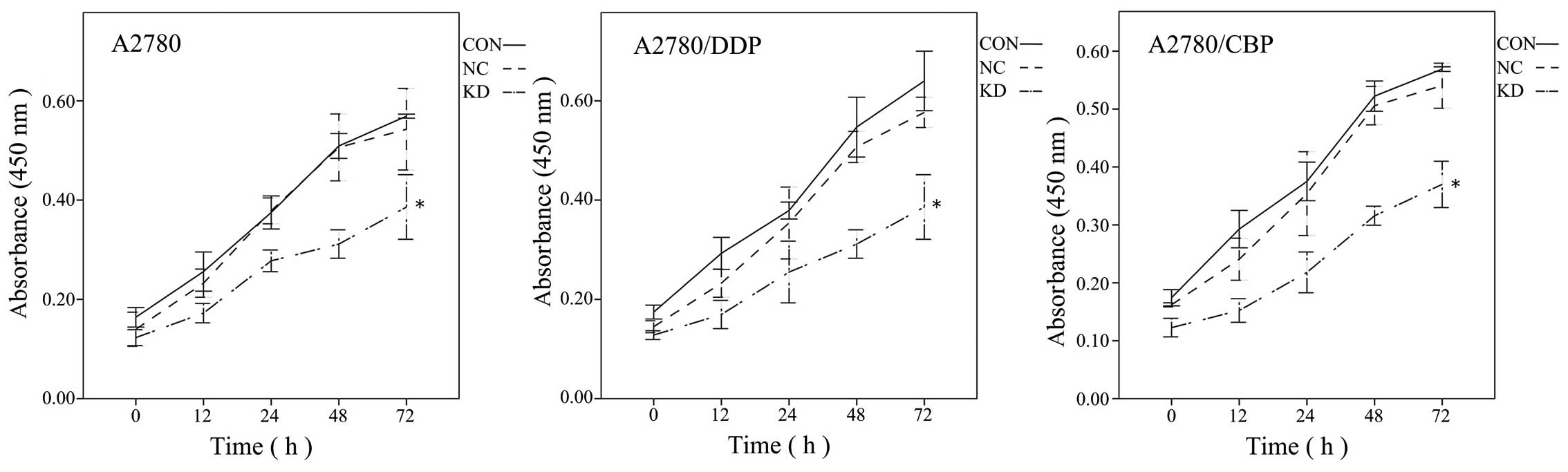
CLN3 knockdown inhibits cell
proliferation and induces G0/G1 cell cycle arrest in the A2780 cell
line and its drug-resistant sub-lines
To evaluate the effects of CLN3 knockdown on ovarian
cancer cell proliferation, a CCK-8 assay was performed with the
modified lentiviral pGCSIL-GFP vector transduced A2780, A2780/DDP
and A2780/CBP cells. As shown in Fig.
3, CLN3 shRNA transfection in the parental A2780 cells
significantly decreased cell viability on days 2 and 3, compared
with the control vector-transduced cells (P<0.05). Similar
results were observed in the drug-resistant A2780/DDP and A2780/CBP
cells. As cell cycle progression is a critical determinant of cell
growth, further cell cycle analysis was performed using flow
cytometry. The results demonstrated that CLN3 knockdown in the
A2780, A2780/DDP and A2780/CBP cells significantly increased the
percentage of G0/G1 phase cells, with a concomitant fall in the
percentage of S phase cells (P<0.05; Fig. 4). These data suggested that growth
inhibition in the CLN3-knockdown drug-resistant ovarian cancer
cells may have been due to the arrest at the G0/G1 transition of
the cell cycle.
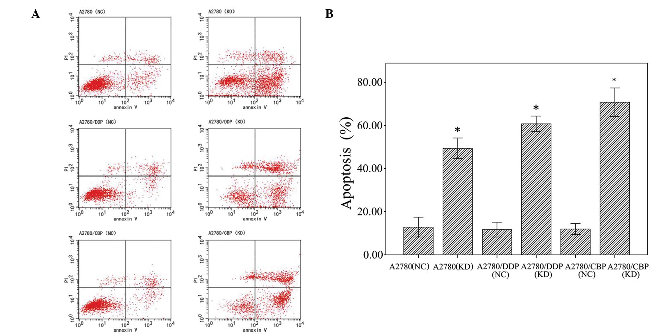
CLN3 knockdown induces cell apoptosis in
the A2780 cell line and its drug-resistant sub-lines
Flow cytometry was used to determine the effects of
CLN3 knockdown on apoptosis in the A2780, A2780/DDP and A2780/CBP
cells. As shown in Fig. 5, CLN3
knockdown significantly increased (35.55%) the percentage of
apoptotic cells in the A2780 cells, compared with the control
cells. Similarly, apoptotic cells were significantly increased by
transfection with the vector expressing CLN3 shRNA in the A2780/DDP
cells, between 11.26±1.05 and 59.17±2.25%, and the A2780/CBP cells,
between 13.18±1.50 and 72.11±2.65%.
CLN3 knockdown enhances the
chemosensitivity of ovarian cancer cells
To assess whether knockdown of CLN3 in ovarian
cancer cells affects their chemosensitivity, the CLN3 shRNA- and
control shRNA-transduced cells were treated with different
concentrations of cisplatin or carboplatin, and a CCK-8 assay was
used to determine cell viability. Compared with cisplatin treatment
alone, combined CLN3 shRNA and cisplatin treatment caused a
significant decrease in the IC50 value of the A2780/DDP
cells, between 0.469±0.009 and 0.196±0.01 µg/ml. Similarly,
in the A2780/CBP cells, the IC50 in the control shRNA
cells was 0.657±0.05 µg/ml, and decreased significantly to
0.343±0.042 µg/ml in CLN3 shRNA cells (P<0.05).
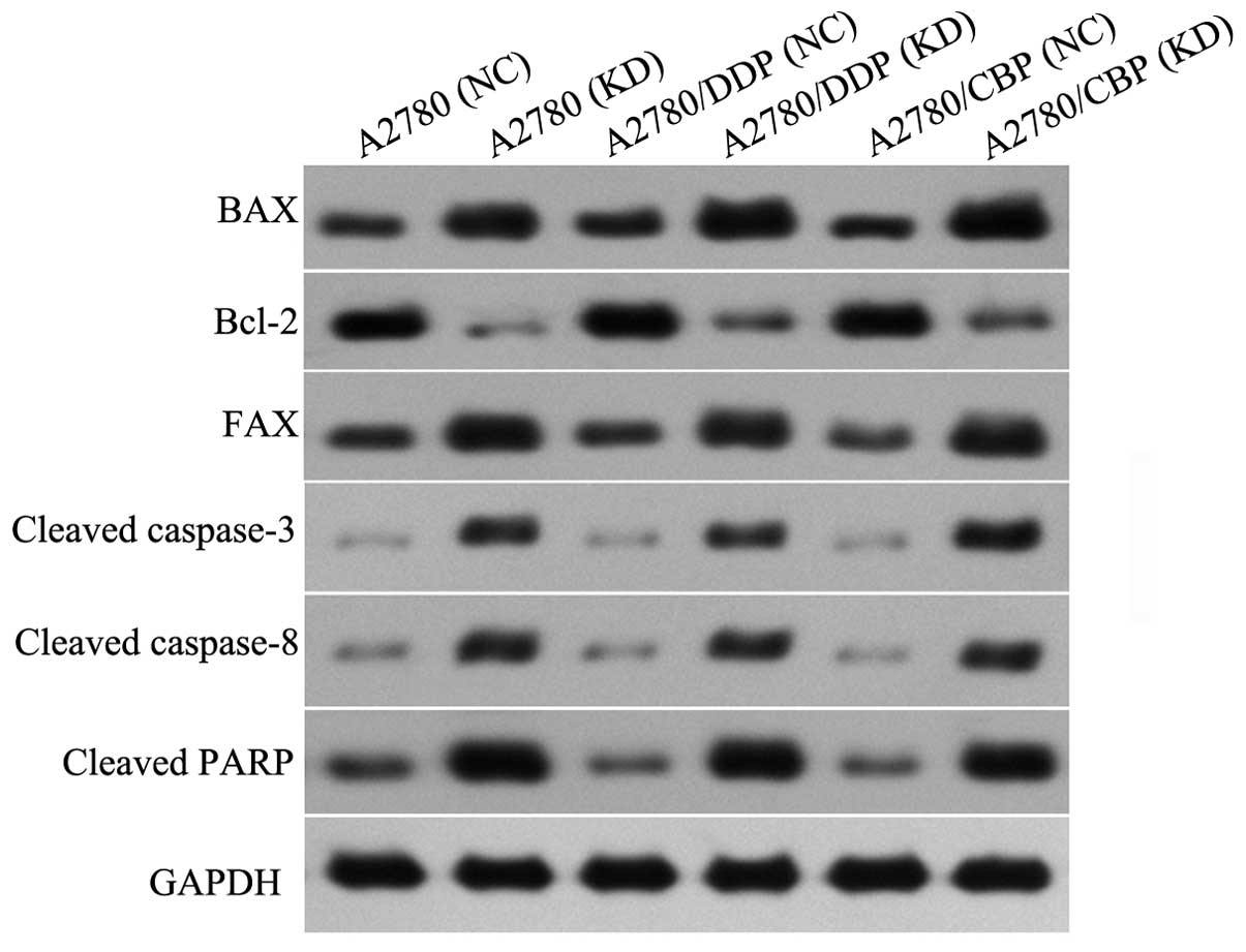
CLN3 knockdown affects
apoptosis-associated gene expression in the A2780 cell line and its
drug-resistant sub-lines
The mRNA and protein expression levels of several
apoptosis-associated factors in the A2780, A2780/DDP and A2780/CBP
cells were analyzed using RT-PCR and western blot analysis,
respectively. Compared with the control cells, the mRNA expression
levels of Bax and FAX were significantly increased, however, the
mRNA expression level of Bcl-2 was markedly reduced in the CLN3
shRNA-transduced cells (P<0.05; Fig. 6). The changes in these
apoptosis-associated factors observed using western blot analysis
were consistent with the results of the RT-qPCR (P<0.05;
Fig. 7). Additionally, the protein
levels of cleaved-caspase 3, cleaved-caspase 8 and cleaved-RARP
were significantly increased in the CLN3 shRNA transduced cells,
compared with the control cells (P<0.05; Fig. 6).
Discussion
Previous evidence indicates that CLN3 is highly
expressed in ovarian cancer cells, however, the potential role of
CLN3 in ovarian cancer cell growth and drug toxicity remains to be
elucidated (11). In the present
study, the expression of CLN3 was examined in the SW626, SK-OV-3,
HO-8910, ES-2, COC1, A2780, OVCaR-3, HO-8910PM human ovarian cancer
cell lines. Compared with normal ovarian cells, the SW626, SK-OV-3,
HO-8910, COC1, A2780 and OVCaR-3 cells exhibited markedly higher
expression levels of CLN3, suggesting a possible role of CLN3 in
the development and progression of ovarian cancer. To further
characterize the role of CLN3 in the chemoresistance of ovarian
cancer, a lentivirus-based RNAi strategy was employed to
transiently inhibit the endogenous expression of CLN3 in ovarian
cancer A2780 cells and their drug-resistant A2780/DDP and A2780/CBP
sub-lines. RT-qPCR and western blot analysis confirmed that the
vector expressing CLN3 shRNA was able to effectively eliminate the
expression of CLN3 in these cells. Further experiments demonstrated
that CLN3 knockdown in the ovarian cancer cells significantly
inhibited cell proliferation. In addition, flow cyto-metric
analysis revealed that CLN3 knockdown in the A2780, A2780/DDP and
A2780/CBP cells significantly increased the percentage of cells in
the G0/G1 phase, with a concomitant fall in the percentage of cells
in the S phase, indicating G0/G1 cell cycle arrest. These data are
consistent with a previous study by Zhu et al, which
reported that CLN3 silencing induces G0/G1 cell cycle arrest in
HCT116 colorectal cancer cells (12). CLN3 has been previously shown to be
an anti-apoptotic gene in NT2 neuronal precursor cells and several
types of cancer (9,10,13).
The present study demonstrated that reduced expression of CLN3
significantly induced apoptotic cell death in the A2780, A2780/DDP
and A2780/CBP cells. In addition, CLN3 knockdown caused increases
in the levels of Bax, FAX, cleaved-caspase 3, cleaved-caspase 8 and
cleaved-RARP, and a decrease in the level of Bcl-2. Concordant with
these results, Zhu et al reported that inhibiting of the
expression of CLN3 promotes cell apoptosis in HCT116 cells
(12). The findings of the present
study, together with previous data, provide further support for
CLN3 as an anti-apoptotic gene in cancer cells. However, the
precise mechanisms by which knockdown of CLN3 causes cancer cell
growth inhibition, cell cycle arrest and apoptosis require further
elucidation.
Currently, resistance to chemotherapy and/or
radiotherapy remains a major obstacle for the successful treatment
of ovarian cancer. CLN3 has been suggested as a potential molecular
target for the development of cancer drugs in the future (11), however, whether CLN3 contributes to
chemoresis-tance in ovarian cancer cells remains to be elucidated.
In the present study, A2780/DDP and A2780/CBP cells transduced with
the CLN3 or scramble control were treated with various
concentrations of cisplatin and carboplatin, respectively. It was
observed that CLN3 knockdown significantly decreased the
IC50 in the two types of cell, suggesting that CLN3
inhibition enhanced chemosensitivity in the drug-resistant ovarian
cancer cells. ATP-binding cassette (ABC) transporters are a large
family of transmembrane proteins, which transport various
substrates across extracellular and intracellular membranes in an
energy-dependent manner (14). In
cancer cells, overexpression of these proteins is frequently
associated with a multidrug resistance (MDR) phenotype (15–18).
P-gp, a glycoprotein encoded by the MDR1 gene, is one of the most
well-characterized ABC transporters, which pumps a variety of
chemotherapeutics across the cell membrane and out of the cells,
thereby contribute to drug resistance (19–21).
A study by Tecedor et al demonstrated that CLN3 is required
for normal trafficking of caveolin-1, syntaxin-6 and MDR1
microdo-main-associated proteins in brain endothelial cells
(22). Thus, it seems reasonable
to suggest that the increased chemosensitivity in drug-resistant
ovarian cancer cells by CLN3 knockdown may be associated with the
decrease in MDR1-mediated drug resistance. However, further
investigation is required.
In conclusion, the present study demonstrated that
knockdown of CLN3 effectively inhibited cell growth, arrested the
cell cycle at the G0/G1 phase, promoted spontaneous apoptosis and
enhanced chemosensitivity in drug-resistant ovarian cancer cells.
These findings suggest that CLN3 may serve as a promising
therapeutic target for the future treatment of ovarian cancer.
Acknowledgments
This study was supported by grants from the Science
Foundation for Youths of Heilongjiang Province (grant. no.
QC2011C122), the Science and Technology Foundation of Department of
Education, Heilongjiang Province (grant. no. 11551161) and the
Foundation of Department of Health, Heilongjiang Province (grant.
no. 2007-429).
References
|
1
|
Jemal A, Bray F, Center MM, Ferlay J, Ward
E and Forman D: Global cancer statistics. CA Cancer J Clin.
61:69–90. 2011. View Article : Google Scholar : PubMed/NCBI
|
|
2
|
Heintz AP, Odicino F, Maisonneuve P,
Beller U, Benedet JL, Creasman WT, Ngan HY and Pecorelli S:
Carcinoma of the ovary. Int J Gynaecol Obstet. 83(Suppl 1):
135–166. 2003. View Article : Google Scholar
|
|
3
|
Jelovac D and Armstrong DK: Recent
progress in the diagnosis and treatment of ovarian cancer. CA
Cancer J Clin. 61:183–203. 2011. View Article : Google Scholar : PubMed/NCBI
|
|
4
|
Isolation of a novel gene underlying
Batten disease, CLN3. The International Batten Disease Consortium.
Cell. 82:949–957. 1995. View Article : Google Scholar : PubMed/NCBI
|
|
5
|
Rakheja D, Narayan SB, Pastor JV and
Bennett MJ: CLN3P, the Batten disease protein, localizes to
membrane lipid rafts (detergent-resistant membranes). Biochem
Biophys Res Commun. 317:988–991. 2004. View Article : Google Scholar : PubMed/NCBI
|
|
6
|
Phillips SN, Benedict JW, Weimer JM and
Pearce DA: CLN3, the protein associated with batten disease:
Structure, function and localization. J Neurosci Res. 79:573–583.
2005. View Article : Google Scholar : PubMed/NCBI
|
|
7
|
Metcalf DJ, Calvi AA, Seaman M, Mitchison
HM and Cutler DF: Loss of the Batten disease gene CLN3 prevents
exit from the TGN of the mannose 6-phosphate receptor. Traffic.
9:1905–1914. 2008. View Article : Google Scholar : PubMed/NCBI
|
|
8
|
Cortese A, Tucci A, Piccolo G, Galimberti
CA, Fratta P, Marchioni E, Grampa G, Cereda C, Grieco G, Ricca I,
et al: Novel CLN3 mutation causing autophagic vacuolar myopathy.
Neurology. 82:2072–2076. 2014. View Article : Google Scholar : PubMed/NCBI
|
|
9
|
Puranam KL, Guo WX, Qian WH, Nikbakht K
and Boustany RM: CLN3 defines a novel antiapoptotic pathway
operative in neurodegeneration and mediated by ceramide. Mol Genet
Metab. 66:294–308. 1999. View Article : Google Scholar : PubMed/NCBI
|
|
10
|
Narayan SB, Rakheja D, Pastor JV,
Rosenblatt K, Greene SR, Yang J, Wolf BA and Bennett MJ:
Over-expression of CLN3P, the Batten disease protein, inhibits
PANDER-induced apoptosis in neuroblastoma cells: Further evidence
that CLN3P has anti-apoptotic properties. Mol Genet Metab.
88:178–183. 2006. View Article : Google Scholar : PubMed/NCBI
|
|
11
|
Rylova SN, Amalfitano A, Persaud-Sawin DA,
Guo WX, Chang J, Jansen PJ, Proia AD and Boustany RM: The CLN3 gene
is a novel molecular target for cancer drug discovery. Cancer Res.
62:801–808. 2002.PubMed/NCBI
|
|
12
|
Zhu X, Huang Z, Chen Y, Zhou J, Hu S, Zhi
Q, Song S, Wang Y, Wan D, Gu W, et al: Effect of CLN3 silencing by
RNA interference on the proliferation and apoptosis of human
colorectal cancer cells. Biomed Pharmacother. 68:253–258. 2014.
View Article : Google Scholar : PubMed/NCBI
|
|
13
|
Wu D, Liu J, Wu B, Tu B, Zhu W and Luo J:
The Batten disease gene CLN3 confers resistance to endoplasmic
reticulum stress induced by tunicamycin. Biochem Biophys Res
Commun. 447:115–120. 2014. View Article : Google Scholar : PubMed/NCBI
|
|
14
|
Dean M, Rzhetsky A and Allikmets R: The
human ATP-binding cassette (ABC) transporter superfamily. Genome
Res. 11:1156–1166. 2001. View Article : Google Scholar : PubMed/NCBI
|
|
15
|
Izquierdo MA, Neefjes JJ, Mathari AE,
Flens MJ, Scheffer GL and Scheper RJ: Overexpression of the ABC
transporter TAP in multidrug-resistant human cancer cell lines. Br
J Cancer. 74:1961–1967. 1996. View Article : Google Scholar : PubMed/NCBI
|
|
16
|
Nooter K and Stoter G: Molecular
mechanisms of multidrug resistance in cancer chemotherapy. Pathol
Res Pract. 192:768–780. 1996. View Article : Google Scholar : PubMed/NCBI
|
|
17
|
Breier A, Gibalova L, Seres M, Barancik M
and Sulova Z: New insight into p-glycoprotein as a drug target.
Anticancer Agents Med Chem. 13:159–170. 2013. View Article : Google Scholar
|
|
18
|
Baguley BC: Multiple drug resistance
mechanisms in cancer. Mol Biotechnol. 46:308–316. 2010. View Article : Google Scholar : PubMed/NCBI
|
|
19
|
Ambudkar SV, Kimchi-Sarfaty C, Sauna ZE
and Gottesman MM: P-glycoprotein: From genomics to mechanism.
Oncogene. 22:7468–7485. 2003. View Article : Google Scholar : PubMed/NCBI
|
|
20
|
Sauna ZE, Kim IW and Ambudkar SV: Genomics
and the mechanism of P-glycoprotein (ABCB1). J Bioenerg Biomembr.
39:481–487. 2007. View Article : Google Scholar : PubMed/NCBI
|
|
21
|
Glavinas H, Krajcsi P, Cserepes J and
Sarkadi B: The role of ABC transporters in drug resistance,
metabolism and toxicity. Curr Drug Deliv. 1:27–42. 2004. View Article : Google Scholar
|
|
22
|
Tecedor L, Stein CS, Schultz ML, Farwanah
H, Sandhoff K and Davidson BL: CLN3 loss disturbs membrane
microdomain properties and protein transport in brain endothelial
cells. J Neurosci. 33:18065–18079. 2013. View Article : Google Scholar : PubMed/NCBI
|