Introduction
Foot-and-mouth disease (FMD) poses a threat to the
livestock industry worldwide, as the disease causes a devastating
effect on the economy (1). The
capacity of the virus to spread, and its ability to change its
antigenic identity, present a major challenge to the beef and dairy
industries in several countries (2). One of the FMD-endemic regions of the
world is the Middle East, where several FMD outbreaks occur
annually (3). In addition,
countries which are declared free of FMD are under a constant
threat from the re-emergence of the disease and emergency measures
are implemented to control its spread, should an outbreak occur, by
slaughtering livestock. FMD-endemic countries regularly vaccinate
their livestock to maintain immunity in the herds. However,
vaccination in isolation is usually insufficient to control FMD,
and the implementation of multiple large ruminant health and
production intervention programmes, in addition to vaccination, to
improve biosecurity, is recommended (4).
Previously, a novel molecular genetics strategy was
implemented to generate transgenic animals, which are genetically
resistant to FMDV infection through the expression of specific
small interfering (si)RNAs. The inhibitory effect of gene-specific
siRNA on FMDV replication in BHK-21 cells and mouse models was
assessed (5). The siRNA targeting
to the highly conserved non-structural protein 2B coding region
resulted in the efficient silencing of four FMDV serotypes
(6). Applying three anti-FMDV
siRNAs in combination, corresponding to proteins 3B and 3D,
resulted in a complete inhibition of FMDV replication in cells
post-transfection (7).
Replication-defective human adenovirus type 5, carrying siRNA
sequences targeted to specifically silence polymerase 3D of FMDV,
completely protected swine and IBR-2 cells from homologous FMDV
infection (8). Mouse models, which
incorporated single short-hairpin (sh)RNAs targeting the protein 3D
region of FMDV, were generated, and the mice exhibited only a
slightly abnormal pathology when infected with the FMDV serotype,
Asia 1. The number of viruses identified in the tissues of the mice
was very low and in certain tissues no virus was detected by
immunohistochemistry (9). These
results suggested that an RNA interference (RNAi) antiviral
strategy may potentially be useful in FMD prevention and
control.
Cows and buffalo are economically important
livestock in Southeast Asia, providing the majority of the milk and
beef, constituting the farming profits for the region. In order to
generate transgenic cows and buffalo genetically resistant to FMDV
infection, a the present study used a strategy to express three
shRNAs simultaneously using the lentivirus vector, LV-3shRNA,
controlled by DNA polymerase III (pol III) promoters from bovine
and buffalo sources. The extent of RNAi against the efficiency of
FMDV infection was determined in vitro (in cultured cells)
and in vivo (in suckling mice).
Materials and methods
Construction of a multi-shRNA expression
vector
The buffalo 7SK/U6 and bovine U6 promoters (bu7SK,
buU6 and boU6) were amplified and cloned into the pMD-18T vector
(Takara Bio, Inc., Shiga, Japan). Three shRNAs were selected and
the target sequences were blasted against the released FMDV genomes
deposited in GenBank (http://www.ncbi.nlm.nih.gov/nuccore/?term=foot+and+mouth+disease+virus).
The shRNA sequences (sense) were as follows: 3B-specific, 5′-TGT
CGC TTT GAA AGT GAAA-3′; 3D-specific, 5′-GAG TAC CGG CGT CTC
TTTG-3′; 3D-specific, 5′-GCT ACA GAT CAC TTT ACCT-3′. The three
shRNAs were synthesized sequentially with additional restriction
sites added between the synthesized fragment to facilitate the
ligation of different promoters (XbaI and SacI for
bu7SK, ClaI and XhoI for buU6, and MluI and
BglII for boU6). Briefly, the bu7SK promoter was inserted at
the front of the first FMDV-specific shRNA, termed construct
(Cons)1, using the XbaI/SacI restriction sites. The
buU6 promoter was excised using ClaI/XhoI and
inserted at the front of Cons2, the second of the FMDV-specific
shRNAs. Lastly, the boU6 promoter was inserted into the
MluI/BglII site of the third shRNA construct, Cons3,
yielding pbu7SK-Cons1-buU6Cons2-boU6Cons3, or p3shRNA. The p3shRNA
cassette was excised with XbaI/NotI and inserted into
the lentiviral vector backbone of pSicoR, resulting in
pSicoR-3shRNA.
Cell culture
Human 293T embryonic kidney adherent cells and the
baby hamster kidney cell line, BHK-21, were maintained in
Dulbecco's modified Eagle's medium (DMEM; Invitrogen Life
Technologies, Carlsbad, CA, USA), containing 1% (w/v) sodium
pyruvate and 10% (v/v) fetal bovine serum (FBS; Gibco Life
Technologies, Grand Island, NY, USA). The cells were maintained in
5% CO2 at 37°C. Subsequently, the cells were harvested
by washing twice with phosphate buffered saline (PBS), followed by
the addition of 0.25% (v/v) PBS-diluted trypsin (MACGENE (Beijing)
Biotechnology Ltd., Beijing, China).
Packaging of the LV-3shRNA
lentivirus
The lentivirus, LV-3shRNA, was produced by
cotransfection of lentiviral vector plasmid and packaging plasmids,
pNRF and pVSV-g, with Lipofectamine 2000 (Invitrogen Life
Technologies), as reported previously (10). Following transfection, the medium
was replaced with 10% FBS-DMEM, and the supernatant containing
lentiviral vector was collected, filtered (0.45 µm; EMD
Millipore, Bedford, MA, USA) and ultracentrifuged at 55,000×g
(Beckman Coulter, Miami, FL, USA), prior to liquidation and storage
at −80°C.
Lentivirus treatment and shRNA detection
in BHK-21 cells
The BHK-21 cells were treated with the collected
lentivirus, LV-3shRNA, together with the enhancing agent, 6 mg/ml
polybrene (Sigma-Aldrich, St. Louis, MO, USA). The expression
levels of enhanced green fluorescent protein (EGFP) were observed
72 h post-treatment. Fluorescence microscopy images were captured
using a fluorescence microscope coupled to a digital camera
(magnification, ×200; Nikon Microsystems; Nikon, Tokyo, Japan). The
transformed cells, BHK-21-LV, were purified by
fluorescence-activated cell sorting and collected at 72 h
post-treatment. The total RNA was subsequently extracted from the
BHK-21-LV cells.
Detection of the expression of the shRNAs in the
BHK-21-LV cells was determined by stem-loop reverse
transcription-quantitative polymerase chain reaction (RT-qPCR),
using primers designed according to previous studies (Table I) (11,12).
The RT reactions (10 µl) contained 5 µg DNA-free
RNAs, 50 nM stem-loop RT primers, 0.25 mM of each of the four
dNTPs, 50 units reverse transcriptase, 1X reverse transcriptase
buffer, 10 mM DTT and 4 units RNase inhibitor (Takara Shuzo, Shiga,
Japan). The reaction parameters were programmed as follows: 16°C
for 30 min, followed by 60 cycles at 30°C for 30 sec, 42°C for 30
sec and 50°C for 1 sec. Reactions were terminated by incubating at
85°C for 5 min. All reverse transcriptase reactions included no
template and minus-RT controls. The mouse U6 gene was used as the
endogenous control.
 | Table IPrimers of stem-loop RT-PCR and
RT-qPCR. |
Table I
Primers of stem-loop RT-PCR and
RT-qPCR.
| Primer | Sequence (5′→3′) |
|---|
| slRT1 |
CTCAACTGGTGTCGTGGAGTCGGCAATTCAGTTGAGTTTCACTT |
| slRT2 |
CTCAACTGGTGTCGTGGAGTCGGCAATTCAGTTGAGAGGTAAAG |
| slRT3 |
CTCAACTGGTGTCGTGGAGTCGGCAATTCAGTTGAGCAAAGAGA |
| QRT1-F |
ACACTCCAGCTGGGTGTCGCTTTGAAAGTG |
| QRT2-F |
ACACTCCAGCTGGGGCTACAGATCACTTTA |
| QRT3-F |
ACACTCCAGCTGGGGAGTACCGGCGTCTCT |
| TYPR |
CTCAACTGGTGTGTCGTGGAGTC |
Viral challenge and kinetic analysis of
FMDV replication in BHK-21-LV cells
The growth, isolation and titration of FMDV was
performed using cultured BHK-21 cells, and the 50% tissue-culture
infective doses (TCID50) was calculated using the
Reed-Muench formula (13). Viral
suspensions of 107 TCID50/ml serotype O FMDV
were used for the following virus challenge experiments. The
purified BHK-21-LV cells were infected with 107
TCID50/ml serotype O FMDV. The viral supernatants of the
infected cells were collected and the titer of FMDV was determined
at 0, 6, 12, 18, 24, 36 and 48 h post-infection. According to the
procedure described by previously (14), one-step RT-qPCR was performed on
each sample. The TaqMan® primers, SA-IR-219-246F and
SA-IR-315-293R, and the probe, SAmulti2-P-IR-292-269R, targeting
conserved sequences in the internal ribosomal-entry site within the
5′-untranslated region of the FMDV genome, were reported previously
(15). The RT-qPCR master mix
comprised 12.5 µl 2X reaction mix (SuperScript III/Platinum
Taq One-Step RT-qPCR kit; Invitrogen Life Technologies), 2
µl each primer (both 10 pmol/µl), 1.5 µl probe
(5 pmol/µl), 1.5 µl nuclease-free H2O
(Promega Corporation, Madison, WI, USA) and 0.5 µl high
fidelity enzyme mix (SuperScript III/platinum Taq one-step
RT-qPCR kit) per reaction. Briefly, 20 µl pre-prepared
RT-qPCR master mix was added to the appropriate number of wells in
a 96-well optical reaction plate (Stratagene, La Jolla, CA, USA),
followed by 5 µl RNA. The reaction was performed in an
Mx4000 Sequence Detection system (Stratagene) using the following
thermal profile: 30 min at 60°C, 10 min at 95°C and 50 cycles of 15
sec at 95°C and 1.06 min at 60°C. The results from all samples were
analyzed using Stratagene® MxPro™ QPCR software version
3.00, and a cycle threshold (Ct) value was assigned to each
reaction, as described previously (15). Analysis of statistically
significant differences between BHK-21 and BHK-21-LV cells at each
time point was performed using a one-sample t-test.
Viral challenge in suckling mice
A total of fourteen specific-pathogen-free 3-day-old
Kunming suckling mice were inoculated by subcutaneous neck
injection with 1.0×10⁷ TCID50/ml purified
LV-3shRNA in 0.2 ml PBS and divided into four groups. Another 14
suckling mice, which were not injected with LV-3shRNA, served as a
negative control. The study was approved by the ethics committee of
Guangxi University Institutional Animal Care and Use Committee. At
72 h post-LV-treatment, challenge of the cells with FMDV was
performed by intradermal injection with 0.1 ml 5, 20, 100 and 1,000
50% lethal dose (LD50) FMDV serotypes O O/HKN/2002 into
the subcutaneous neck region of the mice. Following this time
point, they were immediately challenged. The total viral RNA was
extracted from muscle samples of an identical weight and quantified
using a SuperScript III/Platinum Taq One-Step RT-qPCR
kit.
Statistical analysis
Differences between groups were analyzed by a
one-way analysis of variance and least significant difference post
hoc test. Results are expressed as the mean ± standard error of the
mean from three independent observations. Statistical analysis was
performed using SPSS 17.0 program for Windows (SPSS, Inc., Chicago,
IL, USA). P<0.05 was considered to indicate a statistically
significant difference.
Results
Construction of the anti-FMDV shRNA
expression vector
The buffalo 7SK/U6 and bovine U6 promoters (bu7SK,
JN417658; buU6, JN417659; and boU6, DQ150532) were amplified and
confirmed by sequencing. A total of three shRNAs were designed,
according to sequences located in the protein 3B and 3D coding
regions (Fig. 1A): One shRNA was
targeted to the non-structural protein 3B of FMDV and the other two
were targeted to the viral polymerase protein 3D. From the
alignments of several strains of the A (VIT/2004, IND/17/2009 and
MAY/3/2007), Asia 1 (CHA/06, HN/2006 and CHA/58), C (H595 and
S89p200) and O (ISR/2/2011, TUR/8/2011, BUL/32/2011 and
GZ/CHA/2010) serotype FMDV complete genomes, the three fragments in
proteins 3B [at nucleotide (nt) 4900-nt 4922] and 3D (at nt 6940-nt
6965 and nt 6895-nt 6923) were revealed to be highly conserved
among the commonly occurring strains of FMDV (Fig. 1B). The final anti-FMDV combination,
pSicoR-3shRNA cassette, was constructed using the four expression
combinatorial units, bu7SK-Cons1, buU6-Cons2, boU6-Cons3 and
cytomegalo-virus-EGFP (Fig.
1C).
 | Figure 1FMDV-specific shRNA sequences and
multi-shRNA expression construct design. (A) The sequences of three
FMDV-specific shRNAs (Cons1, Cons2 and Cons3) were selected from
three fragments located in the 3B or 3D protein coding regions,
respectively, which are illustrated featuring the sense, loop,
antisense and DNA polymerase III terminator (TTTTTT) sequences. (B)
The alignment of several strains of the A, Asia I, C and O serotype
FMDV genome are shown. The sequences indicated by different colors
represent the conserved sites. (C) The design of the multi-shRNA
expression construct in the lentiviral plasmid pSicoR is
illustrated schematically. boU6, bovine U6 promoter; bu7SK, buffalo
7SK promoter; buU6, buffalo U6 promoter; Cons, constant; CMV,
cytomegalovirus; EGFP, enhanced green fluorescent protein; FMDV,
foot-and-mouth disease virus; HIV-1 5′-LTR, human immunodeficiency
virus-1 5′-long terminal repeat; RRE, HIV-1 Rev response element;
shRNA, short hairpin RNA; UTR, untranslated region; WPRE, woodchuck
hepatitis virus post-transcriptional regulatory element. |
Expression of multi-shRNAs in
lentivirus-treated cells
To investigate the function of the anti-FMDV
pSicoR-3shRNA cassette, the lentivirus, which included the shRNA
(LV-3shRNA), was packaged by cotransfection with the plasmids,
pNRF, pVSVg and pSicoR-3shRNA, in human 293T embryonic kidney
cells, and the titer of lentivirus obtained measured as much as
107 TCID50/ml. The LV-3shRNA was concentrated
to a titer of 108 TCID50/ml using
ultracentrifugation. The expression of the marker gene, EGFP, was
observed in the LV-3shRNA-treated BHK-21 cells (BHK-21-LV) at 72 h
post-treatment (Fig. 2A). The
subsequent RT-qPCR results revealed that all three shRNAs were
expressed in the transformed BHK-21-LV cells (Fig. 2B), which confirmed that the buffalo
and bovine promoters were able to produce efficient siRNAs in
rodent species.
Inhibition of FMDV replication in
lentivirus-treated cells
The BHK-21-LV cells were challenged with viral
suspensions (107 TCID50/ml) of FMDV serotype
O. Using a TaqMan® probe with RT-qPCR, viral RNA copies
in the cells were determined at 6, 12, 24, 36 and 48 h time points.
To observe the inhibition of viral RNA replication directly, the
quantity of the 5′-long terminal repeat conserved sequence was
measured. At 12 h post-infection, the number of viral RNA copies in
the control BHK-21 cells were ~1.33×10⁸ copies (cp)/ml,
which was significantly higher compared with that of the BHK-21-LV
cells (8.1×10⁷ cp/ml; P<0.05). The number of viral
RNA copies in the transformed BHK-21-LV cells was three times lower
compared with that of the BHK-21 cell group (2.22×10⁷,
vs. 6.82×10⁷ cp/ml; P<0.05) at 24 h post-infection
(Fig. 3 and Table II). These results suggested that
shRNAs expressed in the infected BHK-21-LV cells may effectively
inhibit the replication of FMDV.
| Table IINumber of viral RNA copies in BHK-LV
cells post-infection with FMDV at different time points. |
Table II
Number of viral RNA copies in BHK-LV
cells post-infection with FMDV at different time points.
| Cell line | 12 h | 18 h | 24 h | 36 h | 48 h |
|---|
| BHK-21 |
1.33×10⁸a |
7.09×10⁷ |
6.82×10⁷a |
6.00×10⁷ |
6.62×10⁷ |
|
(±2.38×10⁶) |
(±1.22×10⁷) |
(±2.41×10⁷) |
(±6.82×10⁶) |
(±9.29×10⁶) |
| BHK-21-LV |
8.10×10⁷ |
4.32×10⁷ |
2.22×10⁷ |
3.28×10⁷ |
4.53×10⁷ |
|
(±2.00×10⁷) |
(±3.86×10⁶) |
(±4.99×10⁶) |
(±2.86×10⁶) |
(±1.26×10⁷) |
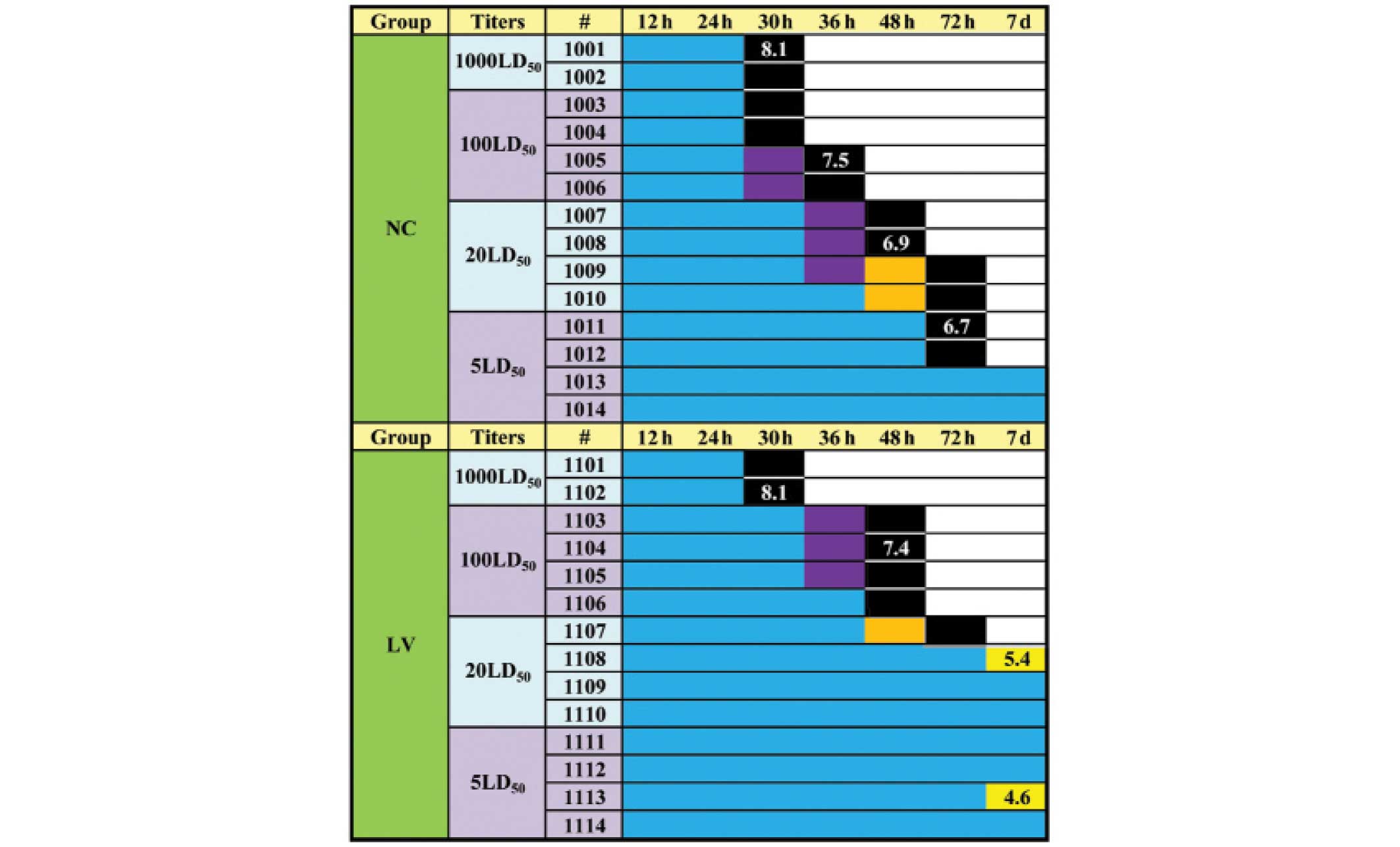
FMDV challenge in LV-3shRNA-pretreated
suckling mice
To examine the anti-FMDV potential of LV-3shRNA
in vivo, suckling mice pretreated with LV-3shRNA were
challenged with four titers of FMDV serotype O (5, 20, 100 and
1,000 times the LD50). No marked increase in the
antiviral effect was observed in any of the 1,000 LD50
groups treated with LV-3shRNA, compared with the PBS-treated groups
(Fig. 4). In the 100
LD50 treatment group, the LV-3shRNA-pretreated mice
exhibited symptoms, including twitching, although not those
associated with mortality, at 36 h, whereas the negative control
mice died. In addition, 75% of the mice challenged with 20
LD50 FMDV survived in the LV-3shRNA-treated group (over
the course of the following 7 days; Fig. 4), whereas mice in the negative
control (NC) group died. One mouse in each group was selected at
random for dissection in order to determine the number of viral
copies present using RT-qPCR. The data revealed that the
log10 of the number of viral RNA copies in the muscles
of the dead LV-treated mice in the 100 and 1,000 LD50
groups was 7.4 and 8.1, respectively, which revealed no significant
difference from the number found in the dead NC mice in
corresponding titers (7.5 and 8.1, respectively). However, the
number of viral RNA copies in the muscles of the surviving mice
treated with LV-3shRNA in the 5 and 20 LD50 groups (4.6
and 5.4, respectively) was far lower compared with the number in
the dead NC mice in the corresponding titers (6.7 and 6.9). These
results indicated that pretreatment with LV-3shRNA may afford the
suckling mice certain protection against FMDV.
Discussion
FMDV is a transmissible disease, which may spread
rapidly over vast areas, and one which causes devastating effects
on the livestock industry. In order to advance towards eradicating
FMDV, one approach emerging over the last decade to address this
problem is to use targeted siRNAs or shRNAs to silence multiple
sites on the viral genome, or to identify siRNAs focused on
conserved target portions of the genes coding for non-structural
proteins of the virus. It was reported that treatment with
shRNA-expressing plasmids directed against the VP1 protein of FMDV
inhibits proliferation of the virus in BHK-21 cells (16,17).
Similar results were obtained in IBRS-2 cells transfected with
plasmids expressing shRNAs targeting the 2B gene (6), and in BHK-21 cells transfected with
synthetic siRNAs or siRNAs produced in vitro using a
cocktail kit (5,18). Kahana et al (7) demonstrated a complete inhibition of
viral growth in BHK-21 cells transfected with a combination of
several anti-FMDV siRNAs. Aiming to improve the anti-FMD strategy
in cows and buffalo, the buffalo 7SK/U6 and bovine U6 promoters
were cloned and used to direct the expression of shRNAs, which have
previously been demonstrated to be efficient in buffalo or mouse
cells (19). In the present study,
a multiple shRNA expression lentiviral vector with different
promoters was used as part of an RNAi antiviral strategy against
FMDV. It was previously reported that the use of multiple shRNAs
exerts more of an anti-viral effect compared with single shRNA
vectors (20). To select the
target site of the RNAi, three fragments from the 3B and 3D protein
coding region of the non-structural protein P3 were selected, and
conserved regions were sought after by multiple alignment of
several viral strains of the A, Asia I, C and O serotypes, in order
to broaden the scope of this strategy across a range of
serotypes.
In view of their ability to infect a broad spectrum
of cell types and their high integration efficiency, lentiviral
vectors have been widely used to deliver siRNAs in vitro and
in vivo. Lentiviral vectors were used as an efficient means
to study the functional effects of antiviral shRNAs in cells
(21), and as an outstanding
method to deliver shRNA-expressing cassettes in vivo,
generating a transgenic mouse model (22). Furthermore, it was reported that
lentiviruses also potentially mediate gene therapy (23). In order to develop a highly
efficient method to knock down the FDMV RNA, the present study
generated an LV-3shRNA vector expressing three 3B-specific (Cons1)
and 3D-specific (Cons2 and Cons3) shRNAs. In previous studies, in
order to assess the function of the siRNAs, the titer of FMDV used
for virus challenge experiments was of the order of ~103
TCID50/ml (5,7,8,18).
In the present study, 107 TCID50/ml FMDV was
used to challenge the LV-3shRNA-transformed BHK-21 cells, which was
~104 times higher compared with that of previous
reports. It was identified that the numbers of viral RNA copies in
the transformed BHK-21-LV cells were three times lower compared
with those of the BHK-21 cell group (2.22×10⁷, vs.
6.82×10⁷ cp/ml) at 24 h post-infection. The subsequent
in vivo experiment, where suckling mice were challenged with
FMDV, demonstrated that 75% of the mice challenged with 20
LD50 FMDV survived in the group pretreated with
LV-3shRNA following a period of 7 days, whereas the mice in the NC
groups were all dead. The lentivirus-treated animals exhibited
markedly more resistance towards FMDV. The present study is the
first, to the best of our knowledge, to demonstrate the protection
of suckling mice, as determined by LD50 measurements,
following pretreatment with lentiviral vector. The LV-3shRNA has
potential to be used in subsequent studies to protect the
FMDV-infected animals by lentiviral-mediated gene therapy. This
will facilitate the timely introduction of novel disease resistance
traits into buffalo or cows via routine injection techniques.
In conclusion, a multiple shRNA-expressing
lentiviral vector with different pol III promoters was used to
great effect in an anti-FMDV study. A marked inhibition of FMDV
sero-type O in vitro and in vivo was identified, and
the present study provided a novel potential strategy in anti-FMDV
buffalo or bovine transgene, and FMDV-targeted therapy for the
future.
Acknowledgments
This work was funded by the China Transgenic Project
(no. 2011ZX08007-003) and the National Natural Science Fund (no.
NSFC 31260552). The authors would like to thank Miss Shuye Qiao and
Miss Meng Wang from Guangxi University, and Mrs Lv Lv, Mr Weijun
Cao, Mrs Ruoqing Mao, Mrs Yan Zhang and Mr Yuanguang Yu from
Lanzhou Veterinary Research Institute for their assistance..
References
|
1
|
Young JR, Suon S, Andrews CJ, Henry LA and
Windsor PA: Assessment of financial impact of foot and mouth
disease on smallholder cattle farmers in Southern Cambodia.
Transbound Emerg Dis. 60:166–174. 2013. View Article : Google Scholar
|
|
2
|
Fasina FO, Connell DR, Talabi OA, Lazarus
DD, Adeleke GA, Olusanya TP and Hernandez JA: Foot-and-mouth
disease virus strains and examination of exposure factors
associated with seropositivity of cattle herds in Nigeria during
2007–2009. Prev Vet Med. 109:334–342. 2013. View Article : Google Scholar
|
|
3
|
Stram Y, Chai D, Fawzy HE, Molad T, Meiri
N, Van-Ham M, el-Kilani S, Fahamy F, Moussa AA and Yadin H:
Molecular epidemiology of foot-and-mouth disease (FMD) in Israel in
1994 and in other Middle-Eastern countries in the years 1992–1994.
Arch Virol. 140:1791–1797. 1995. View Article : Google Scholar
|
|
4
|
Nampanya S, Khounsy S, Phonvisay A, Young
JR, Bush RD and Windsor PA: Financial Impact of Foot and Mouth
Disease on Large Ruminant Smallholder Farmers in the Greater Mekong
Subregion. Transbound Emerg Dis. 6:1–10. 2013.
|
|
5
|
Mohapatra JK, Sanyal A, Hemadri D, Tosh C,
Kumar RM and Bandyopadhyay SK: Evaluation of in vitro inhibitory
potential of small interfering RNAs directed against various
regions of foot-and-mouth disease virus genome. Biochem Biophys Res
Commun. 329:1133–1138. 2005. View Article : Google Scholar : PubMed/NCBI
|
|
6
|
de los Santos T, Wu Q, de Avila Botton S
and Grubman MJ: Short hairpin RNA targeted to the highly conserved
2B nonstructural protein coding region inhibits replication of
multiple serotypes of foot-and-mouth disease virus. Virology.
335:222–231. 2005. View Article : Google Scholar : PubMed/NCBI
|
|
7
|
Kahana R, Kuznetzova L, Rogel A, Shemesh
M, Hai D, Yadin H and Stram Y: Inhibition of foot-and-mouth disease
virus replication by small interfering RNA. J Gen Virol.
85:3213–3217. 2004. View Article : Google Scholar : PubMed/NCBI
|
|
8
|
Chen W, Liu M, Jiao Y, Yan W, Wei X, Chen
J, Fei L, Liu Y, Zuo X, Yang F, Lu Y and Zheng Z:
Adenovirus-mediated RNA interference against foot-and-mouth disease
virus infection both in vitro and in vivo. J Virol. 80:3559–3566.
2006. View Article : Google Scholar : PubMed/NCBI
|
|
9
|
Pengyan W, Jianjun J, Ning L, Jinliang S,
Yan R, Chuangfu C and Zhiru G: Transgenic mouse model integrating
siRNA targeting the foot and mouth disease virus. Antiviral Res.
87:265–268. 2010. View Article : Google Scholar : PubMed/NCBI
|
|
10
|
Kafri T, Blomer U, Peterson DA, Gage FH
and Verma IM: Sustained expression of genes delivered directly into
liver and muscle by lentiviral vectors. Nat Genet. 17:314–317.
1997. View Article : Google Scholar : PubMed/NCBI
|
|
11
|
Chen C, Ridzon DA, Broomer AJ, Zhou Z, Lee
DH, Nguyen JT, Barbisin M, Mahuvakar VR, Andersen MR, et al:
Real-time quantification of microRNAs by stem-loop RT-PCR. Nucleic
Acids Res. 33:e1792005. View Article : Google Scholar : PubMed/NCBI
|
|
12
|
Tang F, Hajkova P, Barton SC, Lao K and
Surani MA: MicroRNA expression profiling of single whole embryonic
stem cells. Nucleic Acids Res. 34:e92006. View Article : Google Scholar : PubMed/NCBI
|
|
13
|
Reed LJ and Muench HA: A simple method of
estimating fifty percent endpoints. Am J Hyg. 27:493–497. 1938.
|
|
14
|
Shaw AE, Reid SM, Ebert K, Hutchings GH,
Ferris NP and King DP: Implementation of a one-step real-time
RT-PCR protocol for diagnosis of foot-and-mouth disease. J Virol
Methods. 143:81–85. 2007. View Article : Google Scholar : PubMed/NCBI
|
|
15
|
Lu Z, Cao Y, Guo J, Qi S, Li D, Zhang Q,
Ma J, Chang H, Liu Z, Liu X and Xie Q: Development and validation
of a 3ABC indirect ELISA for differentiation of foot-and-mouth
disease virus infected from vaccinated animals. Vet Microbiol.
125:157–169. 2007. View Article : Google Scholar : PubMed/NCBI
|
|
16
|
Chen W, Yan W, Du Q, Fei L, Liu M, Ni Z,
Sheng Z and Zheng Z: RNA interference targeting VP1 inhibits
foot-and-mouth disease virus replication in BHK-21 cells and
suckling mice. J Virol. 78:6900–6907. 2004. View Article : Google Scholar : PubMed/NCBI
|
|
17
|
Cong W, Cui S, Chen J, Zuo X, Lu Y, Yan W
and Zheng Z: Construction of a multiple targeting RNAi plasmid that
inhibits target gene expression and FMDV replication in BHK-21
cells and suckling mice. Vet Res Commun. 34:335–346. 2010.
View Article : Google Scholar : PubMed/NCBI
|
|
18
|
Liu M, Chen W, Ni Z, Yan W, Fei L, Jiao Y,
Zhang J, Du Q, Wei X, Chen J, Liu Y and Zheng Z: Cross-inhibition
to heterologous foot-and-mouth disease virus infection induced by
RNA interference targeting the conserved regions of viral genome.
Virology. 336:51–59. 2005. View Article : Google Scholar : PubMed/NCBI
|
|
19
|
Zhang X, Liu Q, Luo C, Deng Y, Cui K and
Shi D: Identification and characterization of buffalo 7SK and U6
pol III promoters and application for expression of short hairpin
RNAs. Int J Mol Sci. 15:2596–2607. 2014. View Article : Google Scholar : PubMed/NCBI
|
|
20
|
ter Brake O, 't Hooft K, Liu YP, Centlivre
M, von Eije KJ and Berkhout B: Lentiviral vector design for
multiple shRNA expression and durable HIV-1 inhibition. Mol Ther.
16:557–564. 2008. View Article : Google Scholar : PubMed/NCBI
|
|
21
|
Luo J, Du J, Gao S, Zhang G, Sun J, Cong
G, Shao J, Lin T and Chang H: Lentviral-mediated RNAi to inhibit
target gene expression of the porcine integrin αv subunit, the FMDV
receptor, and against FMDV infection in PK-15 cells. Virol J.
8:4282011. View Article : Google Scholar
|
|
22
|
Damiri B, Holle E, Yu X and Baldwin WS:
Lentiviral-mediated RNAi knockdown yields a novel mouse model for
studying Cyp2b function. Toxicol Sci. 125:368–381. 2012. View Article : Google Scholar :
|
|
23
|
Liu YP, Westerink JT, ter Brake O and
Berkhout B: RNAi-inducing lentiviral vectors for anti-HIV-1 gene
therapy. Methods Mol Biol. 721:293–311. 2011. View Article : Google Scholar : PubMed/NCBI
|