Introduction
Vascular smooth muscle cells (VSMCs) are highly
differentiated cells maintaining phenotypic plasticity.
Dysregulation of phenotype switching from contractile phenotype to
a synthetic phenotype is associated with vascular disorders,
including hypertension, atherosclerosis and restenosis (1–4).
Thus, VSMC differentiation is an essential component of vascular
development.
NELIN, a gene expressed in cardiac muscles, was
cloned on the basis of heart cDNA library screening in 1999
(5). NELIN protein was shown to be
an actin-binding protein (ABP) by Wang et al (6). According to a previous study by our
group (7), NELIN functions as a
regulatory factor in maintaining the contractile phenotype of VSMCS
and regulating the phenotypic transformation of VSMCs. However, the
mechanism underlying the regulation of the phenotypic
transformation of VSMCs by NELIN has remained elusive.
Ras homolog gene family member A (RhoA) belongs to
the Rho family of small guanine triphosphatases (8) and activates the promotion of F-actin
stress fiber formation and focal adhesion, which link stress fibers
to the plasma membrane. Therefore, it affects the contractility and
adhesion of VSMCs (9). RhoA
regulates numerous cellular functions, including excessive
contraction, phenotypic transformation, migration and proliferation
(10), whose dysregulation is
implicated in cardiovascular disorders, including hypertension,
atherosclerosis and restenosis (11). It has been reported that RhoA/the
Rho kinase signaling pathway participates in the vascular
remodeling process and affects VSMC differentiation by regulating
nuclear translocation of serum response factor (SRF) as well as
actin polymerization (12).
In the present study, NELIN-overexpressing or
-silenced VSMCs were established by infection with lentiviral
vectors (LVs). Smooth muscle (SM)α-actin was used as a marker
protein of different VSMC phenotypes. The association between NELIN
expression and VSMC differentiation was observed and the effect of
NELIN expression levels on VSMCs differentiation was analyzed.
Furthermore, the involvement of the RhoA/SRF signaling pathway in
this process was discussed. The present study provided data which
may serve as a foundation for the use of NELIN in the prevention
and treatment of vascular remodeling disorders.
Materials and methods
Materials
Fetal bovine serum (FBS), Dulbecco's modified
Eagle's medium (DMEM) and pancreatic enzymes were obtained from
Gibco-BRL (Invitrogen Life Technologies, Carlsbad, CA, USA). TRIzol
was purchased from Invitrogen Life Technologies. RNAi-Mate
transfection reagent, interfering (NELIN siRNA forward,
5′-GCCCUGGUAAACUCAAACUTT-3′ and reverse,
5′-AGUUUGAGUUUACCAGGGCTT-3′)/overexpression lentivirus, shuttle
plasmid and packaging plasmid (pGag/Pol, pRev, pVSV-G) were
manufactured by Shanghai Gene Pharma Co. Ltd. (Shanghai, China).
Puromycin, paraformaldehyde, TritonX-100, CBB, xylene, neutral
balsam and glycerol were purchased from Shanghai Haoran Biological
Technology Co., Ltd. (Shanghai, China). PrimeScript® 1st
Strand cDNA Synthesis kit and SYBR® Premix Ex Taq kit
were purchased from Takara Bio Inc. (Otsu, Japan). SDS,
β-mercaptoethanol, bromophenol blue and Tris-buffered saline/Tween
(TBST) were purchased from Shanghai Bogu Biotech Co., Ltd.
(Shanghai, China). Rat polyclonal anti-NELIN (GeneTex Inc., Irvine,
CA, USA; cat. no. GTX46266; 1:100 dilution), rat monoclonal
anti-SMα-actin (Novus Biologicals, Littleton, CO, USA; cat. no.
NB100-74340F; 1:100 dilution), rat monoclonal anti-SRF (GeneTex
Inc.; cat. no. GTX18063; 1:100 dilution), rat monoclonal anti-RhoA
(Novus Biologicals; cat. no. H00000387-M03; 1:100 dilution) and rat
monoclonal anti-GAPDH (cat. no. SAM1003; 1:100 dilution) antibodies
were used. Horseradish peroxidase (HRP) and fluorescently-labeled
sheep anti-rat IgG secondary antibody (cat. no. SE28; 1:100
dilution) was obtained from Beijing Solarbio Science &
Technology Co., Ltd. (Beijing, China). Enhanced chemiluminescence
(ECL) reagent was purchased from Millipore (Billerica, MA, USA).
DAPI was purchased from Beyotime Institute of Biotechnology
(Jiangsu, China). Radioimmunoprecipitation assay (RIPA) lysis
buffer was obtained from Sigma-Aldrich (St. Louis, MO, USA).
Complete Mini was obtained from Roche Diagnostics Co. (Basel,
Switzerland). A bicinchoninic acid (BCA) protein assay kit was
purchased from Pierce (Thermo Fisher Scientific, Waltham, MA, USA).
A nitrocellulose membrane was obtained from BioRad (Hercules, CA,
USA).
Cell culture, lentiviral vector
construction and infection
The present study was approved by the ethics
committee of Shandong University (Jinan, China). Written informed
consent was obtained from the patient, a 59-year-old male who
underwent stripping of great saphenous vein. The VSMCs were
cultured from a human great saphenous vein in DMEM containing 10%
FBS, at 37°C with 5% CO2, as described previously
(13). The cells between passage 2
and 4 were then selected carefully and used. Shuttle plasmid,
packaging plasmid (pGag/Pol, pRev, pVSV-G) and RNAi-Mate were mixed
homogeneously for 20–25 min at room temperature. Subsequently, the
transfection mixture was added into the culture dish with VSMCs
drop by drop. Cell supernatant containing lentivirus particles was
collected after 72 h of culture in an incubator with 5%
CO2 at 37°C. Concentrated lentivirus solution was
obtained from the cell supernatant following centrifugation at 100
× g for 5 min and filtering using a filter (Millipore).
One day prior to infection,
1×10⁵−2×10⁵ selected VSMCs per well were
seeded in a six-well plate. The recombined lentiviral particles
were diluted to a titer of 1×10⁸ TU/l with DMEM
containing 10% FBS. The media in the six-well plate was removed and
replaced with 200 µl diluted viral suspension. Following 24
h of incubation, the viral suspension was replaced with 2 ml DMEM.
The infected cells were selected using 2 µg/ml puromycin for
five days, which was added 72 h following infection. The control
(no infection, treated with medium only) and the VSMCs infected
with NELIN-expression vector only were subjected to the above
procedure. Cells in the NELIN-VSMC group were treated with 0.25%
trypsin and diluted into single cell suspensions
(1.5×10⁴/ml) using 2% FBS medium. These cells were then
inoculated onto a 12-pore plate (200 µl/well) and incubated
with 10 µmol/l Y-27632 (Shanghai Haoran Biological
Technology Co., Ltd.), a Rho kinase inhibitor, for 24 h. All
culture process were performed in an atmosphere of 5%
CO2 at 37°C, and the experiments were performed in
triplicate.
Cell morphology observation
An inverted microscope (IX70; Olympus Corporation,
Tokyo, Japan) was used to observe the morphology of the control
cell group, the LV-NELIN-siRNA-VSMC group and the NELIN-VSMC
group.
Cytoskeleton observation by Coomassie
Brilliant Blue (CBB) staining
Cells were grown on coverslips, pre-fixed using 2%
paraformaldehyde for 10 sec and then washed by phosphate buffered
saline (PBS) for 1 min. The cover slips were then dipped into 1%
TritonX-100 at 4°C for 30 min and washed with PBS for 15 min.
Subsequently, the cells were fixed with 4% paraformaldehyde for 20
min and stained with 0.2% CBB R250 for 30 min. Finally, the cover
slips were washed by distilled water for 15 min, dried naturally,
made transparent by xylene and sealed by neutral balsam for
observation under a microscope (FSX100, Olympus Corporation).
Reverse transcription quantitative
polymerase chain reaction (RT-qPCR)
VSMCs from the control group, the
LV-NELIN-siRNA-VSMCs group and the NELIN-VSMCs group were selected,
respectively. Total RNA was isolated from the cultured cells using
TRIzol and reversely transcribed to cDNA using the
PrimeScript® 1st Strand cDNA Synthesis kit according to
the manufacturer's instructions. PCR was performed on an Mx3000P
real-time PCR instrument (Agilent Stratagene, Amsterdam,
Netherlands) using the manufacturer's instructions of the
SYBR® Premix Ex Taq™ kit. The primers of target genes
were designed and supplied by Invitrogen Life Technologies and
shown in Table I. Each RT-qPCR
reaction mix consisted of 12.5 µl SYBR® Premix Ex
Taq™, 3 µl cDNA, 0.2 µL each primer (10 µM)
and 9.1 µl PCR-grade water, in a final volume of 25
µl. Thermal cycling conditions included an initial step at
95°C for 30 sec, followed by 40 cycles at 95°C for 5 sec and 60°C
for 30 sec. The final extension step included 40 cycles at 95°C for
1 min, 60°C for 30 sec and 95°C for 30 sec. Data were calculated by
the ΔΔCt method and compared to a reference (GAPDH). All
experiments were performed in triplicate and repeated in at least
three separate experiments.
 | Table IPrimer sequences for genes. |
Table I
Primer sequences for genes.
| Gene | Forward primer
(5′-3′) | Reverse primer
(5′-3′) |
|---|
| NELIN |
TCCACGCGGAAAGAAGTACC |
GCAAGCATTTCTTTAATCTCAGCC |
| α-smoth
muscle-actin |
AGCTTTCAGCTTCCCTGAACA |
AGAGCCATTGTCACACACCA |
| GAPDH |
TGTTCGTCATGGGTGTGAAC |
ATGGCATGGACTGTGGTCAT |
Western blot (WB) analysis
Following lysis in cold RIPA lysis buffer containing
the protease inhibitor cocktail Complete Mini, the lysate was
centrifuged at 4,800 ×g for 20 min, and the supernatant was
collected. Total protein was then determined using the BCA protein
assay kit according to the manufacturer's instructions. Protein
samples were incubated with loading buffer containing 10 mM
Tris-HCl (pH 6.8), 1% (w/v) SDS, 25% (v/v) glycerol, 0.1 mM
b-mercaptoethanol and 0.03% (w/v) bromophenol blue. These
preparations were then boiled for 5 min and separated using 12%
(w/v) SDS-PAGE. The protein gels were transferred onto a
nitrocellulose membrane in transfer buffer (25 mM Tris, 192 mM
glycin, 20% methanol) according to a previous method (14). Subsequently, the membrane blots
were blocked with TBST containing 5% (w/v) nonfat dry milk. The
membrane was then incubated with anti-SRF, RhoA, SMα-actin,
anti-NELIN and antiserum as the primary antibody for 1 h at room
temperature, and washed three times with TBST for 10 min. As the
secondary antibody, HRP and fluorescently-labeled antibody was used
for 1 h at room temperature. Membranes were visualized using the
ECL plus kit and exposed to Kodak X-ray film (Eastman Kodak,
Rochester, NY, USA). Finally, the densitometric intensities were
measured and analyzed using an LAS-3000 and Multi Gauge version 3.0
(Fujifilm, Tokyo, Japan) (15).
Immunofluorescence
The selected cells grown on a 48-well plate and
fixed with 4% paraformaldehyde for 20 min and washed sequentially
for 15 min with PBS. Cells were made transparent with 0.2%
TritonX-100 for 30 min and washed with PBS for another 15 min. The
cells were then blocked with 5% goat serum for 30 min and incubated
with SRF primary antibody (1:100) overnight at 4°C. The incubated
cells were washed with PBS for 15 min and subsequently incubated
with the fluorescent labeled secondary antibody (1:100, 37°C, 30
min). The cell nucleus was counterstained with DAPI. Following
washing with PBS, the plate was sealed using glycerin. Finally, the
cells were observed using confocal microscopy (FSX100; Olympus
Corporation) and images were captured.
Statistical analysis
Values are expressed as the mean ± standard error of
the mean and analyzed adopting SPSS 12.0 statistical software
(SPSS, Inc., Chicago, IL, USA). P<0.05 was considered to
indicate a statistically significant difference between values.
One-way analysis of variance was used for data analysis.
Experiments in each group were performed thrice.
Results
Cell morphology
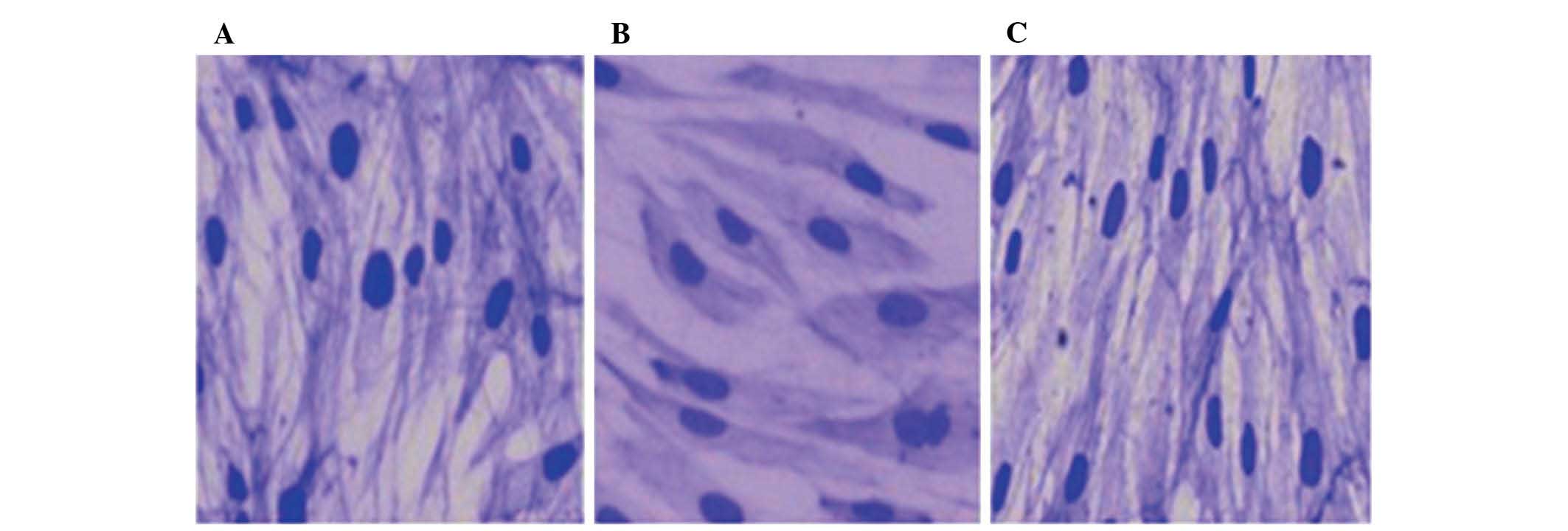
As shown in Fig. 1,
the normal VSMCs grew well in DMEM with 10% FBS and presented a
'valley-peak' form during combination. Cells in the NELIN-VSMC
group were comparatively long and thin with a cyclic cytoplasm of
small volume, and grew in a luminal structure. The cytoskeleton of
these cells exhibited dense grids and fasciculation as shown by CBB
staining. However, cells in the LV-NELIN-siRNA-VSMC group had a
larger volume and exhibited various shapes in the same growing
environment; they had lost polarity and grew in a disordered
fashion. The intensity of CBB staining increased, and the
cytoskeleton was not clearly defined and sparsely latticed.
NELIN expression in VSMCs
Results from real-time quantitative PCR and WB
showed that the expression of NELIN mRNA and NELIN protein
increased in cells from the NELIN-VSMC group, while it decreased
significantly in cells from the LV-NELIN-siRNA-VSMC group compared
with that in the control group (Table
II, Fig. 2).
| Table IIReal-time quantitative polymerase
chain reaction of NELIN. |
Table II
Real-time quantitative polymerase
chain reaction of NELIN.
| Cell group | Ct | Δt | ΔΔ |
2−ΔΔCt |
|---|
| Control | 28.20 | 6.06 | 0 | 1 |
|
LV-NELIN-siRNA-VSMC | 29.26 | 7.77 | 1.70 | 0.31 |
| NELIN-VSMC | 24.69 | 3.48 | −2.58 | 5.98 |
SMα-actin expression in VSMCs
Fig. 3 and Table III show the results of RT-qPCR
and WB analyses of SMα-actin. Compared with that in the control
group, SMα-actin expression in cells from the NELIN-VSMC group was
significantly increased. However, in cells from the
LV-NELIN-siRNA-VSMC group, the expression of SMα-actin was
significantly decreased.
| Table IIIReal-time quantitative polymerase
chain reaction of α-smooth muscle actin. |
Table III
Real-time quantitative polymerase
chain reaction of α-smooth muscle actin.
| Cell group | Ct | Δt | ΔΔ | 2−ΔΔ. |
|---|
| Control | 25.90 | 3.76 | 0 | 1 |
|
LV-NELIN-SiRNA-VSMC | 28.32 | 6.82 | 3.07 | 0.12 |
| NELIN-VSMC | 23.59 | 2.38 | −1.38 | 2.60 |
Effect of NELIN on RhoA and SRF
expression
The WB results in Fig.
4A show that the total RhoA protein expression increased in
cells from the NELIN-VSMC group and decreased in cells from the
LV-NELIN-siRNA-VSMC group compared with that in the control group.
Immunofluorescence (Fig. 4B)
showed that in the control group, SRF was predominantly expressed
in the cytoplasm, while it was mainly expressed in the cell nuclei
of cells in the NELIN-VSMC group, with intra-nuclear translocation.
In cells of the LV-NELIN-siRNA-VSMC group, SRF was moderately
expressed in the cytoplasm. WB analysis of total SRF protein showed
a slight decrease of SRF levels in the LV-NELIN-siRNA-VSMC and a
slight increase in the NELIN-VSMC group as compared with those in
the control group; however, these differences were not
statistically significant (Fig.
4C). In conclusion, the results showed that the overexpression
of NELIN induced an increase of total RhoA protein expression and
intra-nuclear translocation of SRF.
Influence of Y-27632 on NELIN-induced SRF
intra-nuclear translocation
Cells of the NELIN-VSMC group were incubated with 10
µmol/l Y-27632, a Rho kinase inhibitor, for 24 h, and the
effect on protein expression was examined using WB analysis. The
result showed that SRF and SMα-actin expression was decreased,
suggesting that the regulation of VSMC differentiation by NELIN may
be mediated via the RhoA-SRF signaling pathway (Fig. 5).
Discussion
VSMCs are considered to have two phenotypes: The
contractile phenotype and the synthetic phenotype (16). They are not terminally
differentiated and able to modulate their phenotype in response to
changing environmental cues. The phenotypic transformation process
of VSMCs from the contractile to the synthetic phenotype has a
critical role in the pathogenesis of varicosity, atherosclerosis
and restenosis (17).
Previous studies by our group showed that NELIN
expression was lower in varicose vein tissue as compared with that
in normal veins (18,19). Three functional domains, namely the
coiled-coil region, the F-actin biding domain and the I-like domain
of NELIN protein have been reported, suggesting that NELIN may be a
type of F-actin binding protein (6). Furthermore, an association between
NELIN expression and varicosity was found. When the synthetic
phenotype of VSMCs transformed into the contractile phenotype,
NELIN expression increased and the phenotypic transformation of
VSMCs was stopped after NELIN expression was inhibited by
NELIN-siRNA (7). This proved that
NELIN was an important factor in varicose morbidity by affecting
the phenotypic transformation of VSMCs. However, the mechanism
underlying the phenotypic transformation of VSMCs by NELIN remained
elusive. Thus, the present study explored the regulatory mechanism
of NELIN in the phenotypic transformation of VSMCs.
Regulation of phenotype transformation of
VSMCs by NELIN and effect on SMα-actin expression
The results of the present study showed that VSMCs
with NELIN overexpression presented a contractile phenotype
alongside increased SMα-actin expression as well as an increase in
RhoA protein and SRF nucleoprotein expression. Following knockdown
of NELIN expression, VSMCs showed a synthetic phenotype, while the
expression of SMα-actin protein, RhoA protein and SRF nucleoprotein
decreased significantly. These results illustrated that the
overexpression of NELIN induced SMα-actin expression in VSMCs and
promoted the transformation of VSMCs from synthetic to contractile
phenotype. By contrast, VSMCs with knockdown of NELIN had the
synthetic phenotype and had lost the ability to undergo positive
transformation. These results indicated that NELIN may act as a key
regulatory factor for the phenotypic transformation of VSMCs and
may regulate SMα-actin expression.
The RhoA/SRF signaling pathway in NELIN
expression regulation
Upon activation, RhoA combines with its downstream
effector Rho kinase, ROCK, to further phosphorylate its downstream
substrate myosin phosphatase target subunit 1 and regulate
SRF-dependent smooth muscle gene expression (20). VSMC differentiation is influenced
by the RhoA/Rho kinase signaling pathway via regulating SRF nuclear
translocation and actin polymerization (12). SRF was reported to be able to
initiate SMα-actin gene transcription by cooperating with other
specific transcription factors to act on CArG, a regulatory element
in the upstream promoter region of SMα-actin (21).
The results of the present study showed an increase
of RhoA protein expression and SRF intra-nuclear translocation when
NELIN expression was upregulated in VSMCs, while the opposite
result was observed in NELIN-knockdown VSMCs. Following incubation
with the Rho kinase inhibitor Y-27632, SRF and SMα-actin expression
in the cell nucleus decreased significantly. It was demonstrated
that the RhoA/SRF signaling pathway was affected by NELIN, and that
phenotype transformation of VSMCs mediated by NELIN was mediated
via this pathway.
The present study provided a foundation for the
preventive and therapeutic application of NELIN in vascular
remodeling diseases such as varicosity and atherosclerosis.
However, further studies using animal experiments are required to
verify the effect of NELIN in vivo.
References
|
1
|
Rzucidlo EM, Martin KA and Powell RJ:
Regulation of vascular smooth muscle cell differentiation. J Vasc
Surg. 45(Suppl A): A25–A32. 2007. View Article : Google Scholar : PubMed/NCBI
|
|
2
|
Yao QP, Zhang P, Qi YX, et al: The role of
SIRT6 in the differentiation of vascular smooth muscle cells in
response to cyclic strain. Int J Biochem Cell Biol. 49:98–104.
2014. View Article : Google Scholar : PubMed/NCBI
|
|
3
|
Zhao B, Luo X, Shi H and Ma D: Tissue
factor pathway inhibitor-2 is downregulated by ox-LDL and inhibits
ox-LDL induced vascular smooth muscle cells proliferation and
migration. Thromb Res. 128:179–185. 2011. View Article : Google Scholar : PubMed/NCBI
|
|
4
|
Majesky MW: Developmental basis of
vascular smooth muscle diversity. Arterioscl Throm Vas Biol.
27:1248–1258. 2007. View Article : Google Scholar
|
|
5
|
Zhao Y, Wei YJ, Cao HQ and Ding JF:
Molecular cloning of NELIN, a putative human cytoskeleton
regulation gene. Acta Bioch Bioph Sin. 33:19–24. 2001.
|
|
6
|
Wang Z, Cao HQ, Feng Y, et al:
Characteristics of the binding features of Nelin with F-actin and
screening Nelin interactive proteins. Chin Sci Bull. 49:2487–2490.
2004.
|
|
7
|
PEI C, QIN S and CHEN S: Effect of Nelin
on phenotypic transition of human great saphenous vein smooth
muscle cells. Chin J Exp Surg. 30:925–927. 2013.
|
|
8
|
Xu B, Ju Y and Song G: Role of p38,
ERK1/2, focal adhesion kinase, RhoA/ROCK and cytoskeleton in the
adipogenesis of human mesenchymal stem cells. J Biosci Bioeng.
117:624–631. 2014. View Article : Google Scholar
|
|
9
|
Loirand G, Rolli-Derkinderen M and Pacaud
P: RhoA and resistance artery remodeling. Am J Physiol Heart Circ
Physiol. 288:H1051–H1056. 2005. View Article : Google Scholar : PubMed/NCBI
|
|
10
|
Rolfe BE, Worth NF, World CJ, Campbell JH
and Campbell GR: Rho and vascular disease. Atherosclerosis.
183:1–16. 2005. View Article : Google Scholar : PubMed/NCBI
|
|
11
|
Loirand G, Guérin P and Pacaud P: Rho
kinases in cardiovascular physiology and pathophysiology. Circ Res.
98:322–334. 2006. View Article : Google Scholar : PubMed/NCBI
|
|
12
|
Liu HW, Halayko AJ, Fernandes DJ, et al:
The RhoA/Rho kinase pathway regulates nuclear localization of serum
response factor. Am J Resp Cell Mol Biol. 29:39–47. 2003.
View Article : Google Scholar
|
|
13
|
Thormodsson FR and Olafsson IH: Isolation
and culturing of human vascular smooth muscle cells. Methods Mol
Biol. 299:197–210. 2005.PubMed/NCBI
|
|
14
|
Towbin H, Staehelin T and Gordon J:
Electrophoretic transfer of proteins from polyacrylamide gels to
nitrocellulose sheets: procedure and some applications. Proc Natl
Acad Sci USA. 76:4350–4354. 1979. View Article : Google Scholar : PubMed/NCBI
|
|
15
|
Yu HR, Kuo HC, Huang HC, et al:
Glyceraldehyde-3-phosphate dehydrogenase is a reliable internal
control in Western blot analysis of leukocyte subpopulations from
children. Anal Biochem. 413:24–29. 2011. View Article : Google Scholar : PubMed/NCBI
|
|
16
|
Nagel DJ, Aizawa T, Jeon KI, et al: Role
of nuclear Ca2+/calmodulin-stimulated phosphodiesterase 1A in
vascular smooth muscle cell growth and survival. Circ Res.
98:777–784. 2006. View Article : Google Scholar : PubMed/NCBI
|
|
17
|
Hirano K: The roles of
proteinase-activated receptors in the vascular physiology and
pathophysiology. Arterioscl Throm Vas. 27:27–36. 2007. View Article : Google Scholar
|
|
18
|
Pei C and Chen S: Detection of the nelin
in peripheral blood of patients with lower extremity varicose veins
and its significance. J Shandong Univ Health Sci. 51:64–66.
2013.
|
|
19
|
Yu X and Chen S: Expression and roles of
nelin in varicose veins. Chin J Curr Adv Gen Surg. 13:1009–9905.
2010.
|
|
20
|
Morelli A, Chavalmane AK, Filippi S, et
al: Atorvastatin ameliorates sildenafil-induced penile erections in
experimental diabetes by inhibiting diabetes-induced
RhoA/Rho-kinase signaling hyperactivation. J Sex Med. 6:91–106.
2009. View Article : Google Scholar : PubMed/NCBI
|
|
21
|
Owens GK, Kumar MS and Wamhoff BR:
Molecular regulation of vascular smooth muscle cell differentiation
in development and disease. Physiol Rev. 84:767–801. 2004.
View Article : Google Scholar : PubMed/NCBI
|