Introduction
Vascular remodeling of the carotid arteries,
characterized by carotid artery intima-media thickening (IMT), may
be key in the pathogenesis of various cardiovascular diseases
(1,2). Recruitment and infiltration of
inflammatory cells into the vascular walls, followed by the
subsequent secretion of cytokines and growth factors leads to
vascular smooth muscle cell (VSMC) proliferation and migration,
which results in vascular remodeling (3). Abnormal shear stress, as well as
various other stimuli, has been proposed as a critical factor that
leads to vascular remodeling (4,5). To
further investigate this, a mouse model of IMT was developed, which
was induced by partial ligation of the left common carotid artery
(LCA) enabling regulation of the flow rates (6–8).
Rho-associated kinase (ROCK) belongs to the AGC [protein kinase A
(PKA)/protein kinase G/protein kinase C (PKC)] family of
serine/threonine kinases, which is a key downstream effector of the
small GTPase, Ras homolog gene family, member A (RhoA) (9). There are two ROCK isoforms, namely
ROCK1 (also referred to as ROKα or p160ROCK) and ROCK2 (also
referred to as ROKβor Rho-kinase). ROCK regulates actin
cytoskeletal reorganization, smooth muscle contraction and cell
migration (10,11). Previous studies demonstrated that
ROCK participates in numerous cardiovascular diseases, leading to
abnormal contraction of smooth muscle, for example, cerebral and
coronary vasospasm (12,13), hypertension (14) and abdominal aortic aneurysm
(9). Thus, ROCK is regarded as an
important therapeutic target for the treatment of various
cardiovascular conditions, including hypertension, atherosclerosis,
heart failure and ischemic damage (10). However, whether the activity of
ROCK is involved in flow-induced vascular remodeling remains
unclear. In the current study, by investigating the effect of
fasudil (the ROCK inhibitor) on vascular remodeling following a
reduction in blood flow, the aim was to quantify the variations in
ROCK expression in response to flow rate reduction, and investigate
the specific role of fasudil in reducing recruitment of
inflammatory cells, proliferation and migration of VSMCs.
Materials and methods
Animals
A total of 85 male mice (weight, 20–25 g; age,
8-weeks; Academy of Military Medical Sciences) were used in the
current study and all procedures were approved by the Medical
Ethics Committee of Peking University People's Hospital (Beijing;
permit number, 2012-13). The control group consisted of 5 mice. The
remaining 80 mice were randomly divided into 8 groups, with 10 mice
per group. The were fed a standard diet and maintained in a
specific-pathogen free environment in our hospital, with a 12 h
light/dark cycle, and a house temperature of 20–28°C.
Experimental protocol
Blood flow reduction in the LCA and flow
measurements were performed as previously described (6). Briefly, each C57Bl/6J mouse was
anesthetized with ketamine and xylazine (0.2 mg/kg; Sigma-Aldrich,
St. Louis, MO, USA) and were maintained at 37°C on a heating pad.
Antibiotics (50 mg/kg cefazolin; Sigma-Aldrich) and 10 mg/kg
analgesic (pentazocine via intramuscular injection; Sigma-Aldrich)
were delivered subsequent to wound closure and sterilization with
entoiodin (Sigma-Aldrich). The left external carotid artery distal
to the thyroid artery and the left internal carotid/occipital
artery were ligated with 8–0 sutures (Ethicon, Inc., Somerville,
NJ, USA). A partial blood flow was maintained through the superior
thyroid branch (15). In the sham
group, vessel isolation was conducted with suture placed without
ligation. The ROCK inhibitor, fasudil (Tianjin Chase Sun
Pharmaceutical Co., Ltd., Tianjin, China) was dissolved in normal
saline and injected intraperitoneally at 30 mg/kg/day following LCA
partial ligation until day 28. The mice were sacrificed through
acute blood loss on days 3, 7, 14 and 28 for morphology and
immunohistochemical analysis. The blood flow was measured in the
carotid arteries using an ultrasonic flow meter (INC T101;
Transonic Systems, Inc., Ithaca, NY, USA). The systolic blood
pressure and heart rate of the conscious mice were monitored
indirectly via the tail arteries (Visitech Systems, Apex, NC,
USA).
Morphology
On days 3, 7, 14 and 28 following surgery, the
vasculature was fixed by transcardial perfusion at 100 mmHg with 25
ml 10% paraformaldehyde (ZSGB-BIO, Beijing, China) in sodium
phosphate buffer (pH 7.0; Sigma-Aldrich), as previously described
(16). The left and right common
carotid arteries were harvested and embedded in paraffin. Cross
sections were stained with hematoxylin and eosin (H&E) and
elastic-van Gieson (EVG; ZSGB-BIO), and microscopic images were
captured using a Olympus IX83 microscope (Olympus, Tokyo, Japan).
These were analyzed using ImageJ software version 1.48u (National
Institutes of Health, Bethesda, MD, USA). The circumference of the
lumen, and the length of the internal elastic lamina (IEL) and the
external elastic lamina (EEL) were determined by tracing along the
luminal surface. The circumference of the lumen was used to
represent the luminal area. Using ImageJ, the intimal area was
determined as the area defined by the luminal surface and the IEL.
The medial area was defined by the IEL and EEL. The adventitia area
was defined by the EEL and vessel surface.
Immunohistochemical analysis
Immunohistochemical analysis of the
paraffin-embedded vessel cross sections was performed as previously
described (17). Sections
(thickness, 3 µm) from the carotid arteries of the mice that were
sacrificed on days 3, 7, 14 and 28 following ligation were obtained
at 1 mm proximal to the ligature. The common carotid arteries were
cut longitudinally and antibodies against cluster of
differentiation (CD)45 monoclonal antibody (mAb; 1:50; cat. no.
ZM-0213; ZSGB-BIO), CD68 mAb (1:100; cat. no. UM800047; OriGene
Technologies, Inc., Rockville, MD, USA), proliferating cellular
nuclear antigen (PCNA) mAb (1:50; cat. no. TA301137; Cell Signaling
Technology, Inc., Danvers, MA, USA) and smooth muscle cell (SMC)
α-actin mAb (1:100; OriGene Technologies, Inc.) were used. Antigen
retrieval was performed for cross sections with citric acid
monohydrate (BD Pharmingen) at 95°C for 20 min. Subsequently, the
endogenous peroxidase activity was blocked by incubation for 20 min
in methanol with 3% H2O2 (ZSGB-BIO), followed
by incubation with the goat serum to mask any non-specific binding.
For avidin-biotin-peroxidase complex (ABC) staining, the primary
antibodies were incubated at 4°C overnight, followed by secondary
antibody (EnVision™; Dako, Glostrup, Denmark) incubation at room
temperature for 60 min and 30 min with the ABC complex (Vector
Elite; Vector Laboratories, Burlingame, CA, USA). The
peroxidase-binding sites were visualized with 3,3′diaminobenzidine
(Vector Laboratories), followed by hematoxylin counterstaining. En
face preparations were evaluated with a Leica confocal microscope
(TCS SP5 II; Leica, Microsystems GmbH, Wetzlar, Germany).
Western blotting
Vascular tissues were homogenized in ice-cold buffer
(50 mmol/l Tris, pH 7.4; 150 mmol/l NaCl; 0.5% Triton X-100; 1
mmol/l edetic acid; 1 mol/l phenylmeth-ylsulfonyl fluoride; and 5
mg/l aprotinin) and centrifuged at 14,000×g at 4°C for 15 min. The
supernatants were then collected as total proteins, which were
electrophoresed through 8% SDS-PAGE (Sigma-Aldrich) and
electrically transferred to a nitrocellulose membrane. The membrane
was incubated at 4°C overnight in Tris(hydroxymethyl)aminomethane
buffered saline (TBS) containing 5% milk and the primary antibodies
(1:1,000 dilution, Cell Signaling Technology, Inc.) against ROCK1,
myosin light chain (MLC) and p-MLC. Subsequent to washing with TBS,
the membranes were incubated with the secondary antibody (goat
anti-rabbit IgG horseradish peroxidase conjugated) at room
temperature for 1 h. The protein expression levels of ROCK1 mAb
(cat. no. 4035S), MLC mAb (cat. no. 3672S) and p-MLC mAb (cat. no.
3675S) were determined by western blotting where the relative
quantity of protein was determined by densitometric analysis
(Graigar DM3011; Graigar Technology Co., Ltd., Shenzhen, China).
The densitometric intensities of ROCK1, MLC and p-MLC were
normalized to GAPDH values.
Statistical analysis
Data are represented as the mean ± standard error of
the mean. Comparisons for two groups were performed using Student's
t-test. The differences among three or more groups were determined
using one-way analysis of variance with Fisher's exact post-hoc
test (Graphpad Software, Inc., La Jolla, CA, USA). P<0.05 was
considered to indicate a statistically significant difference.
Differences among groups were analyzed by repeated measures three
times.
Results
Physiological and hemodynamic
characteristics of C57B1/6J mice under partial ligation of carotid
arteries
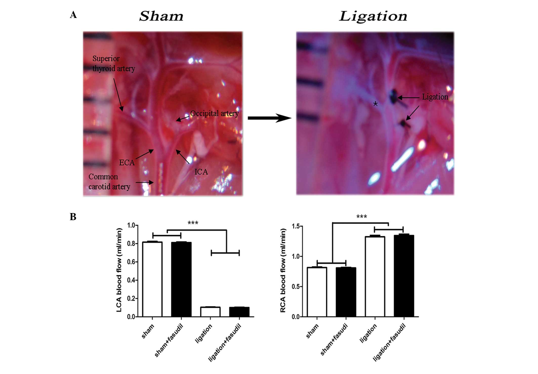
The mouse model used in the current study included
ligation of the internal, external carotid and occipital arteries.
Partial blood flow was maintained through the superior thyroid
branch of the external carotid artery (Fig. 1A). This mouse model demonstrated
significant alterations in blood flow patterns with a reduction of
~80% in the LCA and a 40% increase in the right intact common
carotid arteries (Fig. 1B). This
result was consistent with previous studies (18–20).
For fasudil-treated mice, almost no influence on blood flow was
observed prior or subsequent to LCA ligation (Fig. 1B) and abdominal administration of
fasudil did not influence systemic blood pressure, which was
consistent with previous reports (6). In summary, the LCA blood flow was
significantly reduced in the mouse model, inducing intima-media
thickness and neointima formation with almost no luminal
alterations in LCAs.
ROCK signaling pathway activation in
partially ligated vessels
To determine whether the ROCK signaling pathway
participated in the flow-induced vascular remodeling, western
blotting was used to analyze the protein expression levels of
ROCK1, ROCK2 and their downstream effectors, MLC and p-MLC
(Fig. 2). On days 7 and 28,
significant increases in ROCK1 and p-MLC were observed in ligated
LCA compared with in the sham groups (P<0.001) while no
significant differences in ROCK2 and MLC were identified (Fig. 2A). In addition, fasudil was
demonstrated to attenuate these variations, leading to significant
reductions in protein expression of ROCK1 and p-MLC in the
fasudil-treated group compared with the untreated mice (Fig. 2B; P<0.05 and P<0.01,
respectively).
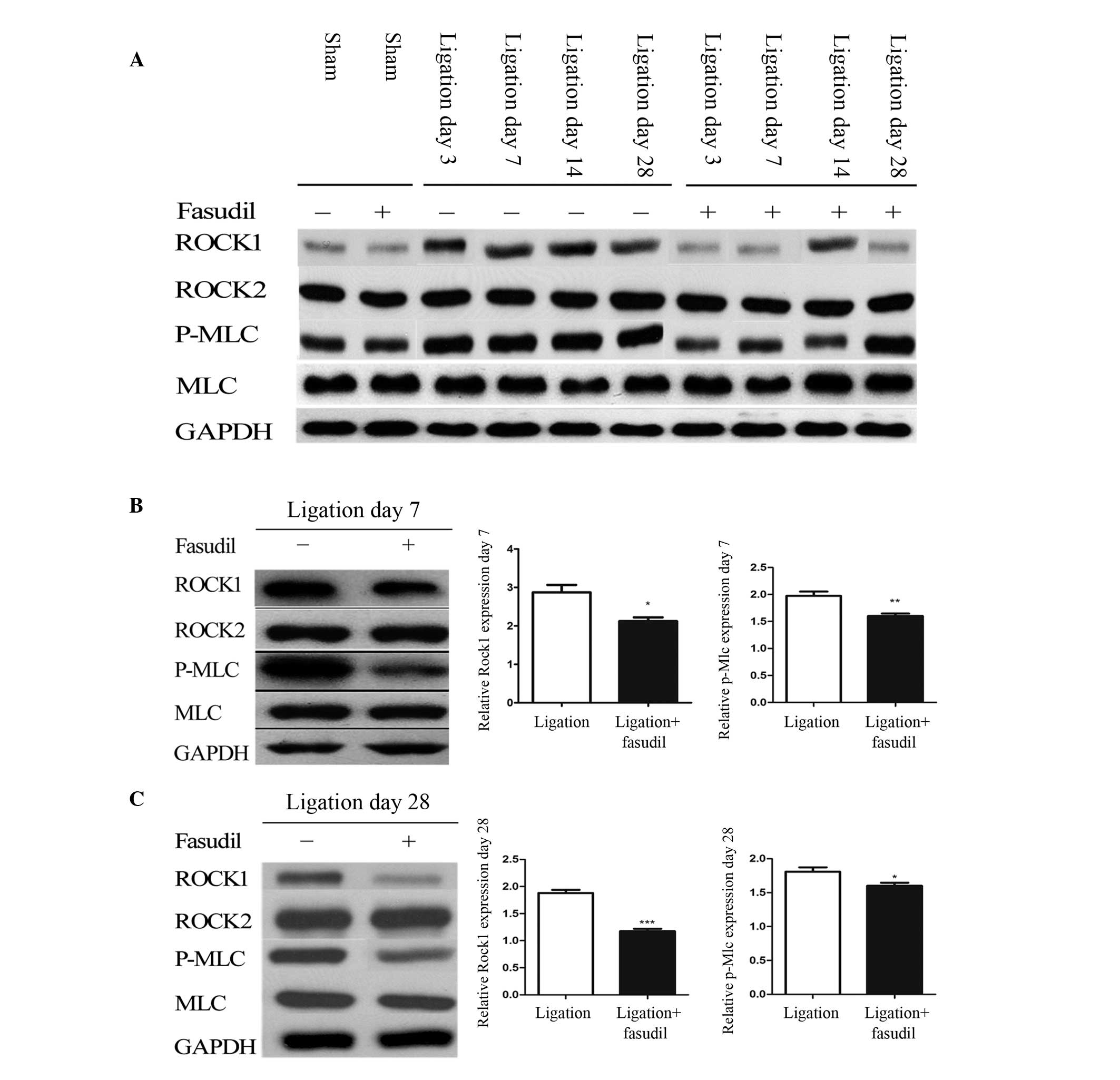 | Figure 2ROCK expression in sham and ligated
mice. (A) Expression of ROCK isoforms and downstream markers of
ROCK in LCA from fasudil-treated and untreated mice, with and
without LCA partial ligation on days 3, 7, 14, and 28.
Quantification of ROCK1, ROCK2, MLC and p-MLC in LCA from
fasudil-treated and untreated mice on (B) day 7 and (C) day 28 [n=8
(number of vessels); *P<0.05, **P<0.01
and ***P<0.001]. ROCK, Rho-associated kinase; LCA,
left common carotid artery; MLC, myosin light chain; p-MLC,
phosphorylated-MLC. |
Fasudil inhibits IMT and neointima
formation in partially ligated LCA
In order to assess how blood flow affects arterial
remodeling in vivo, the LCA partial ligation model was
developed, enabling flow-induced remodeling in the common carotid
arteries of fasudil-treated and untreated mice to be examined and
compared. H&E staining of the LCA vessel areas (Fig. 3A) demonstrated no differences in
the sham fasudil-treated and untreated vessels. Furthermore, in
response to a reduced flow in the LCA, the ligated LCA exhibited
medial thickening and neointima formation. Seven days after
ligation, no significant differences were observed between the
fasudil-treated and untreated vessels. By contrast, 14 days
subsequent to ligation, the areas of the vessel walls in the
fasudil-treated mice were found to be markedly different from the
sham mice. However, the untreated vessels exhibited clear medial
thickening with little neointima formation (Fig. 3A). Twenty-eight days after
ligation, the fasudil-treated mice exhibited a notable reduction in
the media with a small quantity of neointima formation when
compared with the untreated mice (Fig.
3A). Thus, ROCK inhibition may effectively reduce IMT and
neointima formation that is induced by low blood flow in the
carotid arteries. Through EVG staining, no significant alterations
of the elastic lamina were observed, however, an increase in the
media extracellular matrix was identified in the LCA (Fig. 3B). Extracellular matrix increase in
the media was one of the most widespread and clear morphological
features observed. On day 28, the neointima developed in the two
groups, more extensively in the untreated mice than in the
fasudil-treated mice (Fig. 3B).
For fasudil-treated mice, the medial and intima + media areas were
reduced by ~28% (P<0.05) and 45% (P<0.001), respectively,
compared with the untreated group (Fig. 3C). Medial and intima + media
thickness was significantly reduced by 18% (P<0.05) and 32%
(P<0.05), respectively, in the fasudil-treated group when
compared with the untreated group (Fig. 3C). These data indicate that the
ROCK inhibitor is capable of reducing the extent of hyperplasia
following carotid ligation. No significant differences were
observed in the lumenal area of the four groups (Fig. 3D).
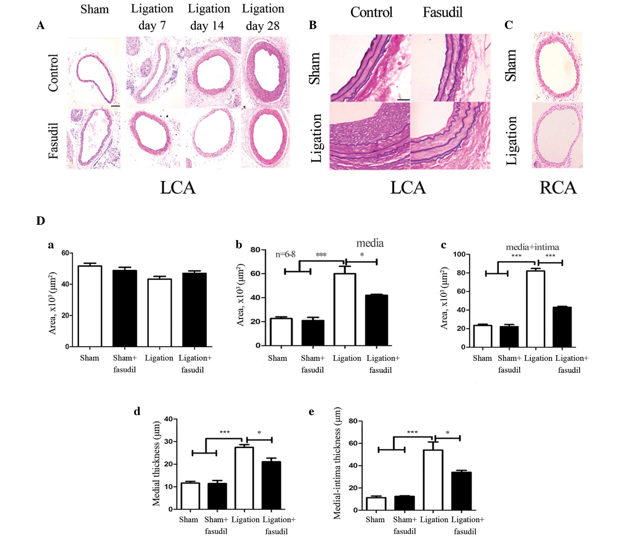 | Figure 3Morphology of the control and
fasudil-treated carotid arteries following ligation. (A)
Representative light microscopy images of hematoxylin and
eosin-stained carotid artery cross sections obtained from the
control and fasudil-treated mice. Medial thickening was observed in
the control and fasudil-treated mice on day 7, and neointima
formation was apparent on days 14 and 28 (magnification, ×40; scale
bar, 20 µm). (B) Elastic-van Gieson staining of carotid arteries
from LCA cross-sections with or without fasudil treatment four
weeks after ligation (sham, sham + fasudil, ligation and ligation +
fasudil; magnification, ×40; scale bar, 20 µm). (C) Representative
light microscopy images of hematoxylin and eosin-stained RCA cross
sections obtained from the sham and ligated mice (magnification,
×40; scale bar, 20 µm). (D) Volumes of vessel components, including
the lumen, media and media + intima, in addition to the vessel wall
thickness of the media and media + intima. Black and white bars
indicate fasudil-treated and untreated groups, respectively. Values
are presented as the mean ± standard error of the mean; n=8 (number
of vessels). *P<0.05, ***P<0.001 (by
analysis of variance). LCA, left common carotid artery; RCA, right
common carotid artery. |
Inhibition effect of fasudil on VSMC
proliferation
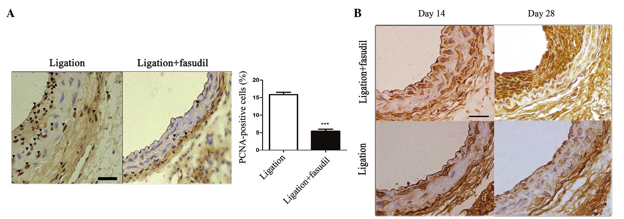
Various studies have demonstrated that VSMC
proliferation is a driving force for vascular remodeling in
response to flow reduction (6,21,22).
To gain further insights into the mechanisms of ROCK signaling
pathway-based vascular remodeling, the level of VSMC proliferation
in the vascular wall was assessed by staining for PCNA. As
presented in Fig. 4A, PCNA
staining in the vascular wall was absent in the early stage
following carotid artery ligation. On day 28, subsequent to
ligation, in the untreated ligated mice the PCNA-positive cells had
significantly increased and were observed to migrate into the
neointima, when compared with the fasudil-treated ligated mice
(P<0.001; Fig. 4A).
To characterize the phenotype of VSMCs, staining for
SMC α-actin expression was performed. On day 14 subsequent to
ligation, α-actin-positive cells were observed to migrate to the
intima-media area in the fasudil-treated and untreated vessels. On
day 28, subsequent to ligation, a markedly increased intensity of
α-actin-positive cells accumulated in the neointima area of the
untreated ligated mice, whereas there was a reduced intensity of
α-actin staining in the media and neointima of the fasudil-treated
mice (Fig. 4B).
Fasudil treatment induced a reduction of
inflammatory cell infiltration to the vascular wall, in the
partially carotid ligated mouse model
Previous studies have demonstrated that the
inflammatory response is important during vascular remodeling
(6). Inflammatory cell interaction
and recruitment to the vascular wall contributes to neointima
formation and vascular inflammation (5). In the current study, inflammatory
cells were identified by staining for the leukocyte antigen, CD45,
which was followed by quantitative analysis using ImageJ software
(23).
In the sham group, quantitative evaluation
demonstrated that there were almost no CD45+ cells
within the mouse intima-media area and few in the adventitia
(Fig. 5A). Ligation notably
increased the number of leukocytes in the vessel walls, where the
majority of CD45+ cells were infiltrated to the
neointima area and a small portion remained in the media area
(Fig. 5A). For fasudil-treated
mice, the number of CD45+ cells infiltrated into the
vessel wall was markedly reduced (Fig.
5A).
Macrophages in the vessel wall were identified by
staining for antigen CD68 (a macrophage marker). In the sham group,
there were almost no CD68+ cells observed in the mouse
intima-media area and adventitia (Fig.
5B). Ligation significantly increased the number of
CD68+ cells present in the vessel walls, with a
significant increase in the intima-media area, particularly in the
neointima. For fasudil-treated mice, infiltration of
CD68+ cells to the vessel wall was markedly reduced
(Fig. 5B).
The graph in Fig.
5C demonstrates that the number of CD45+ cells
infiltrated to the intima-media area in the ligated untreated
vessel walls was ~5-fold that of the fasudil-treated vessels
(Fig. 5C). The percentage of
CD45+ cells among all the cells in the ligated vessel
walls was ~3-fold that of the fasudil-treated vessels (Fig. 5D). In addition, the number of
CD68+ cells infiltrated into the intima-media area in
the ligated untreated vessel walls was ~3-fold that of
fasudil-treated vessels (Fig. 5E).
The percentage of CD68+ cells among all the cells in the
ligated vessel walls was ~2.5-fold that of the fasudil-treated
vessels (Fig. 5F). In summary,
inhibition of the ROCK signaling pathway may reduce inflammatory
cell infiltration to the vascular walls.
Discussion
Flow-induced vascular remodeling is a complex
process, involving the expression of multiple genes and the
interaction of multiple cell types (24). Previous studies based on inbred
mouse or rat strains have demonstrated the role of genetic factors
in vascular remodeling and IMT (21,25).
The observations of the current study provide the first evidence
that ROCK is required for flow-induced remodeling and IMT. The
important conclusions of the current study include: i) Protein
expression levels of ROCK1 and p-MLC significantly increased in
response to the blood flow reduction in LCA. ii) Fasudil prevents
the process of flow-induced vascular remodeling by reducing IMT and
neointima formation. iii) Fasudil inhibits proliferation of VSMCs
in vascular remodeling. iv) Fasudil alleviates inflammatory cell
infiltration into the vascular walls during the process of vascular
remodeling.
The current study aimed to investigate the ROCK
effects on flow-induced vascular remodeling based on partial
ligation of the LCA in C57Bl/6J mice. The left external carotid,
internal carotid and occipital arteries were ligated leaving the
superior thyroid artery intact. Sham procedures consisted of
suturing without ligation. This ligation reduces ~80% of the flow
rate in the LCA and increases the flow rate in the right common
carotid arteries by ~40% with the endothelium intact (18–20).
The current study is, to the best of our knowledge, the first to
investigate the effects of ROCK on flow-induced vascular
remodeling. The positive effect of fasudil was previously
demonstrated in animal models associated with vascular injury and
arteriosclerosis in general (25–28).
Previously, fasudil (136 or 213 mg/kg/day) was demonstrated to
attenuate angiotensin II-induced abdominal aortic aneurysm in
apolipo-protein E (apoE)-KO mice without affecting blood pressure
(9). Furthermore, fasudil (30
mg/kg/day) was reported to prevent early atherosclerotic plaque
formation and terminate lesion progression in apoE-KO mice
(29). In the current study,
fasudil (30 mg/kg/day) was injected intraperitoneally into mice,
and almost no influence on blood pressure was noted (Fig. 1B). In the current study, abdominal
administration of fasudil (the inhibitor of ROCK) was demonstrated
to reduce IMT and neointima formation, inhibit cell proliferation
and alleviate inflammation to the vessel walls. Thus, it was
proposed that the inhibition of ROCK is able to attenuate
flow-induced vascular remodeling.
Previous studies have demonstrated that the ROCK/MLC
signaling pathway is extensively described in VSMCs (30–33)
and ROCK1, rather than ROCK2, mediates leukocyte recruitment and
neointima formation following vascular injury (34). In the present study, western blot
analysis was used to quantify the relative quantity of proteins of
partially ligated LCA with and without fasudil treatment. ROCK1
protein expression levels significantly increased in untreated
ligated vessels when compared with vessels of the sham group.
Subsequent to ligation, the expression levels of ROCK1 protein
significantly reduced in fasudil-treated mice compared with
untreated mice. A similar trend was observed for the expression of
p-MLC. Thus it was concluded that ROCK is involved in the
pathogenesis of vascular remodeling following flow reduction, and
that ROCK inhibitors effectively attenuate flow-induced vascular
remodeling.
In the current study, the mice subjected to 28 days
of LCA partial ligation with or without fasudil treatment were
observed to exhibit IMT and neointima formation in the vessel
walls. Evaluated by H&E and EVG staining, 28 days subsequent to
ligation, fasudil-treated mice exhibited a significant reduction in
media with a lower quantity of neointima formation when compared
with untreated ligated mice (Fig. 3A
and B). In addition, significantly reduced intima-media
thickness and neointima formation in fasudil-treated mice were
observed. These results indicated that inhibition of ROCK results
in the attenuation of IMT and neointima formation following flow
reduction, which is consistent with previous studies (34).
Migration and proliferation of VSMCs are important
factors involved in flow-induced vascular remodeling. In the
current study, the number of PCNA-positive cells in the medial and
intimal areas was markedly reduced in the fasudil-treated mice when
compared with their untreated counterparts (Fig. 4A). In addition, VSMCs in
fasudil-treated vessels exhibited lower migration and proliferation
capabilities compared with untreated vessels. These observations
indicate that the inhibition of the ROCK signaling pathway reduces
VSMC migration and proliferation. Furthermore, the reductions in
VSMC proliferation in the neointima observed in fasudil-treated
mice may result from the reduction in the inflammatory response of
the vessel wall (for example, reduction in leukocyte recruitment,
and the subsequent reduction in the release of inflammatory and
growth stimuli) (Fig. 4A).
Vascular inflammation participates in flow-induced
vascular remodeling, while ROCK1 has been proposed to serve a
critical role in vascular inflammatory diseases (35). Upregulation of the active form of
RhoA leads to a significant increase in inflammatory cell
accumulation in the endothelial cell monolayer in the present
study, which was prevented by administration of fasudil. Similarly,
fasudil has been demonstrated to inhibit MCP-1-induced migration of
leukocytes (36). In the current
study, CD45+ cells were measured on days 14 and 28
subsequent to ligation, a greater number of CD45+ cells
were observed to accumulate in the medial-intimal areas of the
untreated mice, making up ~50% of the total cell count. In the
fasudil-treated mice, the CD45+ cell counts were
significantly reduced to ~15% of the total cell count (P<0.001;
Fig. 5D). Furthermore,
CD68+ cells were almost absent in the sham vessels, were
significantly increased in the partially ligated vessels, and this
increase was partially reversed by fasudil treatment. Thus, ROCK
signaling pathway inhibition prevents inflammatory cells from
infiltrating the vessel walls in flow-induced vascular
remodeling.
Concerning the limitations of the current study, as
fasudil is a non-selective ROCK inhibitor, it may have additional
non-selective effects (36–38),
and at higher concentrations, fasudil may additionally inhibit
serine/threonine kinases, such as PKA and PKC (39). In addition, no dose comparison was
included in the present study.
In conclusion, the present study identified the
involvement of the ROCK signaling pathway in flow-induced vascular
remodeling, observing significant ROCK1 expression level increases
in ligated LCA. p-MLC, as the activated downstream effector of
ROCK, was also significantly increased in ligated mouse LCA.
Fasudil, as a ROCK inhibitor, was demonstrated to reduce ROCK1 and
p-MLC expression levels in ligated mice and attenuate the process
of flow-induced vascular remodeling (for example, IMT, VSMC
migration, and proliferation and lymphocyte accumulation). The
current study elucidated the role of ROCK signaling in the
regulation of vascular remodeling, and highlighted the role of a
ROCK inhibitor, fasudil, in the attenuation of the process of
flow-induced vascular remodeling. Therefore, further
characterization of ROCK signaling and its pharmacological
modulation may lead to the development of novel therapeutic
strategies for targeting flow-induced vascular remodeling.
Acknowledgments
This study was supported by the Peking University
People's Hospital Research and Development Funds (grant no.
2118000537 to Dr. Wei Li), Specialized Research Fund for the
Doctoral Program of Higher Education of China (grant no. 2109000075
to Dr Xiaoming Zhang) and the National Natural Science Foundation
of China (grant no. 81470574 to Dr Wei Li).
References
|
1
|
Davis PH, Dawson JD, Riley WA and Lauer
RM: Carotid intimal-medial thickness is related to cardiovascular
risk factors measured from childhood through middle age: The
Muscatine Study. Circulation. 104:2815–2819. 2001. View Article : Google Scholar : PubMed/NCBI
|
|
2
|
Cheng KS, Mikhailidis DP, Hamilton G and
Seifalian AM: A review of the carotid and femoral intima-media
thickness as an indicator of the presence of peripheral vascular
disease and cardiovascular risk factors. Cardiovasc Res.
54:528–538. 2002. View Article : Google Scholar : PubMed/NCBI
|
|
3
|
Libby P: Inflammation in atherosclerosis.
Nature. 420:868–874. 2002. View Article : Google Scholar : PubMed/NCBI
|
|
4
|
Pasterkamp G, Galis ZS and de Kleijn DP:
Expansive arterial remodeling: Location, location, location.
Arterioscler Thromb Vasc Biol. 24:650–657. 2004. View Article : Google Scholar : PubMed/NCBI
|
|
5
|
Bobik A and Tkachuk V: Metalloproteinases
and plasminogen activators in vessel remodeling. Curr Hypertens
Rep. 5:466–472. 2003. View Article : Google Scholar : PubMed/NCBI
|
|
6
|
Korshunov Va and Berk BC: Flow-induced
vascular remodeling in the mouse: A model for carotid intima-media
thickening. Arterioscler Thromb Vasc Biol. 23:2185–2191. 2003.
View Article : Google Scholar : PubMed/NCBI
|
|
7
|
Post MJ, Borst C and Kuntz RE: The
relative importance of arterial remodeling compared with intimal
hyperplasia in lumen renarrowing after balloon angioplasty. A study
in the normal rabbit and the hypercholesterolemic Yucatan micropig.
Circulation. 89:2816–2821. 1994. View Article : Google Scholar : PubMed/NCBI
|
|
8
|
Wentzel JJ, Gijsen FJ, Stergiopulos N,
Serruys PW, Slager CJ and Krams R: Shear stress, vascular
remodeling and neointimal formation. J Biomech. 36:681–688. 2003.
View Article : Google Scholar : PubMed/NCBI
|
|
9
|
Wang YX, Martin-McNulty B, da Cunha V,
Vincelette J, Lu X, Feng Q, Halks-Miller M, Mahmoudi M, Schroeder
M, Subramanyam B, et al: Fasudil, a Rho-kinase inhibitor,
attenuates angiotensin II-induced abdominal aortic aneurysm in
apolipo-protein E-deficient mice by inhibiting apoptosis and
proteolysis. Circulation. 111:2219–2226. 2005. View Article : Google Scholar : PubMed/NCBI
|
|
10
|
Shimokawa H and Takeshita A: Rho-kinase is
an important therapeutic target in cardiovascular medicine.
Arterioscler Thromb Vasc Biol. 25:1767–1775. 2005. View Article : Google Scholar : PubMed/NCBI
|
|
11
|
Noma K, Oyama N and Liao JK: Physiological
role of ROCKs in the cardiovascular system. Am J Physiol Cell
Physiol. 290:C661–C668. 2006. View Article : Google Scholar : PubMed/NCBI
|
|
12
|
Sato M, Tani E, Fujikawa H and Kaibuchi K:
Involvement of Rho-kinase-mediated phosphorylation of myosin light
chain in enhancement of cerebral vasospasm. Circ Res. 87:195–200.
2000. View Article : Google Scholar : PubMed/NCBI
|
|
13
|
Masumoto A, Mohri M, Shimokawa H, Urakami
L, Usui M and Takeshita A: Suppression of coronary artery spasm by
the Rho-kinase inhibitor fasudil in patients with vasospastic
angina. Circulation. 105:1545–1547. 2002. View Article : Google Scholar : PubMed/NCBI
|
|
14
|
Uehata M, Ishizaki T, Satoh H, Ono T,
Kawahara T, Morishita T, Tamakawa H, Yamagami K, Inui J, Maekawa M,
et al: Calcium sensitization of smooth muscle mediated by a
Rho-associated protein kinase in hypertension. Nature. 389:990–994.
1997. View Article : Google Scholar : PubMed/NCBI
|
|
15
|
Chen Z and Tzima E: PECAM-1 is necessary
for flow-induced vascular remodeling. Arterioscler Thromb Vasc
Biol. 29:1067–1073. 2009. View Article : Google Scholar : PubMed/NCBI
|
|
16
|
Szendröi M, Labat-Robert J, Godeau G and
Robert AM: Immunohistochemical detection of fibronectin using
different fixatives in paraffin embedded sections. Pathol Biol
(Paris). 31:631–636. 1983.
|
|
17
|
Erami C, Zhang H, Tanoue A, Tsujimoto G,
Thomas SA and Faber JE: Adrenergic catecholamine trophic activity
contributes to flow-mediated arterial remodeling. Am J Physiol
Heart Circ Physiol. 289:H744–H753. 2005. View Article : Google Scholar : PubMed/NCBI
|
|
18
|
Chatzizisis YS and Giannoglou GD:
Pulsatile flow: A critical modulator of the natural history of
atherosclerosis. Med Hypotheses. 67:338–340. 2006. View Article : Google Scholar : PubMed/NCBI
|
|
19
|
Stone PH, Coskun AU, Yeghiazarians Y,
Kinlay S, Popma JJ, Kuntz RE and Feldman CL: Prediction of sites of
coronary atherosclerosis progression: In vivo profiling of
endothelial shear stress, lumen and outer vessel wall
characteristics to predict vascular behavior. Curr Opin Cardiol.
18:458–470. 2003. View Article : Google Scholar : PubMed/NCBI
|
|
20
|
Silver AE and Vita JA:
Shear-stress-mediated arterial remodeling in atherosclerosis: too
much of a good thing? Circulation. 113:2787–2789. 2006. View Article : Google Scholar : PubMed/NCBI
|
|
21
|
Korshunov VA and Berk BC: Strain-dependent
vascular remodeling: The 'Glagov phenomenon' is genetically
determined. Circulation. 110:220–226. 2004. View Article : Google Scholar : PubMed/NCBI
|
|
22
|
Korshunov VA, Mohan AM, Georger MA and
Berk BC: Axl, a receptor tyrosine kinase, mediates flow-induced
vascular remodeling. Circ Res. 98:1446–1452. 2006. View Article : Google Scholar : PubMed/NCBI
|
|
23
|
Phillips JW, Barringhaus KG, Sanders JM,
Hesselbacher SE, Czarnik AC, Manka D, Vestweber D, Ley K and
Sarembock IJ: Single injection of P-selectin or P-selectin
glycoprotein ligand-1 monoclonal antibody blocks neointima
formation after arterial injury in apolipoprotein E-deficient mice.
Circulation. 107:2244–2249. 2003. View Article : Google Scholar : PubMed/NCBI
|
|
24
|
Korshunov VA, Schwartz SM and Berk BC:
Vascular remodeling: Hemodynamic and biochemical mechanisms
underlying Glagov's phenomenon. Arterioscler Thromb Vasc Biol.
27:1722–1728. 2007. View Article : Google Scholar : PubMed/NCBI
|
|
25
|
Ibrahim J, Miyashiro JK and Berk BC: Shear
stress is differentially regulated among inbred rat strains. Circ
Res. 92:1001–1009. 2003. View Article : Google Scholar : PubMed/NCBI
|
|
26
|
Matsumoto Y, Uwatoku T, Oi K, Abe K,
Hattori T, Morishige K, Eto Y, Fukumoto Y, Nakamura K, Shibata Y,
et al: Long-term inhibition of Rho-kinase suppresses neointimal
formation after stent implantation in porcine coronary arteries:
involvement of multiple mechanisms. Arterioscler Thromb Vasc Biol.
24:181–186. 2004. View Article : Google Scholar
|
|
27
|
Miyata K, Shimokawa H, Kandabashi T, Higo
T, Morishige K, Eto Y, Egashira K, Kaibuchi K, Takeshita A, et al:
Rho-kinase is involved in macrophage-mediated formation of coronary
vascular lesions in pigs in vivo. Arterioscler Thromb Vasc Biol.
20:2351–2358. 2000. View Article : Google Scholar : PubMed/NCBI
|
|
28
|
Pearce JD, Li J, Edwards MS, English WP
and Geary RL: Differential effects of Rho-kinase inhibition on
artery wall mass and remodeling. J Vasc Surg. 39:223–228. 2004.
View Article : Google Scholar : PubMed/NCBI
|
|
29
|
Wu DJ, Xu JZ, Wu YJ, Jean-Charles L, Xiao
B, Gao PJ and Zhu DL: Effects of fasudil on early atherosclerotic
plaque formation and established lesion progression in
apolipoprotein E-knockout mice. Atherosclerosis. 207:68–73. 2009.
View Article : Google Scholar : PubMed/NCBI
|
|
30
|
Grimm M, Haas P, Willipinski-Stapelfeldt
B, Zimmermann WH, Rau T, Pantel K, Weyand M and Eschenhagen T: Key
role of myosin light chain (MLC) kinase-mediated MLC2a
phosphorylation in the alpha 1-adrenergic positive inotropic effect
in human atrium. Cardiovasc Res. 65:211–220. 2005. View Article : Google Scholar
|
|
31
|
Rajashree R, Blunt BC and Hofmann PA:
Modulation of myosin phosphatase targeting subunit and protein
phosphatase 1 in the heart. Am J Physiol Heart Circ Physiol.
289:H1736–H1743. 2005. View Article : Google Scholar : PubMed/NCBI
|
|
32
|
Davis JS, Hassanzadeh S, Winitsky S, Lin
H, Satorius C, Vemuri R, Aletras AH, Wen H and Epstein ND: The
overall pattern of cardiac contraction depends on a spatial
gradient of myosin regulatory light chain phosphorylation. Cell.
107:631–641. 2001. View Article : Google Scholar : PubMed/NCBI
|
|
33
|
Wettschureck N and Offermanns S:
Rho/Rho-kinase mediated signaling in physiology and
pathophysiology. J Mol Med (Berl). 80:629–638. 2002. View Article : Google Scholar
|
|
34
|
Noma K, Rikitake Y, Oyama N, Yan G,
Alcaide P, Liu PY, Wang H, Ahl D, Sawada N, Okamoto R, et al: ROCK1
mediates leukocyte recruitment and neointima formation following
vascular injury. J Clin Invest. 118:1632–1644. 2008. View Article : Google Scholar : PubMed/NCBI
|
|
35
|
Ming XF, Viswambharan H, Barandier C,
Ruffieux J, Kaibuchi K, Rusconi S and Yang Z: Rho GTPase/Rho kinase
negatively regulates endothelial nitric oxide synthase
phosphorylation through the inhibition of protein kinase B/Akt in
human endothelial cells. Mol Cell Biol. 22:8467–8477. 2002.
View Article : Google Scholar : PubMed/NCBI
|
|
36
|
Ohashi K, Nagata K, Maekawa M, Ishizaki T,
Narumiya S and Mizuno K: Rho-associated kinase ROCK activates
LIM-kinase 1 by phosphorylation at threonine 508 within the
activation loop. J Biol Chem. 275:3577–3582. 2000. View Article : Google Scholar : PubMed/NCBI
|
|
37
|
Davies SP, Reddy H, Caivano M and Cohen P:
Specificity and mechanism of action of some commonly used protein
kinase inhibitors. Biochem J. 351:95–105. 2000. View Article : Google Scholar : PubMed/NCBI
|
|
38
|
Bain J, Plater L, Elliott M, Shpiro N,
Hastie CJ, McLauchlan H, Klevernic I, Arthur JS, Alessi DR and
Cohen P: The selectivity of protein kinase inhibitors: A further
update. Biochem J. 408:297–315. 2007. View Article : Google Scholar : PubMed/NCBI
|
|
39
|
Liao JK, Seto M and Noma K: Rho kinase
(ROCK) inhibitors. J Cardiovasc Pharmacol. 50:17–24. 2007.
View Article : Google Scholar : PubMed/NCBI
|