Introduction
MicroRNAs (miRs) are a class of small non-coding
RNAs that regulate gene expression by targeting messenger (m)RNA. A
previous study suggested that an estimated third of all genes are
regulated by miRs (1). miRs
usually bind to the 3′-untranslated region (UTR) of mRNA sequences
with imperfect complementarity. Consensus sequences of miR are also
located in 5′-UTRs, however their regulatory roles differ (2,3).
Numerous miRs have been identified to date. A previous study
demonstrated that miR participated in cellular differentiation,
proliferation and survival in various tissue types. In addition,
aberrant miR expression was identified in a number of different
pathologies (4). The specific role
of miRs has been identified in numerous tumor types, including
suppressor or oncogenic functions (5). However, few studies have focused on
the role of miRs in acute myeloid leukemia (AML). In chronic
lymphocytic leukemia, miRs have been suggested to possess important
roles (6,7). Furthermore, miR-223 and -155 have
been reported to be involved in the pathogenesis of AML (8,9).
These studies suggested that miRs may be involved in myeloid
transformation, which leads to an increase in genomic alterations
that result in consecutive transformation events in the leukemic
clones during leukemic development (10). At present, abnormal signaling
molecules, such as abnormal growth factor receptors and
transcription factors, have been identified in specific leukemic
phenotypes and treatment strategies (11).
In addition to miRs, AU-rich element (ARE)-mediated
transcript degradation is significant in gene regulation at the
post-transcriptional level (12).
Numerous cancer-associated transcripts contain AREs in the 5′ or
3′-UTR, including cytokines, growth factors and invasion factors,
and regulate growth and proliferation (13). Alterations to the stability and
translation efficiency of mRNAs result in the disruption of gene
expression patterns (13). Human
antigen R (HuR) is a human embryonic lethal abnormal vision-like
(ELAV) RNA-binding protein, which belongs to the Hu family
(14). In mammalian cells, HuR
binds to AREs to adjust the instability of mRNAs (15). To regulate protein translation, HuR
recognizes and binds to AREs in the 3′-UTR of mRNAs. Furthermore,
HuR was reported to increase the stability of the majority of these
mRNAs (16–18). A previous study demonstrated that
HuR localizes primarily to the nucleus, and was able to move
between the nucleus and cytoplasm. This trans-location enables HuR
to efficiently stabilize the mRNAs (19). Increasing evidence has suggested
that HuR is important in carcinogenesis and cancer progression by
regulating the expression of numerous target genes, such as p53,
p21, p27, and B cell lymphoma 2 (Bcl-2) (20).
A previous study reported that miR-519 may regulate
the protein expression levels of HuR by binding to specific regions
in the 3′ or 5′-UTR (21). In the
present study, the main aim was to examine the role of miR-519 in
AML. Furthermore, the biological role of HuR in AML cell
proliferation and migration was investigated.
Materials and methods
Cell culture and human tissue
samples
The HL60 human AML cell line [American Type Culture
Collection (ATCC), Manassas, VA, USA] was cultured in Dulbecco's
modified Eagle's medium (DMEM; Invitrogen Life Technologies,
Carlsbad, CA, USA) supplemented with 10% fetal bovine serum (FBS;
Invitrogen Life Technologies). The NOMO1 human myeloid cell line
(ATCC) was cultured in RPMI-1640 (Invitrogen Life Technologies)
supplemented with 10% FBS.
The bone marrow tissue samples from 60 patients (29
males and 31 females with an average age of 32±4.4 years) with
newly diagnosed pediatric AML and 60 patients (30 males and 30
females with an average age of 36±4.3 years) with healthy pediatric
bone marrow were collected between January 2013 and January 2014.
The leukemia diagnoses were made according to standard
morphological criteria based on immunohistochemistry,
immunophenotyping and cytogenetic studies, according to the
AIEOP-2002 AML pediatric protocol (22). Informed written consent in
compliance with the Helsinki protocol was obtained from the
patients. The study was approved by the ethics committee of The
Third Hospital of Chinese People's Liberation Army (Baoji,
China).
Transient transfection
Prior to transfection, the cells were seeded in
6-well plates at a density of 1×10⁵ cells/well with 2 ml
DMEM, containing 10% FBS, and 100 U/ml penicillin and 100 U/ml
streptomycin (Beijing Solarbio Science & Technology Co., Ltd.,
Beijing, China). miR-519 mimic, miR-519 inhibitor or negative
control (Shanghai GenePharma Co., Ltd., Shanghai, China) were
pre-incubated with HiPerFect transfection reagent (Qiagen China
Co., Ltd., Shanghai, China) at room temperature for 10 min.
Briefly, 12 µl Hiperfect transfection reagent was incubated
with 100 µl L-DMEM without serum for 5 min. Meanwhile,
miR-519 mimics, inhibitor or negative control was incubated with
L-DMEM without serum for 5 min. Subsequently, they were mixed
together for 10 min. The complex was then transfected into the HL60
cells at a final concentration of 50 nM. The transfected cells were
incubated in RPMI-1640 supplemented with 10% FBS for a further 48
h.
RNA extraction and miR quantification by
reverse transcription-quantitative polymerase chain reaction
(RT-qPCR)
The RNA from the cell lines and from the bone marrow
tissue samples was isolated following homogenization using a
mirVana miRNA Isolation kit (Ambion Life Technologies, Carlsbad,
CA, USA) according to the manufacturer's instructions. RNA quality
was checked using an Agilent 2100 Bioanalyzer (Agilent
Technologies, Inc., Santa Clara, CA, USA), which was then used to
conduct the RT-qPCR. Taqman® MicroRNA Reverse
Transcription kit (Applied Biosystems Life Technologies, Foster
City, CA, USA) were conducted using the stem-loop method to detect
the expression levels of mature miRs (23). Total RNA (10 ng) was reverse
transcribed and mixed with specific stem-loop primers (Applied
Biosystems Life Technologies). All RT-qPCRs were conducted in
triplicate and the miR expression levels relative to U6 small
nuclear (sn)RNA (RNU6B) were calculated using the comparative Ct
(ΔΔCt) method (24).
Protein extraction, western blotting and
antibodies
Proteins were extracted from the HL60 cells using
radio-immunoprecipitation assay buffer containing 1% Triton X-100,
150 mmol/l NaCl, 5 mmol/l EDTA and 10 mmol/l Tris-HCl (pH 7.0;
Beijing Solarbio Science & Technology Co., Ltd.) supplemented
with a protease inhibitor cocktail (cat. no. P4830; Sigma-Aldrich,
St. Louis, MO, USA). The cell lysates were separated by 10%
SDS-PAGE and transferred onto polyvinylidene difluoride membranes
(Qiagen China Co., Ltd.). Following blocking with 8% milk in
phosphate-buffered saline (PBS; pH 7.5; Beijing Zhongshan Jinqiao
Biotechnology Co., Ltd., Beijing, China), the membranes were
incubated with the following primary antibodies: Anti-HuR (cat. no.
12582; Cell Signaling Technology, Inc., Danvers, MA, USA; 1:1,000),
anti-Bcl-2 (cat. no. 2876; Cell Signaling Technology, Inc.;
1:1,000), anti-Bcl-2-like protein 4 (Bax; cat. no. 2772; Cell
Signaling Technology, Inc.; 1:1,000), and anti-β-actin (cat. no.
H-190; Santa Cruz Biotechnology, Inc., Dallas, TX, USA; 1:3,000).
Following overnight incubation at 4°C, the membranes were incubated
with the appropriate horseradish peroxidase-conjugated anti-rabbit
immunoglobulin (Ig) G secondary antibodies (all at a 1:5,000; cat.
no. ZB-2301; Zhongshan Golden Bridge Biotechnology Co., Ltd.,
Beijing, China), and immunodetection was achieved using the
Electrochemiluminescence Plus Detection system (EMD Millipore,
Billerica, MA, USA) according to the manufacturer's instructions.
β-actin served as an internal control.
MTT assay
To investigate the effects of miR-519 on cell
viability, 5×10³ cells/well were seeded in 96-well
plates in 100 µl RPMI-1640 supplemented with 10% FBS, and
transfected with 50 nM miR-519 mimics or 50 nM negative control miR
mimics (Genepharma Co., Ltd.) for 24, 48 and 72 h, as described
above. MTT reagent (20 µl; Beijing Solarbio Science &
Technology Co., Ltd.) was added to the wells 24 h
post-transfection, prior to a 4-h incubation in darkness. The
medium was then carefully discarded and blue formazan was dissolved
with 200 µl dimethyl sulfoxide (Beijing Solarbio Science
& Technology Co., Ltd.), and absorbance was measured at 550 nm
(xMark, Bio-Rad Laboratories, Inc., Hercules, CA, USA). Wells
containing only HL60 cells served as controls.
Hoechst 33258 staining
The HL60 cells were cultured in 6-well plates at a
density of 1×10⁵ cells/well with DMEM (10% FBS). After
48 h transfection with miR-519 mimics, miR-519 inhibitor or
negative control, the cells were washed with PBS and stained with
10 µg/ml Hoechst 33258 (Beijing Zhongshan Jinqiao
Biotechnology Co., Ltd.) for 5 min prior to being washed three
times with PBS.
Immunofluorescence
The HL60 cells were cultured in 6-well chamber
slides and fixed with 4% paraformaldehyde at a density of
1×10⁵ cells/well for 10 min at −20°C. The slides were
washed three times in PBS and incubated with a polyclonal antibody
targeting HuR (cat. no. 12582) diluted in PBS (1:50) supplemented
with 1% bovine serum albumin (BSA; Sigma-Aldrich) at 50
µl/slide for 2 h at room temperature. Following three washes
with PBS (5 min each), the slides were incubated with
tetramethylrhodamine-conjugated anti-rabbit IgG (1:100; cat. no.
AP132R; Chemicon, Temecula, CA, USA). diluted in PBS (1:100)
supplemented with 1% BSA at 50 µl/slide, for 1 h at room
temperature. Following three washes with PBS, the slides were
incubated with 10 µg/ml Hoechst 33258 for 5 min. The slides
were washed again and examined using a fluorescence microscope
(Leica CM3000; Leica Microsystems GmbH, Wetzlar, Germany).
Quantification of apoptotic cells
To quantify the number of apoptotic cells, flow
cytometry was performed using an Annexin
V-Fluorescein-5-Isothiocyanate (FITC) Apoptosis Detection kit
(BioVision, Inc. Milpitas, CA, USA). A total of 48 h after
transfection with 50 nM miR-519 mimics and 50 nM negative control,
the HL60 cells were harvested in a 5-ml tube (Beijing Solarbio
Science & Technology Co., Ltd.). The cells were then washed
with cold PBS and resuspended at a final concentration of
1×10⁶ cells/ml. FITC-Annexin V (5 µl) and
propidium iodide (PI; 5 µl) were gently added and incubated
with the cells for 15 min at a room temperature. Following
incubation, the samples were analyzed by flow cytometry
(EPICS® ALTRA™; BD Biosciences, Franklin Lakes, NJ, USA)
within 1 h.
Inhibition of HuR by small interfering
(si)RNA
HuR-specific siRNA (siHuR;
5′-aaggacgtagaagacatgt-3′) and negative control
(5′-aagctctaattctgcaactct-3′) were purchased from Genepharma Co.,
Ltd. The cells were seeded at 1×10⁵ cells/well in 6-well
plates, and transfected with 50 nM siHuR or negative control for 48
h using a HiperFect transfection reagent (Qiagen China Co., Ltd.)
as described above.
Cell migration assay
The cells were grown to confluence mono-layer in
6-well plates. To initiate migration, the cell layer was scratched
using a pipette tip. The cells were subsequently transfected with
antago-miR-519 (5′-CCAGAGGGAAGCGCCG-3′) or negative control
(5′-CCATCAGTCCCAAATCCA-3′). Cell migration was captured under a
microscope (CX21BIM-SET5; Olympus, Tokyo, Japan).
Statistical analysis
The data are presented as the mean ± standard error
of the mean from three independent experiments. Statistical
analysis was conducted with Student's t-test using Graphpad Prism 5
(GraphPad Software, Inc., San Diego, CA, USA). P<0.05 was
considered to indicate a statistically significant difference.
Results
miR-519 is downregulated in ALM cells and
tissue samples
RT-qPCR was used to quantify the expression levels
of miR-519 in the HL60 human AML cells. U6 snRNA served as an
internal control. miR-519 expression was significantly
downregulated in >60% of HL60 cells, as compared with the NOMO1
cells (Fig. 1A; P<.0.05). The
expression levels of miR-519 were then investigated in the tissue
samples of 60 patients with AML, and 60 healthy control patients.
The mean miR-519 expression levels were 0.39±0.17 and 0.96±0.21 for
the AML and non-tumorigenic tissue samples, respectively.
Therefore, miR-519 was downregulated in AML tissues compared with
non-tumor tissues (Fig. 1B;
P<.01).
MiR-519 regulates HL60 cell
viability
In order to determine the effects of miR-519 on cell
viability, the HL60 cells were transfected with miR-519 mimics,
miR-519 inhibitors or negative control for 24, 48, 72 h. As shown
in Fig. 2A, upregulation of
miR-519 expression decreased cell viability by 25 and 30% at 48 and
72 h, respectively, whereas downregulation of miR-519 expression
increased cell viability in the HL60 cells by 20 and 30% at 48 and
72 h, respectively (Fig. 2B).
These results suggest that miR-519 modulates HL60 cell
viability.
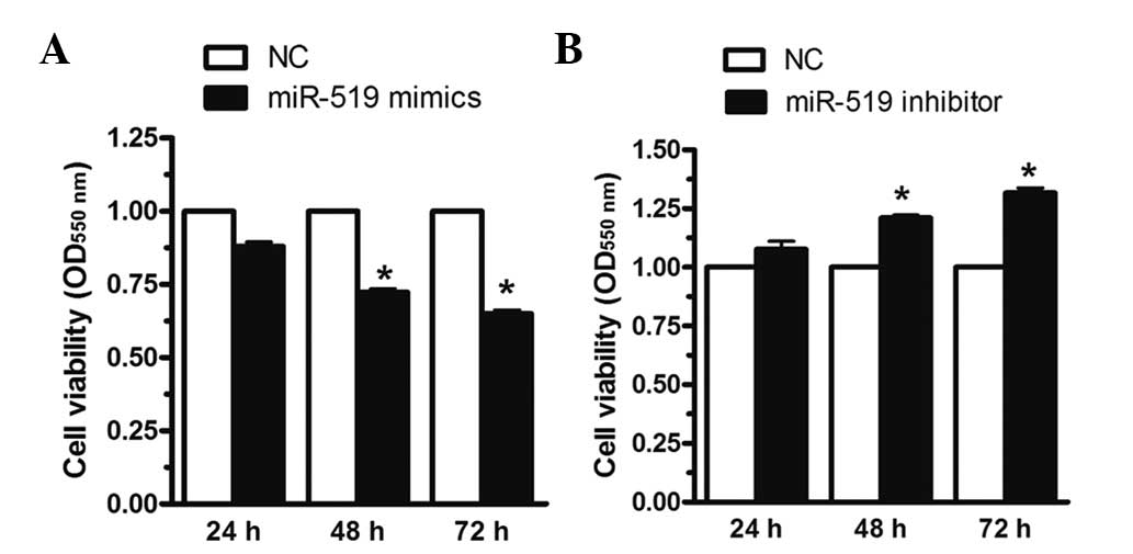 | Figure 2miR-519 regulates HL60 human AML cell
viability. The HL60 cells were transfected with miR-519 mimics,
miR-519 inhibitors or NCs for 24, 48 and 72 h. (A) Upregulation of
miR-519 expression decreased cell viability by 25 and 30%, at 48
and 72 h, respectively, whereas (B) downregulation of miR-519
expression increased cell viability in the HL60 cells by 20 and 30%
at 48 and 72 h, respectively, as determined by MTT assay. The data
are presented as the mean ± standard error of the mean (n=6) from
independent experiments. *P<0.05 vs. NC. miR,
microRNA; AML, acute myeloid leukemia; NC, negative control. |
Upregulation of miR-519 induced HL60 cell
apoptosis
The present study investigated the effects of
miR-519 on cell apoptosis. Upregulation of miR-519 expression
increased cell apoptosis by ~4-fold in the HL60 cells, as compared
with the negative control (Fig.
3A). Cell morphology was also examined by Hoechst 33342
staining. As shown in Fig. 3B, the
number of apoptotic cells increased in the HL60 cells transfected
with miR-519 mimics, as compared with the negative control. These
data suggest that upregulation of miR-519 induces HL60 cell
apoptosis. Furthermore, western blot analysis was used to identify
the molecular signaling pathway of cell apoptosis induced by
miR-519 mimics. Upregulation of miR-519 expression significantly
decreased the expression levels of HuR, and significantly increased
the expression levels of Bax and Bcl-2-like protein 11 (Bim;
Fig. 3C). miR-519 inhibitor was
used to suppress miR-519 expression. As shown in Fig. 3D, suppression of miR-519 expression
significantly increased the expression levels of HuR, and
significantly decreased the expression levels of Bax and Bim. These
results suggest that miR-519 may regulate HL60 cell apoptosis via
modulating HuR, Bax and Bim expression.
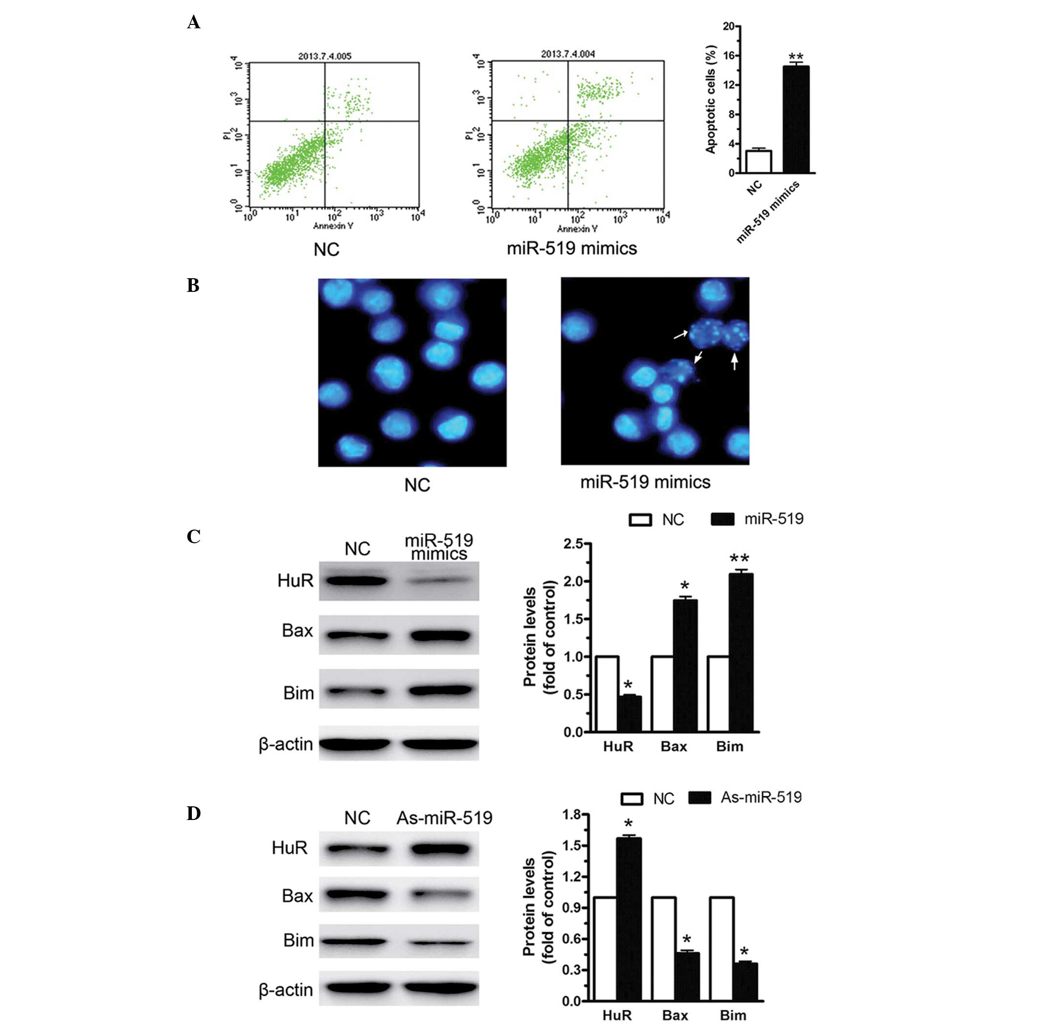 | Figure 3Upregulation of miR-519 expression
induces HL60 human AML cell apoptosis. miR-519 mimics or miR-519
inhibitors were transfected into the HL60 cells for 48 h. (A)
Upregulation of miR-519 expression significantly increased the
levels of cell apoptosis by 130% in the HL60 cells, as compared
with the NC. (B) The number of apoptotic cells increased in the
HL60 cells transfected with miR-519 mimics, as determined by
Hoechst 33342 staining. The white arrows indicate the apoptotic
cells (magnification, ×40). (C) As determined by western blotting,
upregulation of miR-519 decreased the expression levels of HuR, and
increased the expression levels of Bax and Bim, (D) whereas
suppression of miR-519 expression elevated the expression levels of
Bcl-2, and reduced the levels of Bax and Bim. The data are
presented as the mean ± standard error of the mean (n=3) of
independent experiments. *P<0.05 and
**P<0.01 vs. NC. AML, acute myeloid leukemia; miR,
microRNA; NC, negative control; HuR, human antigen R; Bcl-2, B cell
lymphoma 2; Bax, Bcl-2-like protein 4; Bim, Bcl-2-like protein
11. |
Overexpression of miR-519 inhibits cell
growth, enhances apoptosis and decreases invasion of AML cancer
cells
To investigate the role of miR-519 in AML, a cell
migration assay was conducted by scratching the cell layer prior to
transfection with antoga-miR-519 or negative control.
Downregulation of miR-519 expression markedly enhanced cell
migration (Fig. 4A). In addition,
HuR-specific siRNA and Annexin V/PI were used to investigate the
levels of cell apoptosis. Increased levels of cell apoptosis were
detected in the HL60 cells transfected with HuR-specific siRNA, as
compared with the control. Cell migration was also significantly
reduced following HuR suppression (P<0.01; Fig. 4C).
Discussion
The function and tissue specificity of miRs are
under investigation in numerous diseases to further elucidate the
underlying mechanisms of tumorigenesis. Previous studies suggested
that miRs participate in numerous cell signaling pathways by
targeting specific genes (25,26).
In addition to miRs, ARE-mediated transcript degradation is an
important post-transcriptional gene regulatory mechanism (12). AREs are cis-elements that control
cell growth and proliferation, and numerous cancer-associated
transcripts contain AREs in the 5′ or 3′-UTR, such as cytokines,
growth factors and invasion factors (27). Via recognition of the AREs, RNA
binding proteins regulate gene expression by altering the stability
and translation efficiency of mRNA. The human ELAV protein, HuR, is
an RNA-binding protein in the Hu family. HuR is widely expressed in
mammalian cells and binds to AREs in order to regulate the
stability of mRNAs; HuR is predominantly located in the nucleus of
resting cells (28). A previous
study reported that HuR binds to ARE-containing mRNAs and is
transported into the cytoplasm. The translocation of HuR from the
nucleus to the cytoplasm is an important mechanism underlying
target mRNA stabilization (29).
A previous study demonstrated that miR-519 reduced
cell proliferation by directly regulating HuR expression (21). The present study focussed primarily
on miR-519, and investigated its ability to target HuR in AML.
miR-519 is expressed at lower levels in AML cell lines, compared
with healthy bone marrow cell lines. Overexpression of miR-519 was
previously reported to decrease cell growth in murine ovarian
tumors, and to control cell proliferation and clonogenic potential
in ovarian surface epithelial cells (26), suggesting that miR-519 expression
is significant in the underlying mechanisms of tumorigenesis in
various cell types, potentially via regulation of HuR
expression.
The present study establishes HuR as a target of
miR-519 in AML. Furthermore, the restoration of miR-519 expression
altered the leukemia phenotype, indicating the possible role of
miR-519 as a tumor suppressor. To date, to the best of our
knowledge, no genomic evidence has demonstrated miR-519
downregulation, other than observation of a decrease in functional
HuR, which regulates miR-519 transcription in numerous types of
cancer cell. These data lead to the hypothesis that myeloid
leukemia cells may downregulate miR-519 to sustain HuR protein
overexpression, resulting in leukemia progression.
The molecular mechanism underlying tumor suppression
by miR-519 has been investigated in numerous types of cancer
(21,26). Gene expression analyses have yet to
be performed, suggesting that the cause of tumor suppression may be
the ability of miRs to target genes associated with the cell cycle
signaling pathway. To the best of our knowledge, HuR was the first
direct miR-519 target gene to be identified (21). As a transcription factor, HuR
regulates numerous genes that are known to contribute to normal
cell life. The observed decrease in the expression levels of HuR
target genes, such as Bax and Bim, in the present study may
elucidate the cell cycle abnormalities exhibited by myeloid cell
lines.
The results of the present study demonstrated that
miR-519 targets HuR, mediating biological activity in normal and
leukemic tissue samples. The reduced expression levels of miR-519
that deregulate HuR expression in hematopoietic development result
in pathologic outcomes (primarily via upregulation of HuR protein
expression), which leads to significant cell proliferation and
survival. Therefore, restoration of miR-34b expression is
potentially a fundamental step in treating AML and may provide a
novel therapeutic strategy.
References
|
1
|
Esquela-Kerscher A and Slack FJ:
Oncomirs-microRNAs with a role in cancer. Nat Rev Cancer.
6:259–269. 2006. View
Article : Google Scholar : PubMed/NCBI
|
|
2
|
Garzon R, Calin GA and Croce CM: MicroRNAs
in cancer. Annu Rev Med. 60:167–179. 2009. View Article : Google Scholar : PubMed/NCBI
|
|
3
|
Borralho PM, Simões AE, Gomes SE, Lima RT,
Carvalho T, Ferreira DM, Vasconcelos MH, Castro RE and Rodrigues
CM: miR-143 overexpression impairs growth of human colon carcinoma
xenografts in mice with induction of apoptosis and inhibition of
proliferation. PLoS One. 6:e237872011. View Article : Google Scholar : PubMed/NCBI
|
|
4
|
Lima RT, Busacca S, Almeida GM, Gaudino G,
Fennell DA and Vasconcelos MH: MicroRNA regulation of core
apoptosis pathways in cancer. Eur J Cancer. 47:163–174. 2011.
View Article : Google Scholar
|
|
5
|
Ciafrè SA, Galardi S, Mangiola A, Ferracin
M, Liu CG, Sabatino G, Negrini M, Maira G, Croce CM and Farace MG:
Extensive modulation of a set of microRNAs in primary glioblastoma.
Biochem Biophys Res Commun. 334:1351–1358. 2005. View Article : Google Scholar : PubMed/NCBI
|
|
6
|
Mi S, Lu J, Sun M, Li Z, Zhang H, Neilly
MB, Wang Y, Qian Z, Jin J, Zhang Y, et al: MicroRNA expression
signatures accurately discriminate acute lymphoblastic leukemia
from acute myeloid leukemia. Proc Natl Acad Sci U S A.
104:19971–19976. 2007. View Article : Google Scholar : PubMed/NCBI
|
|
7
|
Marcucci G, Radmacher MD, Maharry K,
Mrózek K, Ruppert AS, Paschka P, Vukosavljevic T, Whitman SP,
Baldus CD, Langer C, et al: MicroRNA expression in cytogenetically
normal acute myeloid leukemia. N Engl J Med. 358:1919–1928. 2008.
View Article : Google Scholar : PubMed/NCBI
|
|
8
|
Weiss GJ, Bemis LT, Nakajima E, Sugita M,
Birks DK, Robinson WA, Varella-Garcia M, Bunn PA Jr, Haney J,
Helfrich BA, et al: EGFR regulation by microRNA in lung cancer:
Correlation with clinical response and survival to gefitinib and
EGFR expression in cell lines. Ann Oncol. 19:1053–1059. 2008.
View Article : Google Scholar : PubMed/NCBI
|
|
9
|
Evangelisti C, Florian MC, Massimi I,
Dominici C, Giannini G, Galardi S, Buè MC, Massalini S, McDowell
HP, Messi E, et al: MiR-128 up-regulation inhibits Reelin and DCX
expression and reduces neuroblastoma cell motility and
invasiveness. FASEB J. 23:4276–4287. 2009. View Article : Google Scholar : PubMed/NCBI
|
|
10
|
Godlewski J, Nowicki MO, Bronisz A,
Williams S, Otsuki A, Nuovo G, Raychaudhury A, Newton HB, Chiocca
EA and Lawler S: Targeting of the Bmi-1 oncogene/stem cell renewal
factor by microRNA-128 inhibits glioma proliferation and
self-renewal. Cancer Res. 68:9125–9130. 2008. View Article : Google Scholar : PubMed/NCBI
|
|
11
|
Zhang Y, Chao T, Li R, Liu W, Chen Y, Yan
X, Gong Y, Yin B, Liu W, Qiang B, et al: MicroRNA-128 inhibits
glioma cells proliferation by targeting transcription factor E2F3a.
J Mol Med (Berl). 87:43–51. 2009. View Article : Google Scholar
|
|
12
|
Sharma S, Verma S, Vasudevan M, Samanta S,
Thakur JK and Kulshreshtha R: The interplay of HuR and miR-3134 in
regulation of AU rich transcriptome. RNA Biol. 10:1283–1290. 2013.
View Article : Google Scholar :
|
|
13
|
Wang J, Guo Y, Chu H, Guan Y, Bi J and
Wang B: Multiple functions of the RNA-binding protein HuR in cancer
progression, treatment responses and prognosis. Int J Mol Sci.
14:10015–10041. 2013. View Article : Google Scholar : PubMed/NCBI
|
|
14
|
Sahlberg AS, Ruuska M, Granfors K and
Penttinen MA: Altered regulation of ELAVL1/HuR in
HLA-B27-expressing U937 monocytic cells. PLoS One. 8:e703772013.
View Article : Google Scholar : PubMed/NCBI
|
|
15
|
Lu L, Zheng L, Si Y, et al: Hu antigen R
(HuR) is a positive regulator of the RNA-binding proteins TDP-43
and FUS/TLS: implications for amyotrophic lateral sclerosis. J Biol
Chem. 289:31792–31804. 2014. View Article : Google Scholar : PubMed/NCBI
|
|
16
|
Chen CY, Xu N and Shyu AB: Highly
selective actions of HuR in antagonizing AU-rich element-mediated
mRNA destabilization. Mol Cell Biol. 22:7268–7278. 2002. View Article : Google Scholar : PubMed/NCBI
|
|
17
|
Yaman I, Fernandez J, Sarkar B, Schneider
RJ, Snider MD, Nagy LE and Hatzoglou M: Nutritional control of mRNA
stability is mediated by a conserved AU-rich element that binds the
cytoplasmic shuttling protein HuR. J Biol Chem. 277:41539–41546.
2002. View Article : Google Scholar : PubMed/NCBI
|
|
18
|
Wang W, Furneaux H, Cheng H, Caldwell MC,
Hutter D, Liu Y, Holbrook N and Gorospe M: HuR regulates p21 mRNA
stabilization by UV light. Mol Cell Biol. 20:760–769. 2000.
View Article : Google Scholar : PubMed/NCBI
|
|
19
|
Zou T, Mazan-Mamczarz K, Rao JN, Liu L,
Marasa BS, Zhang AH, Xiao L, Pullmann R, Gorospe M and Wang JY:
Polyamine depletion increases cytoplasmic levels of RNA-binding
protein HuR leading to stabilization of nucleophosmin and p53
mRNAs. J Biol Chem. 281:19387–19394. 2006. View Article : Google Scholar : PubMed/NCBI
|
|
20
|
Ishimaru D, Ramalingam S, Sengupta TK,
Bandyopadhyay S, Dellis S, Tholanikunnel BG, Fernandes DJ and
Spicer EK: Regulation of Bcl-2 expression by HuR in HL60 leukemia
cells and A431 carcinoma cells. Mol Cancer Res. 7:1354–1366. 2009.
View Article : Google Scholar : PubMed/NCBI
|
|
21
|
Abdelmohsen K, Srikantan S, Kuwano Y and
Gorospe M: miR-519 reduces cell proliferation by lowering
RNA-binding protein HuR levels. Proc Natl Acad Sci USA.
105:20297–20302. 2008. View Article : Google Scholar : PubMed/NCBI
|
|
22
|
Locatelli F, Masetti R, Rondelli R, et al:
Outcome of children with high-risk acute myeloid leukemia given
autologous or allogeneic hematopoietic cell transplantation in the
aieop AML-2002/01 study. Bone Marrow Transplant. 50:181–188. 2015.
View Article : Google Scholar
|
|
23
|
Yang LH, Wang SL, Tang LL, et al:
Universal stem-loop primer method for screening and quantification
of microRNA. PLoS One. 9:e1152932014. View Article : Google Scholar : PubMed/NCBI
|
|
24
|
Bartel DP: MicroRNAs: Genomics,
biogenesis, mechanism and function. Cell. 116:281–297. 2004.
View Article : Google Scholar : PubMed/NCBI
|
|
25
|
Krek A, Grün D, Poy MN, Wolf R, Rosenberg
L, Epstein EJ, MacMenamin P, da Piedade I, Gunsalus KC, Stoffel M
and Rajewsky N: Combinatorial microRNA target predictions. Nat
Genet. 37:495–500. 2005. View
Article : Google Scholar : PubMed/NCBI
|
|
26
|
Abdelmohsen K, Kim MM, Srikantan S,
Mercken EM, Brennan SE, Wilson GM, Cabo RD and Gorospe M: miR-519
suppresses tumor growth by reducing HuR levels. Cell Cycle.
9:1354–1359. 2010. View Article : Google Scholar : PubMed/NCBI
|
|
27
|
Von Roretz C, di Marc S, Mazroui R and
Gallouzi IE: Turnover of AU-rich-containing mRNAs during stress: A
matter of survival. Wiley Interdiscip Rev RNA. 2:336–347. 2011.
View Article : Google Scholar : PubMed/NCBI
|
|
28
|
Abdelmohsen K and Gorospe M:
Posttranscriptional regulation of cancer traits by HuR. Wiley
Interdiscip Rev RNA. 1:214–229. 2010. View
Article : Google Scholar
|
|
29
|
Hope NR and Murray GI: The expression
profile of RNA-binding proteins in primary and metastatic
colorectal cancer: relationship of heterogeneous nuclear
ribonucleoproteins with prognosis. Hum Pathol. 42:393–402. 2013.
View Article : Google Scholar
|


