Introduction
Osteosarcoma (OS) is the most common type of
malignant bone tumor, with a morbidity of ~5 cases per million
worldwide (1). Due to its
resistance to radiotherapy, chemotherapy and adjuvant therapies,
the median survival rate of OS has not markedly improved over the
past few decades (1,2). Previous studies have demonstrated
that aberrant upregulation of oncogenes has a key role in the
development and progression of OS (3,4).
Therefore, identification of novel oncogenes may have potential as
therapeutic targets for OS.
It has previously been reported that aberrant
activation of phosphoinositide 3-kinase (PI3K) signaling is tightly
associated with numerous types of human cancer (5,6).
Whole-exome, whole-genome and RNA sequencing have highlighted the
PI3K pathway as a common vulnerability in OS, and inhibition of
PI3K signaling attenuates the malignant progression of OS (7–9).
Phosphatase and tensin homolog deleted on chromosome ten (PTEN) is
able to inhibit PI3K signaling activity, and is thus considered an
important tumor suppressors (10).
Phosphatidylinositol 3,4,5-trisphosphate-dependent Rac exchange
factor 2a (PREX2a), which is a regulator of the small guanosine
triphosphatase Rac, has recently been reported to act as an
inhibitor of PTEN activity via directly binding to PTEN through its
guanine nucleotide exchange factor (GEF) domains (11,12).
PREX2a is able to inhibit the lipid phosphatase activity of PTEN,
leading to the accumulation of phosphatidylinositol (3,4,5)-trisphosphate and phosphorylation of
Akt/mammalian target of rapamycin (13). Previous studies have suggested that
PREX2a may have an oncogenic role in numerous types of human
cancer, including gastric cancer, neuroblastoma and melanoma
(14–16). However, the exact role of PREX2a in
OS has yet to be elucidated.
The present study aimed to investigate the role of
PREX2a in the regulation of the proliferation, migration and
invasion of OS cells. In addition, the underlying molecular
mechanisms were examined.
Materials and methods
Reagents
Fetal bovine serum (FBS), TRIzol®
reagent, Lipofectamine® 2000,
3-(4,5-dimethylthiazol-2-yl)-2,5-di-phenyltetrazolium bromide (MTT)
and RevertAid First Strand cDNA Synthesis kit were purchased from
Thermo Fisher Scientific, Inc. (Waltham, MA, USA). SYBR Green
quantitative polymerase chain reaction (qPCR) assay kit was
purchased from TOYOBO (Shanghai) Co., Ltd. (Shanghai, China). Mouse
polyclonal anti-PREX2a (1:100 dilution; cat. no. ab169027), rabbit
monoclonal anti-phosphorylated (p)-PTEN (1:200; cat. no. ab109454),
mouse monoclonal anti-PTEN (1:100 dilution; cat. no. ab79156),
rabbit monoclonal anti-p-PI3K (1:200 dilution; cat. no. ab182651),
mouse monoclonal anti-PI3K (1:200 dilution; cat. no. ab86714),
mouse monoclonal anti-glyceraldehyde 3-phosphate dehydrogenase
(GAPDH; 1:100 dilution; cat. no. ab8245), mouse monoclonal
anti-MMP-2 antibody (1:200 dilution; cat. no. ab86607) and mouse
monoclonal anti-MMP-9 (1:100 dilution; cat. no. ab58803 antibodies,
and rabbit anti-mouse IgG (1:20,000 dilution; cat. no. ab6728) and
goat anti-rabbit IgG (1:10,000 dilution; cat. no. ab6721) secondary
antibodies were purchased from Abcam (Cambridge, MA, USA). Enhanced
chemiluminescence (ECL) kit was purchased from Pierce Protein
Biology; Thermo Fisher Scientific, Inc. (Rockford, IL, USA). Cell
Invasion Assay kit was purchased from Merck Millipore (Darmstadt,
Germany).
Cell culture
The MG63, Saos-2 and U2OS human OS cell lines, and
the hFOB1.19 human osteoblast cell line were obtained from the Cell
Bank of Central South University (Changsha, China). The cells were
cultured in RPMI-1640 (Thermo Fisher Scientific, Inc.) medium
supplemented with 10% FBS at 37°C in a humidified incubator
containing 5% CO2.
Transfection
Lipofectamine® 2000 was used to transfect
the cells, according to the manufacturer's protocol. Briefly, cells
were cultured to 70% confluence, and resuspended in serum-free
medium. Small interfering (si)RNAs (PREX2a specific siRNA and
negative control siRNA), plasmids (pcDNA3.1-PREX2a plasmid and
pcDNA3.1 blank vector; Auragene Biosciences, Changsha, China) and
Lipofectamine® 2000 were diluted with serum-free medium.
The diluted Lipofectamine® 2000 was added to the diluted
siRNA or plasmid, and the mixture was incubated for 20 min at room
temperature. Subsequently, the mixture was added to the cell
suspension. Following a 6 h incubation at 37°C and 5%
CO2, the medium was replaced with normal
serum-containing medium.
Reverse transcription (RT)-qPCR
analysis
Total RNA was extracted from the cells using
TRIzol® reagent, according to the manufacturer's
protocol. Total RNA was reverse transcribed into cDNA using the
RevertAid First Strand cDNA Synthesis kit, according to the
manufacturer's protocol. mRNA expression levels were examined using
the SYBR Green qPCR assay kit, in accordance with the
manufacturer's protocol. The sequences of the specific primer
(Sangon Biotech Co., Ltd., Shanghai, China) pairs used were as
follows: PREX2a, sense 5′-TGG GAG GGG TCC AAC ATCA-3′, anti-sense
5′-TCT TCA ACC GTC TGT GTT TTC TT-3′; GAPDH, sense 5′-CTC CTC CTG
TTC GAC AGT CAGC-3′ and anti-sense: 5′-CCC AAT ACG ACC AAA TCC
GTT-3′. An ABI 7500 thermal cycler was used (Applied Biosystems,
Foster City, CA, USA). The reaction conditions were as follows:
95°C for 3 min and 40 cycles of denaturation at 95°C for 15 sec and
annealing/elongation at 60°C for 30 sec. GAPDH was used as an
internal control. Independent experiments were repeated three
times. The relative mRNA expression levels were analyzed using the
2−ΔΔCq method (17).
Western blotting
Cells were solubilized in cold
radioimmunoprecipitation lysis buffer (Beyotime Institute of
Biotechnology, Shanghai, China). Protein concentration was
determined using a BCA Protein Assay kit (Beyotime Institute of
Biotechnology). Subsequently, protein (20 µg per lane) was
separated by 10% sodium dodecyl sulfate-poly-acrylamide gel
electrophoresis, and was transferred from the gel to nitrocellulose
membranes (Thermo Fisher Scientific, Inc.). The membranes were
blocked in 5% nonfat dried milk in phosphate-buffered saline (PBS)
containing 0.1% Tween for 3 h and were then incubated overnight at
4°C with mouse anti-PREX2a monoclonal antibody (1:100), mouse
anti-matrix metalloproteinase (MMP)2 monoclonal antibody (1:100),
mouse anti-MMP9 monoclonal antibody (1:100), mouse anti-p-PI3K
monoclonal antibody (1:100), mouse anti-PI3K monoclonal antibody
(1:100), mouse anti-p-PTEN monoclonal antibody (1:100), mouse
anti-PTEN monoclonal antibody (1:100) and mouse anti-GAPDH
monoclonal antibody (1:400). Following washing twice with
Dulbecco's phosphate-buffered saline (Thermo Fisher Scientific,
Inc.; 5 min/wash), the membranes were incubated with rabbit
anti-mouse secondary antibody (1:5,000) for 40 min at room
temperature. Subsequently, immune complexes were detected using an
ECL kit. The membranes were scanned in grayscale by Image-Pro Plus
software 6.0 (Media Cybernetics, Inc., Rockville, MD, USA), in
order to determine the relative protein expression levels. The
relative protein expression levels are presented as the density
ratio vs. GAPDH.
Cell proliferation assay
The MTT assay was used to measure cell
proliferation. Cells (1×10⁵ cells/well) in each group
were cultured in a 96-well plate, each well was filled with 100
µl fresh serum-free medium and 0.5 g/l MTT. The plate was
incubated at 37°C for 0, 24, 48 and 72 h, after which the medium
was removed by aspiration and 50 µl dimethyl sulfoxide was
added to each well. Following a further 10 min incubation at 37°C,
the absorbance of each sample was measured at a wavelength of 492
nm using a plate reader (CX22; Olympus, Tokyo, Japan).
Cell invasion assay
Cells (1×10⁶ cells/well) were starved in
serum-free medium for 24 h and were subsequently resus-pended in
serum-free medium. The Transwell was coated in Matrigel. Cells were
added to the upper chamber of a Transwell system, whereas the lower
chamber was filled with medium supplemented with 10% FBS. Following
a 24 h incubation, the cells attached to the bottom were stained
with crystal violet for 20 min, and were washed and air-dried.
Invasive cells were observed under a microscope.
Wound scratch assay
Wound scratch assay was performed in order to
determine the cell migratory capacity of each group. Cells were
cultured to full confluence and a wound of ~1 mm width was created
in the cell layer using a plastic scriber. Subsequently, the cells
were washed with PBS and were incubated for 48 h at 37°C and 5%
CO2. The cells in each group were fixed with 90% ethanol
and observed under a microscope.
Statistical analysis
Data are presented as the mean ± standard deviation
of three independent experiments. The results were analyzed using
SPSS 17.0 statistical software (SPSS, Inc., Chicago, IL, USA).
Differences between the groups were determined using one-way
analysis of variance. P<0.05 was considered to indicate a
statistically significant difference.
Results
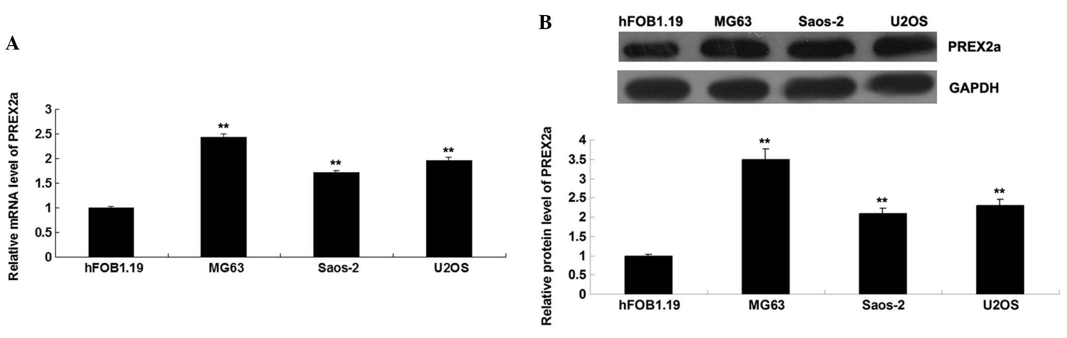
PREX2a is upregulated in OS cells
To determine the role of PREX2a in OS, the
expression levels of PREX2a were detected in OS cell lines: MG63,
Saos-2 and U2OS. The normal human osteoblast cell line hFOB1.19 was
used as a control. As shown in Fig. 1A
and B, the mRNA and protein expression levels of PREX2a were
significantly increased in the OS cells, as compared with in the
hFOB1.19 cells. Since MG63 cells exhibited the most significant
upregulation in PREX2a expression, this cell line was used in all
subsequent experiments. The MG63 OS cells were transfected with
PREX2a-specific siRNA or pcDNA3.1-PREX2a plasmid. Non-specific
siRNA and blank pcDNA3.1 vector were used as negative controls.
Post-transfection, the protein and mRNA expression levels of PREX2a
were detected in each group. The expression levels of PREX2a were
significantly reduced post-transfection with PREX2a siRNA (Fig. 2A and B); however, the expression
levels were markedly increased post-transfection with the
pcDNA3.1-PREX2a plasmid (Fig. 2C and
D). Transfection with non-specific siRNA or blank pcDNA3.1
vector did not affect the expression levels of PREX2a. These data
indicate that the transfection was successful.
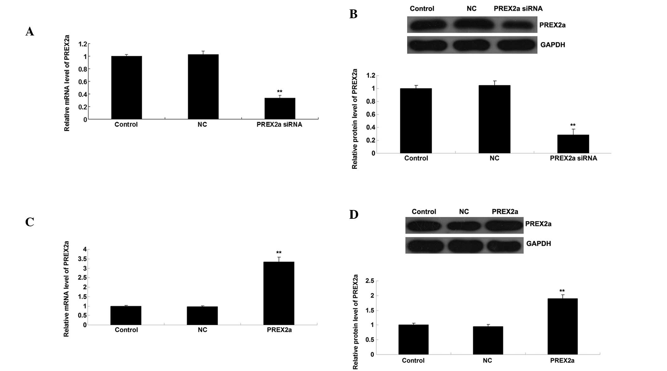 | Figure 2(A) mRNA and (B) protein expression
levels of PREX2a in MG63 cells transfected with PREX2a siRNA or
non-specific siRNA (NC) as detected by RT-qPCR and western
blotting, respectively. (C) mRNA and (D) protein expression levels
of PREX2a in MG63 cells transfected with pcDNA3.1-PREX2a plasmid or
blank pcDNA3.1 vector (NC), as detected by RT-qPCR and western
blotting, respectively. Control group: Untransfected cells. Data
are presented as the mean ± standard deviation.
**P<0.01 vs. the control cells. PREX2a,
phosphatidylinositol-3,4,5-trisphosphate-dependent Rac exchange
factor 2a; GAPDH, glyceral-dehyde 3-phosphate dehydrogenase; siRNA,
small interfering RNA; NC, negative control; RT-qPCR, reverse
transcription-quantitative polymerase chain reaction. |
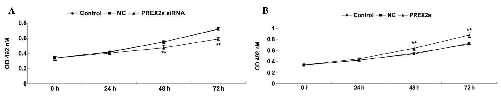
PREX2a enhances OS cell
proliferation
The present study determined the proliferative
capacity of OS cells in each group. As shown in Fig. 3A and B, knockdown of PREX2a
significantly inhibited OS cell proliferation. Conversely,
overexpression of PREX2a notably promoted OS cell proliferation.
These results suggest that PREX2a may promote the proliferation of
MG63 OS cells.
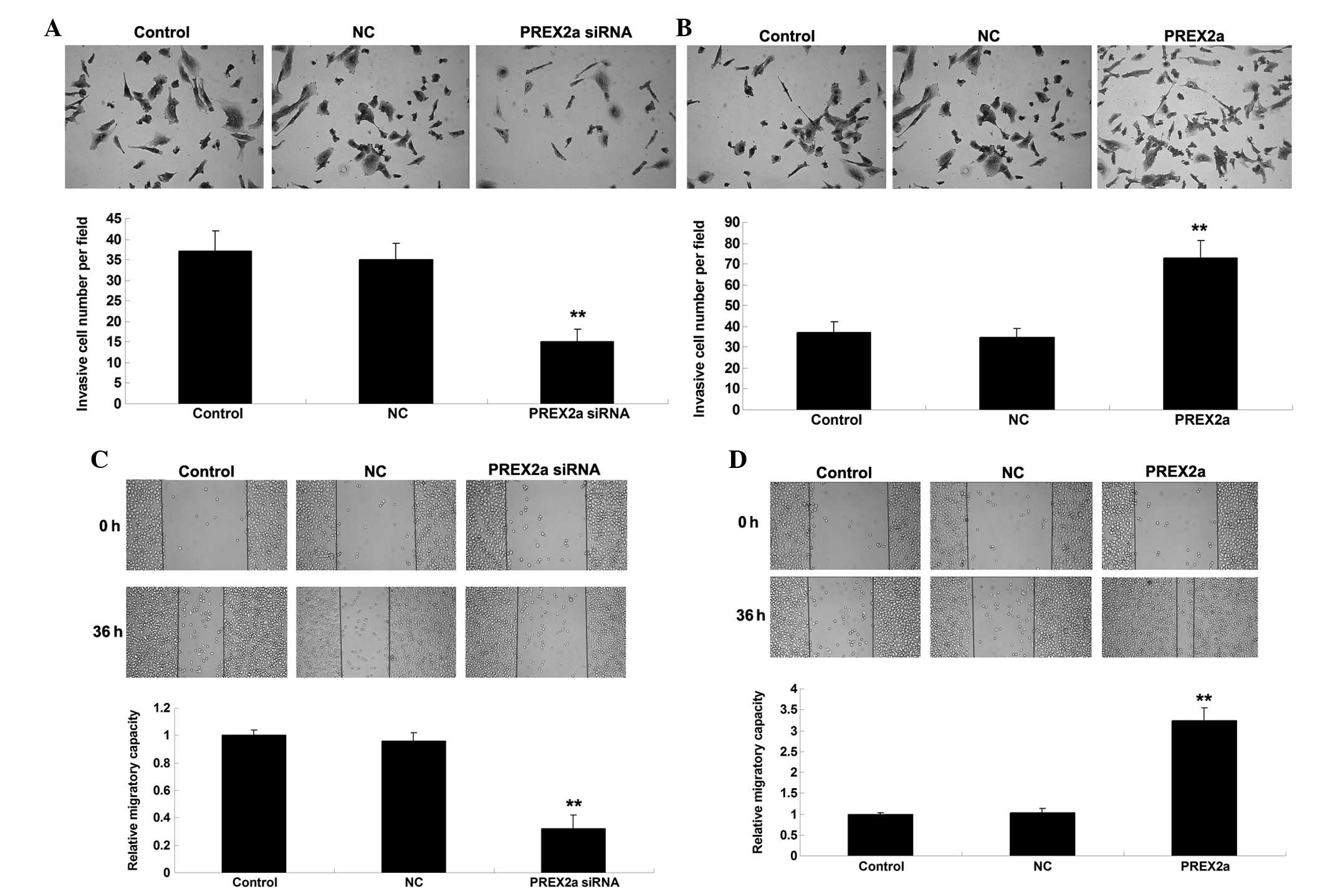
PREX2a promotes the invasion and
migration of OS cells
Since tumor cell invasion and migration have
essential roles in cancer metastasis, the role of PREX2a was
investigated in the regulation of OS cell invasion and migration,
as determined by Transwell and wound healing assays, respectively.
As shown in Fig. 4A and B, the
invasive capacity of MG63 OS cells was significantly reduced
following knockdown of PREX2a; however, PREX2a overexpression
increased the invasion of MG63 cells. Similarly, cell migration was
downregulated following knockdown of PREX2a expression, whereas it
was increased following overexpression of PREX2a in MG63 cells
(Fig. 4C and D). In addition,
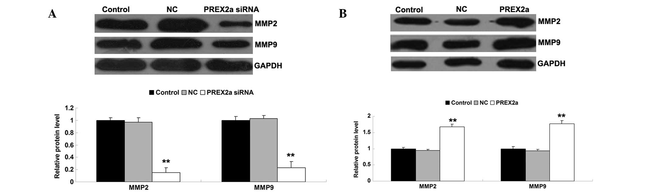
since MMP2 and MMP9 have important roles in mediating tumor cell
invasion and migration, the expression levels of these proteins
were detected in each group. Consistent with the results of the
cell invasion and migration assays, knockdown of PREX2a inhibited
the protein expression levels of MMP2 and MMP9, whereas
overexpression of PREX2a enhanced their protein expression levels
(Fig. 5A and B). These results
indicate that PREX2a may have a promoting role in the regulation of
OS cell invasion and migration, at least partly via modulation of
MMP2 and MMP9 expression.
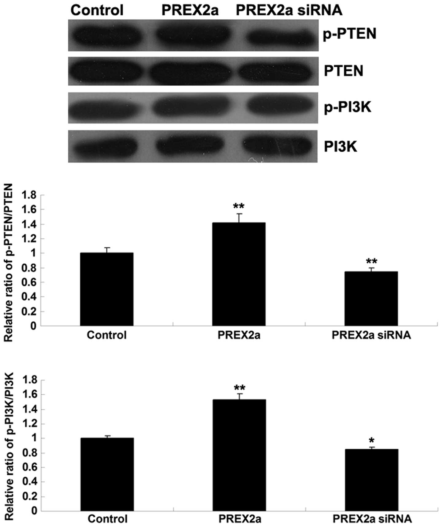
siRNA-mediated knockdown of PREX2a
inhibits PI3K signaling activity in OS cells
PTEN and PI3K activity was further investigated in
the MG63 OS cells following knockdown or overexpression of PREX2a.
In MG63 cells overexpressing PREX2a, the phosphorylation levels of
PTEN were increased, which was accompanied by upregulated
phosphorylation of PI3K, thus indicating that PI3K signaling was
activated (Fig. 6). Conversely,
the phosphorylation levels of PTEN were reduced in MG63 cells
transfected with PREX2a-specific siRNA, indicating that the
activity of PTEN was upregulated, which was accompanied by
downregulated phosphorylation of PI3K, thus indicating that PI3K
signaling activity was reduced (Fig.
6). These results suggest that PTEN and PI3K signaling activity
may be modulated by PREX2a in MG63 cells.
Discussion
The identification of novel oncogenes may aid in the
development of therapeutic strategies against OS. The present study
is the first, to the best of our knowledge, to investigate the role
of PREX2a in OS in vitro. The results demonstrated that
PREX2a was significantly upregulated in OS cells, as compared with
in normal human osteoblast cells. Further investigations revealed
that PREX2a was able to promote OS cell proliferation, invasion and
migration, potentially via upregulation of MMP2 and MMP9. In
addition, the present study suggested that the oncogenic role of
PREX2a in OS may be realized via direct inhibition of PTEN activity
and subsequent activation of PI3K signaling.
PREX2a contains an N-terminal Dbl homology and
pleckstrin homology domain, which confers GEF activity; pairs of
PDZ and Dishevelled, Egl-10 and Pleckstrin domains; and a
C-terminus with weak similarity to inositol 4-polyphosphate
phosphatase (12). Through its GEF
domains, PREX2a is able to directly bind to PTEN, and act as an
inhibitor of PTEN activity (18,19).
Furthermore, the genetic sequence of PREX2a is similar to that of
PREX1, which has been demonstrated to have an oncogenic role in
human cancer. Qin et al (20) reported that upregulation of PREX1
enhanced prostate cancer metastasis; the expression of recombinant
PREX1 in non-metastatic prostate cancer cells increased cell
migration and invasion via Rac-dependent lamellipodia formation. In
addition, using a mouse xenograft model, the expression of
recombinant PREX1 was shown to induce lymph node metastasis of
non-metastatic prostate cancer cells without an effect on primary
tumor growth (20). Furthermore,
Montero et al (21)
identified a significant correlation between high PREX1 expression
and poor patient outcome in breast cancer; knockdown of PREX1
expression suppressed breast cancer cell migration and invasion,
and tumorigenic potential in vivo.
It has previously been reported that loss of PTEN
function frequently occurs in human cancer; therefore, the binding
partners of PTEN may also be involved in tumorigenesis via
modulation of PTEN activity (10).
Similar to PREX1, PREX2a has been demonstrated to act as an
oncogene in human cancer via regulation of biological processes,
including cell proliferation, cell cycle progression, migration and
invasion (14,15,22).
Whole-genome sequencing identified PREX2 as a significantly mutated
gene in melanoma (16). In
addition, overexpression of PREX has been shown to be associated
with poor patient outcome in breast cancer (23). PREX2a has also been identified as a
direct target of microRNA (miR)-338-3p, and knockdown of PREX2a
inhibited cell proliferation and clonogenicity, and induced a
G1/S arrest and apoptosis in gastric cancer cells,
potentially via mediation of PTEN-PI3K signaling (14). Chen et al (15) also reported similar findings in
neuroblastoma; knockdown of PREX2a inhibited neuroblastoma cell
proliferation, migration and invasion via the PTEN-PI3K pathway.
Furthermore, PREX2a was identified as a target gene of miR-338-3p,
and overexpression of PREX2a reversed the inhibitory effects on the
proliferation and invasion of neuroblastoma cells (15).
In the present study, PREX2a was shown to be
significantly upregulated in OS cell lines, as compared with in a
normal osteoblast cell line. In addition, silencing PREX2a
expression suppressed OS cell proliferation, migration and
invasion. MMP2 and MMP9 have been suggested to participate in the
metastasis of OS (24), and the
expression and activity of MMP2 and MMP9 are tightly associated
with OS cell migration and invasion (25–27).
The present study demonstrated that suppression of PREX2a notably
inhibited the invasion and migration of OS cells, at least partly
via suppressing the protein expression of MMP2 and MMP9.
In conclusion, the present study indicated that
PREX2a had an oncogenic role in the regulation of proliferation,
invasion and migration of OS cells via mediation of PI3K
signaling.
Acknowledgments
The present study was supported by the Fundamental
Research Funds for the Central Universities of Central South
University (grant no. 2013zzts092).
References
|
1
|
Thompson LD: Osteosarcoma. Ear Nose Throat
J. 92:288–290. 2013.PubMed/NCBI
|
|
2
|
PosthumaDeBoer J, Witlox MA, Kaspers GJ
and van Royen BJ: Molecular alterations as target for therapy in
metastatic osteosarcoma: A review of literature. Clin Exp
Metastasis. 28:493–503. 2011. View Article : Google Scholar : PubMed/NCBI
|
|
3
|
Yang J and Zhang W: New molecular insights
into osteosarcoma targeted therapy. Curr Opin Oncol. 25:398–406.
2013. View Article : Google Scholar : PubMed/NCBI
|
|
4
|
Namløs HM, Meza-Zepeda LA, Barøy T,
Østensen IH, Kresse SH, Kuijjer ML, Serra M, Bürger H,
Cleton-Jansen AM and Myklebost O: Modulation of the osteosarcoma
expression phenotype by microRNAs. PLoS One. 7:e480862012.
View Article : Google Scholar : PubMed/NCBI
|
|
5
|
Braccini L, Ciraolo E, Martini M, Pirali
T, Germena G, Rolfo K and Hirsch E: PI3K keeps the balance between
metabolism and cancer. Adv Biol Regul. 52:389–405. 2012. View Article : Google Scholar : PubMed/NCBI
|
|
6
|
Wu P and Hu YZ: PI3K/Akt/mTOR pathway
inhibitors in cancer: A perspective on clinical progress. Curr Med
Chem. 17:4326–4341. 2010. View Article : Google Scholar : PubMed/NCBI
|
|
7
|
Burris HA III: Overcoming acquired
resistance to anticancer therapy: Focus on the PI3K/AKT/mTOR
pathway. Cancer Chemother Pharmacol. 71:829–842. 2013. View Article : Google Scholar : PubMed/NCBI
|
|
8
|
Perry JA, Kiezun A, Tonzi P, Van Allen EM,
Carter SL, Baca SC, Cowley GS, Bhatt AS, Rheinbay E, Pedamallu CS,
et al: Complementary genomic approaches highlight the PI3K/mTOR
pathway as a common vulnerability in osteosarcoma. Proc Natl Acad
Sci USA. 111:E5564–E5573. 2014. View Article : Google Scholar : PubMed/NCBI
|
|
9
|
Yang L, Shu T, Liang Y, Gu W, Wang C, Song
X, Fan C and Wang W: GDC-0152 attenuates the malignant progression
of osteosarcoma promoted by ANGPTL2 via PI3K/AKT but not p38MAPK
signaling pathway. Int J Oncol. 46:1651–1658. 2015.PubMed/NCBI
|
|
10
|
Eng C: PTEN: One gene, many syndromes. Hum
Mutat. 22:183–198. 2003. View Article : Google Scholar : PubMed/NCBI
|
|
11
|
Hodakoski C, Hopkins BD, Barrows D, Mense
SM, Keniry M, Anderson KE, Kern PA, Hawkins PT, Stephens LR and
Parsons R: Regulation of PTEN inhibition by the pleckstrin homology
domain of P-REX2 during insulin signaling and glucose homeostasis.
Proc Natl Acad Sci USA. 111:155–160. 2014. View Article : Google Scholar :
|
|
12
|
Donald S, Hill K, Lecureuil C, Barnouin R,
Krugmann S, John Coadwell W, Andrews SR, Walker SA, Hawkins PT,
Stephens LR and Welch HC: P-Rex2, a new guanine-nucleotide exchange
factor for Rac. FEBS Lett. 572:172–176. 2004. View Article : Google Scholar : PubMed/NCBI
|
|
13
|
Fine B, Hodakoski C, Koujak S, Su T, Saal
LH, Maurer M, Hopkins B, Keniry M, Sulis ML, Mense S, et al:
Activation of the PI3K pathway in cancer through inhibition of PTEN
by exchange factor P-REX2a. Science. 325:1261–1265. 2009.
View Article : Google Scholar : PubMed/NCBI
|
|
14
|
Guo B, Liu L, Yao J, Ma R, Chang D, Li Z,
Song T and Huang C: miR-338-3p suppresses gastric cancer
progression through a PTEN-AKT axis by targeting P-REX2a. Mol
Cancer Res. 12:313–321. 2014. View Article : Google Scholar : PubMed/NCBI
|
|
15
|
Chen X, Pan M, Han L, Lu H, Hao X and Dong
Q: miR-338-3p suppresses neuroblastoma proliferation, invasion and
migration through targeting PREX2a. FEBS Lett. 587:3729–3737. 2013.
View Article : Google Scholar : PubMed/NCBI
|
|
16
|
Berger MF, Hodis E, Heffernan TP, Deribe
YL, Lawrence MS, Protopopov A, Ivanova E, Watson IR, Nickerson E,
Ghosh P, et al: Melanoma genome sequencing reveals frequent PREX2
mutations. Nature. 485:502–506. 2012.PubMed/NCBI
|
|
17
|
Livak KJ and Schmittgen TD: Analysis of
relative gene expression data using real-time quantitative PCR and
the 2(-Delta Delta C(T)) Method. Methods. 25:402–408. 2001.
View Article : Google Scholar
|
|
18
|
Rosenfeldt H, Vázquez-Prado J and Gutkind
JS: P-REX2, a novel PI-3-kinase sensitive Rac exchange factor. FEBS
Lett. 572:167–171. 2004. View Article : Google Scholar : PubMed/NCBI
|
|
19
|
Leslie NR: P-REX2a driving tumorigenesis
by PTEN inhibition. Sci Signal. 2:pe682009. View Article : Google Scholar : PubMed/NCBI
|
|
20
|
Qin J, Xie Y, Wang B, Hoshino M, Wolff DW,
Zhao J, Scofield MA, Dowd FJ, Lin MF and Tu Y: Upregulation of
PIP3-dependent Rac exchanger 1 (P-Rex1) promotes prostate cancer
metastasis. Oncogene. 28:1853–1863. 2009. View Article : Google Scholar : PubMed/NCBI
|
|
21
|
Montero JC, Seoane S, Ocaña A and
Pandiella A: P-Rex1 participates in Neuregulin-ErbB signal
transduction and its expression correlates with patient outcome in
breast cancer. Oncogene. 30:1059–1071. 2011. View Article : Google Scholar
|
|
22
|
Donald S, Humby T, Fyfe I, Segonds-Pichon
A, Walker SA, Andrews SR, Coadwell WJ, Emson P, Wilkinson LS and
Welch HC: P-Rex2 regulates Purkinje cell dendrite morphology and
motor coordination. Proc Natl Acad Sci USA. 105:4483–4488. 2008.
View Article : Google Scholar : PubMed/NCBI
|
|
23
|
Pandiella A and Montero JC: Molecular
pathways: P-Rex in cancer. Clin Cancer Res. 19:4564–4569. 2013.
View Article : Google Scholar : PubMed/NCBI
|
|
24
|
Wang JY, Wu PK, Chen PC, Yen CC, Hung GY,
Chen CF, Hung SC, Tsai SF, Liu CL, Chen TH and Chen WM:
Manipulation therapy prior to diagnosis induced primary
osteosarcoma metastasis - from clinical to basic research. PLoS
One. 9:e965712014. View Article : Google Scholar
|
|
25
|
Fromigué O, Hamidouche Z and Marie PJ:
Blockade of the RhoA-JNK-c-Jun-MMP2 cascade by atorvastatin reduces
osteosarcoma cell invasion. J Biol Chem. 283:30549–30556. 2008.
View Article : Google Scholar : PubMed/NCBI
|
|
26
|
Chueh FS, Chen YY, Huang AC, Ho HC, Liao
CL, Yang JS, Kuo CL and Chung JG: Bufalin-inhibited migration and
invasion in human osteosarcoma U-2 OS cells is carried out by
suppression of the matrix metalloproteinase-2, ERK, and JNK
signaling pathways. Environ Toxicol. 29:21–29. 2014. View Article : Google Scholar
|
|
27
|
Jin J, Cai L, Liu ZM and Zhou XS:
miRNA-218 inhibits osteosarcoma cell migration and invasion by
down-regulating of TIAM1, MMP2 and MMP9. Asian Pac J Cancer Prev.
14:3681–3684. 2013. View Article : Google Scholar : PubMed/NCBI
|