Introduction
Spinal manipulative therapy (SMT) has been
universally applied in the treatment of lumbar degenerative
diseases and injuries, with a definite curative effect. Based on
the mechanical characteristics of the manipulation, SMT is divided
into two main methods (1): i)
Mobilization, which has a low speed and low amplitude force; and
ii) thrust manipulation, which has a high speed and low amplitude
force. It is generally accepted that the purpose of SMT is to
correct the mechanical disturbance of the intervertebral joint and
to restore intervertebral joint function (2). The lumbar intervertebral joint
consists of three joints, including the intervertebral disc and two
facet joints (3). The
intervertebral disc functions to bear and transmit load, allowing
the lumbar vertebra to move in all directions, while limiting
excessive movement of the waist (4). The facet joint controls lumbar motion
segment activity and limits lumbar spine load. Therefore,
determining the characteristics of intradiscal pressure (IDP) and
pressure of facet joints is important in distinguishing the
mechanical effects of the two techniques, which may provide an
experimental basis for their use. Our previous study analyzed the
pressure alteration characteristics of the facet joints under
simulation of the two techniques (5). In the present study, the
characteristics of IDP under simulation of the two techniques were
further investigated.
In a previous report, the maximum IDP in the lumbar
spine was measured under simulation of the two techniques, but a
comparison of the speed of IDP was not clarified (6). Furthermore, due to hydrostatic
properties of the nucleus pulposus, the IDP in only a single point
is measured (6). However, during
SMT, the lumbar vertebrae is in a complex process of movement.
Because different parts of the disc are compressed to different
degrees, the IDP is also different in the nucleus (7). The finite element study (8) demonstrated that the stress of the
rotation side of the lumbar intervertebral disc was significantly
greater than the contralateral side during lumbar thrust
manipulation. In order to further elaborate the characteristics of
the IDP under two simulated techniques, a micro-pressure sensor
measurement was performed in the present study to further analyze
the differences in velocity, as well as the differences in IDP
between the rotating and contralateral side. This may aid the
selection of techniques applied in clinical practice provide
insight into the underlying mechanism of SMT.
Materials and methods
Experimental specimens
A total of 12 adult male fresh cadaver lumbar sacral
segments (T12-S2) were provided by the Department of Human Anatomy
of Southern Medical University. Specimens with anatomical variation
and osteopathy were excluded. Samples were dissected free of
superficial musculature to expose the facet joint capsule. The
samples were sealed with double layer plastic wrap at −20°C. The
specimens were placed overnight in the refrigerator 1 day prior to
experimentation. The present study was approved by the Medical
Ethics Committee of Wang Jing Hospital (Beijing, China). Informed
consent to donate the body to medical research was obtained from
all donators or their next of kin.
Experimental instruments
Electroforce 3510 test instrument (Bose
Corporation-Electroforce Systems Group; TA Instruments, Inc., New
Castle, DE, USA). A micro-pressure sensor (060 type; range, 0–6.895
Mpa) with a diameter of 1.5 mm and thickness of 0.3 mm was obtained
from Precision Measurements, Inc. (Virginia Beach, VA, USA). The
USB7360 data acquisition system was developed by Mingtong Century
Science and Technology Co., Ltd. (Tianjin, China).
Specimen preparation
Once thawed, 2 screws were set from the ventral side
of S2 with a 1.5 cm exposure, and 2 screws were also set from the
ventral side of T12 with 1.5 cm exposure. Specimens were potted at
S2 and at T12 using quick-setting methacrylate (Shanghai New
Century Dental Material Co., Ltd., Shanghai, China) and the L1-S1
segment was exposed. During the experimental process, the specimens
were intermittently sprayed with PBS (pH=7.4) to keep the specimens
moist, and wrapped in gauze soaked intermittently with PBS. Lumbar
vertebrae (n=12) were randomly divided into two groups, with six
specimens each. Simulated mobilization was used in one group and
thrust manipulation was used in the other group.
Simulation of SMT
During the experimental process, the room
temperature was maintained at ~25°C. The prepared lumbar specimens
were fixed in the Electroforce3510 test instrument with a square
head fixture, and the end was fixed in a vice. A 2 mm diameter
drill hole was punched at the L5 spinous process. A thin steel wire
was tied and fixed through the hole and the wire end was fixed. The
other end of the wire was used for hanging weights through a fixed
pulley, which enabled the L5 spike to be subjected to a horizontal
pulling force, and the finger pointing force of the thumb was
simulated (Fig. 1). To minimize
the viscoelastic effect of the specimens, a small scale
loading/unloading pre-treatment of the specimens was performed
prior to the experiment. Torque (10 Nm) was loaded for 10 sec, and
subsequently unloaded. The rotation angle was recorded. To restore
the specimen to its physiological state, there was a 3 min interval
between each measurement (9).
Torque (10 Nm) loading/unloading was repeated until three
consecutive loads were at the same rotation angle (+0.1°).
The angle control mode was adopted and the specific
parameters were as follows.
Mobilization (rotate to the right): The pre-loading
angle was 15° at a speed of 3°/sec. The maximum loading angle was
20° at a speed of 1°/sec, and the return speed was 3°/sec. The L5
spike level was loaded with 9 N, and 300 N was down-loaded to
simulate body weight.
Thrust manipulation (rotate to the right): The
pre-loading angle was 15° at a speed of 3°/sec. The maximum loading
angle was 20° at a speed of 33°/sec and the return speed was
3°/sec. The L5 spike level was loaded with 22 N, and 300 N was
down-loaded to simulate body weight.
Measurement of the IDPs
Calibrated micro-pressure sensors were located at
the right front and left rear of the L4-5 and L5-S1 discs. Using
the placement of micro-pressure sensors on the right front of the
L5-S1 intervertebral disc as an example, the L5-S1 intervertebral
disc was punctured (1.5, 2 and 3 mm diameter one by one) at a depth
of 1–1.2 cm. A stainless steel sleeve was fixed to prevent the
sensor from being extruded from the intervertebral disc. A 2 mm
diameter Kirschner wire was punctured into the sleeve by 2.3 cm, so
that the intervertebral disc tissue in the sleeve could be pushed
out.
The sensor was pushed in the disc through the
sleeve, and then the sleeve was slowly withdrawn. To keep the disc
relatively closed and to stabilize the sensor, 502 glue was applied
to the puncture entrance. In the left posterior part of the L5-S1
intervertebral disc (from the left of the anterior longitudinal
ligament; depth, 2–2.2 cm), the right posterior part of the L4-5
intervertebral disc (from the right side of the anterior
longitudinal ligament; depth, 1–1.2 cm) and the left posterior part
of the L4-5 intervertebral disc (from the left of the anterior
longitudinal ligament; depth, 2–2.2 cm), the #2, #3, and #4
micro-pressure sensors were inserted (Fig. 1). The sensor was connected to the
USB7360 data acquisition system.
Main outcome index
During the simulation process, the load torque of
the specimen was recorded, as well as the IDP of the left and right
sides of the L5-S1 and L4-5 intervertebral discs.
Statistical analysis
SPSS 19.0 software (IBM Corp., Armonk, NY, USA) was
used for statistical analysis of the collected data. Each specimen
was retested twice using the same protocol, and measurements were
reproducible. Data were presented as the mean ± standard error of
the mean. All data were tested for normality and homogeneity of
variance. P<0.05 was considered to indicate a statistically
significant difference. A t-test was used to compare the maximum
load torque between the manual groups. In each group, the IDP at
different stages and different sides was compared by two way
analysis of variance followed by the Fisher's Least Significant
Difference post-hoc test. Then, a paired t-test was used to compare
the IDP of each stage between the left and right sides. An
independent sample t-test was used to compare the velocity of IDP
between the two groups.
Results
Experimental loading parameters
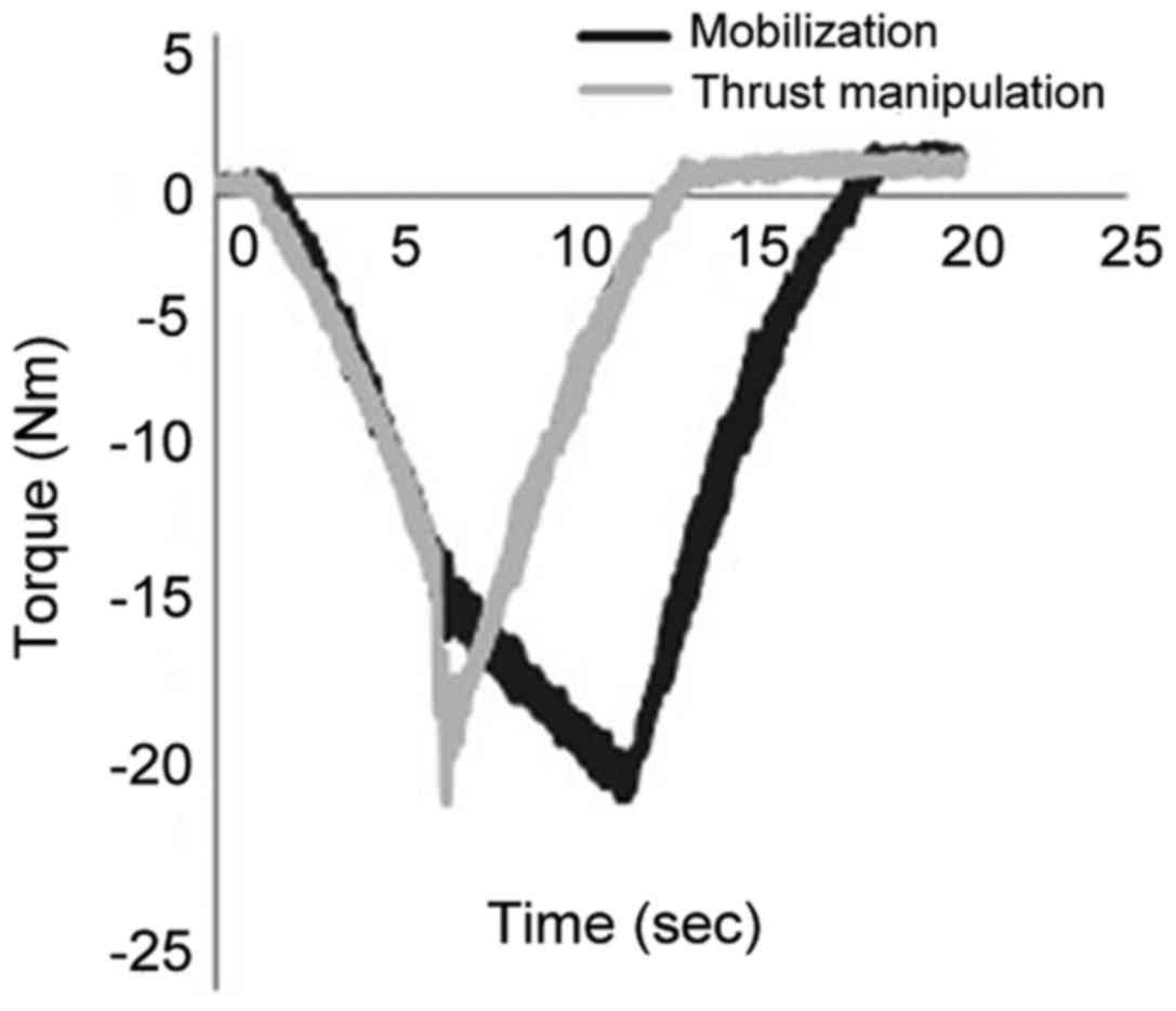
Loading torque: The load torque during simulated
mobilization increased slowly. Once the peak was reached, the load
torque decreased. During simulated thrust manipulation, the loading
torque increased slowly at first, suddenly reached a peak, then
decreased gradually (Fig. 2).
There was no significant difference in the maximum load torque
(P>0.05; Table I). The results
suggested the main difference in loading was torque velocity
between the two simulated techniques.
 | Table I.Comparison of maximum torque of two
simulated spinal manipulative therapies under loading (x±s,
Nm). |
Table I.
Comparison of maximum torque of two
simulated spinal manipulative therapies under loading (x±s,
Nm).
| Group | n | Maximum loading
torque |
|---|
| Mobilization | 6 | 18.71±1.25 |
| Thrust
manipulation | 6 | 17.61±1.20 |
| T-value | – | 1.556 |
| P-value | – | 0.151 |
Distribution characteristics of IDP
during simulated SMT
In the simulated mobilization group, IDP alteration
was not apparent. The rotation angle increased and the IDP
gradually increased to the maximum value. In the thrust
manipulation group, IDP alteration was not apparent in the preload
phase, and then suddenly reached a peak during the thrust phase
(Fig. 3).
The maximum IDP of L4-5 and L5-S1 in the two groups
was greater than the initial and end IDP (P<0.01), and there was
no significant difference between the initial and end IDPs
(P>0.05). The same situation occurred on the rotation and
contralateral sides of the intervertebral disc (Fig. 4; Tables II–V). The maximum IDP on the rotating side
of L4-5 and L5-S1 was greater than on the contralateral side in two
groups (P<0.05), which suggested that the IDP increased
transiently during the two techniques, particularly on the rotating
side.
| Table II.Comparison of the IDP on the rotating
right sides and contralateral left sides of the L5-S1
intervertebral disc in different stages of simulated mobilization
(x±s, MPa). |
Table II.
Comparison of the IDP on the rotating
right sides and contralateral left sides of the L5-S1
intervertebral disc in different stages of simulated mobilization
(x±s, MPa).
| Position | n | 300 N pressure | Maximum pressure | End pressure | F-value | P-value |
|---|
| Right | 6 | 0.793±0.134 | 1.078±0.084 | 0.783±0.129 | 24.211 |
<0.0001b |
| Left | 6 | 0.659±0.047 | 0.804±0.009 | 0.662±0.050 |
|
|
| T-value | – | 2.317 | 7.249 | 1.892 | – | – |
| P-value | – | 0.068 | 0.001a | 0.117 | – | – |
| Table V.Comparison of the IDP on the rotating
and contralateral sides of the L4-5 intervertebral disc in
different stages of simulated thrust manipulation (x±s, MPa). |
Table V.
Comparison of the IDP on the rotating
and contralateral sides of the L4-5 intervertebral disc in
different stages of simulated thrust manipulation (x±s, MPa).
| Site | n | 300 N | Maximum
pressure | End pressure | F-value | P-value |
|---|
| Right | 6 | 0.761±0.121 | 1.204±0.121 | 0.777±0.131 | 11.789 |
<0.0001b |
| Left | 6 | 0.650±0.205 | 0.827±0.258 | 0.648±0.190 |
|
|
| T-value | – | 1.147 | 3.243 | 1.367 | – | – |
| P-value | – | 0.278 | 0.009a | 0.202 | – | – |
Comparison of IDP between two
groups
There was no significant difference (P>0.05) in
the maximum IDP between the two groups on the L5-S1 or L4-5
rotating side (Fig. 5A and C;
Table VI). There was no
significant difference in the maximum IDP on the contralateral side
between the two groups (P>0.05; Fig. 5B and D).
| Table VI.Comparison of the intradiscal
pressure of the L5-S1 intervertebral disc on rotating side between
the two groups (x±s, MPa). |
Table VI.
Comparison of the intradiscal
pressure of the L5-S1 intervertebral disc on rotating side between
the two groups (x±s, MPa).
| Technique | n | 300 N | Maximum
pressure |
|---|
| Mobilization | 6 | 0.793±0.134 | 1.078±0.084 |
| Thrust
manipulation | 6 | 0.779±0.098 | 1.050±0.130 |
| T-value | – | 0.206 | 0.450 |
| P-value | – | 0.841 | 0.663 |
Comparison of the speed of change of
IDP in two groups
The speed of IDP increase on both sides of the L4-5
and L5-S1 intervertebral discs in the simulated thrust manipulation
group was higher than the simulated mobilization group (P<0.01),
and the speed of IDP decrease was not significantly different
(P>0.05; Fig. 6). The results
indicated that simulated thrust manipulation may have more of an
instant impact to discs than simulated mobilization.
Discussion
A large number of spinal manipulation parameters
have been determined in vivo (10–14),
and the following characteristics have been identified: When the
treatment site differs, the force is significantly different. When
the operator is different, the force also differs. In the current
thrust manipulation experiments, the average level was selected
from several studies. The pre-loading force was 88 N for 5 sec, and
the peak force was 323 N for 150 msec. Mechanical data of grade IV
of mobilization were used as a reference in the present study.
According to the results of previous research (6,15),
in mobilization the positioning thumb force is 9 N, and in thrust
manipulation the positioning thumb force is 22 N. In order to
facilitate the comparison of the two methods, the minimum force of
mobilization was set to 88 N. Therefore, the mechanical parameters
of mobilization in this experiment were as follows: The minimum
force was 88 N; the duration was 5 sec; the maximum force was 135
N; and the peak time was 5 sec. The vertical distance from the
shoulder to the spine was 25 cm (16). In the present study, the pre-loaded
torque was calculated to be 22 Nm in thrust manipulation. The
thrust torque was 80.8 Nm. While the minimal torque was 22 Nm, the
maximum torque was 33.8 Nm in mobilization. In the preliminary
experiment, when the torque reached at 23 Nm, the specimen rotated
23° and could not endure more rotation. Therefore, the parameters
from in vivo investigations could not be set in the in
vitro experiment of the present study. Combining previous
research and pre-experimental conditions (15,16),
the torque control was adjusted to the angle control, in which the
features of low speed and low amplitude in mobilization were still
reflected, and also the characteristics of high speed and low
amplitude in thrust manipulation.
In the present study, entire lumbar spine specimens
were used to simulate the SMT, which better reflected the
biomechanical properties of the spine and the role of
macro-mechanical effects. During rotation, lateral bending and
rotating angles from the upper to lower vertebral bodies gradually
decrease (16). The rotation and
lateral bending motion of the L5 vertebral body was the smallest,
but the L5 vertebral body and adjacent vertebral body had relative
displacement following the application of horizontal thrust
(17). Thus, the aim of correcting
the mechanical state of the intervertebral joint may be achieved,
and the function of the intervertebral joint restored (2). Therefore, IDP of L4-5 and L5-S1
intervertebral disc (L5 vertebral adjacent segments) were observed
in the present study.
The IDP time curve demonstrated a transient increase
under simulation of the two techniques. The peak pressure was
increased by 33–58%. The IDP was produced by the extrusion of the
upper and lower vertebral bodies. There was no axial compression
load in the lumbar rotation process; however, in the rotation
process, the coupling motion of the lateral bending may be a
certain extrusion to the intervertebral disc. Therefore, the
pressure alteration was relatively smaller. In addition, it was
revealed that the maximum IDP on the rotating side was higher than
the contralateral side (30.6–45.6%) during the simulation of the
two techniques. This finding is different from a previous report
that the IDP in different sites of the nucleus pulposus were equal
under SMT (16). This indicated
that the IDPs varied in different sites of the nucleus pulposus
during the process of SMT. The difference of IDP distribution
between the left and right sides may be associated with the
coupling motion of the lumbar spine. In present study, when the
lumbar spine was rotated to the right side, it was also accompanied
by bending to the right side. The right side of the intervertebral
disc was compressed heavier by the upper and lower vertebrae so
that the right side of the disc pressure was higher than the left
side. Xu et al (8,18) reported the phenomenon of variations
in IDP distribution during SMT. An L4-5 three-dimensional finite
element model was adopted to analyze IDP during SMT in these
studies (8,18); the IDP on the rotation side was
significantly greater than on the contralateral side. The findings
of the present study confirmed the results of the finite element
study by means of biomechanics. The IDP may be uneven in the state
of intervertebral joint dysfunction (19), and disturbed stress distribution
has been detected in degenerated discs (20). Providing that SMT is performed to
both left and right sides in practice, during SMT, the IDP
difference between the two sides appears twice, which may
transiently affect the disturbed stress distribution in discs. By
magnetic resonance myelography (MRM), Feng et al (21) demonstrated that following spinal
manipulation treatment in patients with lumbar disc herniation, the
diameter of the nerve root sleeve was significantly increased. The
volume of cerebrospinal fluid to the nerve root sleeve increased;
however, the morphology of the nucleus pulposus was not altered,
which indicated that the stress of the nerve root was decreased
after manual treatment. The underlying mechanisms may be associated
with adjustment of IDP distribution.
In the current study, there was no difference in the
peak IDP between the two methods. It may be related to that there
was no difference in the maximum torque between the two methods.
Guo et al (15) reported
that there was no significant difference in the maximum IDP between
thrust manipulation and mobilization, which drew a similar
conclusion to the present study. However, the present study
initially determined that in the thrust manipulation group, the
rate of the increase of the IDP was significantly higher than the
mobilization group. From the aspect of material safety, compared
with mobilization, thrust manipulation may have more of an instant
impact to discs, which indicated that attention should be paid to
the use of thrust manipulation, particularly the intensity and
speed, especially for annular-injured patients.
The results of the present study revealed that IDP
was increased, and the maximum IDP on the rotating side was higher
than that of the contralateral side. There was no significant
difference in the maximum IDP between the two methods, but the rise
speed of IDP during the simulated thrust manipulation was higher
than the simulated mobilization. The solution to this problem will
be helpful to understand the mechanism and the security of the two
methods from biomechanics.
In this study, the isolated specimens were used as
the research object, so the mechanical state of the intervertebral
joint could not be simulated completely. Thus, the results may vary
in clinical practice. In the future, modern measuring and virtual
simulation technologies will be further combined to measure the IDP
distribution of these two techniques in vivo. A more
detailed understanding of the similarities and differences between
the two methods in the field of biomechanics is required, to
provide an experimental basis for methods selection.
Acknowledgements
The authors would like to thank Dr Peidong Sun and
Mr. Dongzhu Liang of the Medical Biomechanics Laboratory of
Southern Medical University for help in mechanical experiment. We
also thank Professor Yikai Li of Southern Medical University for
guidance in the study design.
Funding
The present study was supported by National Natural
Science Foundation of China (grant no. 81373657).
Availability of data and materials
The datasets used and/or analyzed during the current
study are available from the corresponding author on reasonable
request.
Authors' contributions
JZ, QL and PZ made substantial contributions to the
design of the present study. FW, XY, HZ and LH performed the
experiments. WF and YM made substantial contributions to the
analysis of the data. FW and JZ wrote the manuscript; WF revised
the manuscript. All authors read and approved the final version of
the manuscript.
Ethics approval and consent to
participate
The present study was approved by the Medical Ethics
Committee of Wang Jing Hospital, Beijing, China. Informed consent
to donate the body to medical research was obtained from all
donators or their next of kin.
Patient consent for publication
Not applicable.
Competing interests
The authors declare that they have no competing
interests.
References
|
1
|
Hurley DA, McDonough SM, Baxter GD,
Dempster M and Moore AP: A descriptive study of the usage of spinal
manipulative therapy techniques within a randomized clinical trial
in acute low back pain. Man Ther. 10:61–67. 2005. View Article : Google Scholar : PubMed/NCBI
|
|
2
|
Henderson CN: The basis for spinal
manipulation: Chiropractic perspective of indicationsand theory. J
Electromyogr Kinesiol. 22:632–642. 2012. View Article : Google Scholar : PubMed/NCBI
|
|
3
|
Liu Q, Wang F and Zhang J: A biomechanical
progress of facet joints in loads. J Trad Chin Orthop Trauma.
28:72–75. 2016.(In Chinese).
|
|
4
|
Wei XN, Wang Y and Pei F: A biomechanical
progress of lumbar intervertebral disk in terms of structural
features, internal pressure and different loads. Zhongguo Zuzhi
Gongcheng Yanjiu. 19:3242–3247. 2015.
|
|
5
|
Zhang J, Wang F, Liu Q, Zhang H, Sun PD,
Liang DZ and Zhao P: Conduction characteristics of human lumbar
facet joint pressures during simulated spinal manipulation versus
spinal mobilization. Zhongguo Zuzhi Gongcheng Yanjiu. 20:2546–2554.
2016.
|
|
6
|
Guo W, Gong C, Han L, et al: The curative
effect analysis and mechanics measurement of the thrusting
manipulation and mobilization. Med J Air Force. 31:226–229.
2009.
|
|
7
|
Xu HT, Xu DC, Li YG, Zhang MC, Li YK and
Wang GL: Analyses of intra-stress and displacement of degenerate
lumbar disc during simulating rotatory manipulaiton by finite
element. Chin J Rehabil Med. 22:769–771. 2007.(In Chinese).
|
|
8
|
Xu HT, Li S, Liu L, Lai QL, Luo XW, Xu DC
and Li YK: Finite element analysis of the intervertebral disc
during lumbar obligue-pulling manipulation. Chin J Clin Rehabil
Tissue Eng Res. 15:2335–2338. 2011.
|
|
9
|
Avramov AI, Cavanaugh JM, Ozaktay CA,
Getchell TV and King AI: The effects of controlled mechanical
loading on group-II, III and IV afferent units from the lumbar
facet joint and surrounding tissue: An in vitro study. J Bone Joint
Surg Am. 74:1464–1471. 1992. View Article : Google Scholar : PubMed/NCBI
|
|
10
|
Herzog W: The biomechanics of spinal
manipulation. J Bodyw Mov Ther. 14:280–286. 2010. View Article : Google Scholar : PubMed/NCBI
|
|
11
|
Downie AS, Vemulpad S and Bull PW:
Quantifying the high-velocity, low-amplitude spinal manipulative
thrust: A systematic review. J Manipulative Physiol Ther.
33:542–553. 2010. View Article : Google Scholar : PubMed/NCBI
|
|
12
|
Triano J and Schultz AB: Loads transmitted
during lumbosacralspinal manipulative therapy. Spine. 22:1955–1964.
1997. View Article : Google Scholar : PubMed/NCBI
|
|
13
|
Van Zoest GG and Gosselin G:
Three-dimensionality of direct contact forces in chiropractic
spinal manipulative therapy. J Manipulative Physiol Ther.
26:549–556. 2003. View Article : Google Scholar : PubMed/NCBI
|
|
14
|
Herzog W, Conway PJ, Kawchuk GN, Zhang Y
and Hasler EM: Forces exerted during spinal manipulative therapy.
Spine (Phila Pa 1976). 18:1206–1212. 1993. View Article : Google Scholar : PubMed/NCBI
|
|
15
|
Guo W: The clinical curative effect
analysis and biomechanical study of lumbar joint manipulation. Chin
Acad Trad Chin Med. 153:2014.(In Chinese).
|
|
16
|
Liu Q: Simulation of pulling effect on
lumbar intradiscal pressure under cyclic loading. Chin Acad Trad
Chin Med. 57–63. 2013.(In Chinese).
|
|
17
|
Maigne JY and Guillon F: Highlighting of
intervertebral movements and variations of intradiskal pressure
during lumbar spine manipulation: A feasibility study. J
Manipulative Physiol Ther. 23:531–535. 2000. View Article : Google Scholar : PubMed/NCBI
|
|
18
|
Xu HT, Zhang MC, Li YK, Wang GL and Qiu
GC: Real-time measure of displacement and intra-stress of normal
lumbar disc during simulating rotatory manipulation in sitting
position. Chin J Rehabil Med. 20:563–565. 2005.(In Chinese).
|
|
19
|
Feng Y, Gao Y, Zhang GR, Jia GX and Wang
SQ: Experimental study on correlation between vertebral
displacement and pressure change of intervertebral disc. China J
Orthop Trauma. 14:83–84. 2001.(In Chinese).
|
|
20
|
Adams MA and Roughley PJ: What is
intervertebral disc degeneration and what causes it? Spine (Phila
Pa 1976). 31:2151–2161. 2006. View Article : Google Scholar : PubMed/NCBI
|
|
21
|
Feng Y, Gao Y, Yang WD and Feng TY:
Reduction in nerve root compression by the nucleus pulposus after
Feng's Spinal Manipulation. Neural Regen Res. 8:1139–1145.
2013.PubMed/NCBI
|