Introduction
Brucella spp. are facultative intracellular
bacteria that cause brucellosis, which is a globally occurring
zoonotic disease (1). The disease
is characterized by abortion in domestic animals, and undulant
fever, arthritis, endocarditis and meningitis in humans (2). Brucella normally colonize the
host reticuloendothelial system and occasionally other target
organs, including the joints and placenta (3). There are currently no licensed
Brucella vaccines for human use, and only a few licensed
live Brucella vaccines are available for use in animals.
However, the available animal vaccines may cause abortion and are
associated with lower protection rates in animals and higher
virulence in humans. An increasing amount of research has been
recently performed to develop novel Brucella vaccines for
the prevention and control of animal brucellosis.
Vaccination is a crucial component of brucellosis
eradication programs worldwide (4). Efforts to develop an effective,
stable and non-reactogenic vaccine against brucellosis have been
ongoing in a number of laboratories, and the use of a live,
attenuated platform has become the established benchmark through
the use of the B. abortus rough strain RB51 (5). To improve the safety of the vaccine,
researchers have conducted studies of genetically engineered
vaccines, particularly recombinant live vector vaccines. Poxvirus
is a widely used live vector (6);
however, it may additionally infect specific types of animals,
including humans, thus Capripoxvirus (CaPV) are now more frequently
used because of their more restricted host specificity. The family
Poxviridae includes numerous viruses of medical and veterinary
importance. Goat pox virus (GTPV) is member of the CaPV genus of
the Poxviridae, and causes the disease of goat pox (7). The genome is 150 kbp double-stranded
DNA, which encodes at least 147 open reading frames, including
conserved replicative and structural genes, and genes that are
likely involved in virulence and host range (8). The GTPV is an ideal recombinant live
vaccine vector for the principal infectious diseases of sheep. To
select a recombinant virus, researchers have taken advantage of the
recent characterization of the vaccinia virus thymidine kinase (TK)
gene (9). Therefore, the present
study aimed to construct plasmids containing a Brucella
protective antigen inserted within the vaccinia virus TK gene.
I1L is a promoter of GTPV that is highly active. The
activity of the I1L promoter may be amenable to high-level
expression of target genes. This promoter is a useful tool for
poxvirus expression and does not require a specialized virus
expressing a heterologous protein (10).
Therefore, the present study used intergenic regions
around the central genomic region of GTPV for the insertion of
foreign genes, based on the highly homologous genomic structures of
the CaPVs. These regions were used to insert the guanine
phosphoribosyl-transferase (GPT) gene of Escherichia coli
(E. coli) as a dominant selectable marker (11). In the present study, a recombinant
(r)GTPV vector was used, which selected the TK gene as an insertion
site for an exogenous gene and the vaccine virus I1L promoter to
regulate the expression of a foreign gene. As a result, the
Brucella outer membrane protein (OMP)25 was expressed. These
foreign genes were successfully expressed and demonstrated
immunogenicity.
Materials and methods
Ethics statement
The present study was approved by the Institutional
Committee of Post-Graduate Studies and Research at Shihezi
University (Shihezi, China). All efforts were made to minimize
animal suffering.
Virus and cells
The GTPV strain G14-STV44-55, an attenuated live
vaccine, was provided by Xinjiang Tecon Animal Husbandry
Biotechnology Co., Ltd. (Urumqi, China). Sheep fetus fibroblast
(SFF) cells and lamb testis (LT) cells (obtained from Cell Resource
Center, Institute of Basic Medical Sciences, Chinese Academy of
Medical Sciences, Beijing, China) were cultured in Dulbecco's
modified Eagle's medium (DMEM; Sigma-Aldrich; Merck KGaA,
Darmstadt, Germany) supplemented with 10% fetal bovine serum (FBS;
Gibco; Thermo Fisher Scientific, Inc., Waltham, MA, USA) at 37°C in
5% CO2. The GTPV was propagated and titrated on SFF or
LT cells in DMEM supplemented with 2% FBS. The plasmid pLSEG-GPT
includes the GPT gene and 7.5K early/late promoter (P7.5). The
plasmid pGM-TK-I1L-GPT1 was synthesized by the Key Laboratory of
Xinjiang Endemic and Ethnic Disease, Shihezi University.
Mice
In total, 30 female six-week-old BALB/c mice
(weight, 25 g) were obtained from the Experimental Animal Center of
the Academy Military Medical Science (Beijing, China). Animals were
maintained in barrier housing with filtered inflow air in a
restricted-access room in pathogen-limited conditions. The
light/dark cycle was 12/12 h, the temperature was 26°C and the
humidity was 40–60%. They were acclimatized for a minimum of 1 week
prior to experimentation. Water and commercial food were provided
ad libitum. All experimental procedures and animal care were
performed in compliance with institutional animal care
regulations.
Construction of the GTPV G14-STV44-55
strain expression vector and expression of B. melitensis OMP25 by
rGTPV
Total genomic DNA was extracted from infected LT
cells using a commercial virus DNA extraction kit (Bioteke
Corporation, Beijing, China). Polymerase chain reaction (PCR)
amplification of the conserved TK gene was successfully performed
on all isolates using Taq DNA polymerase (Takara Biotechnology,
Co., Ltd., Dalian, China) for PCR with specific primers (Table I). The PCR reaction system
contained the following: 1.5 µl 10X buffer, 0.2 µl dNTP (10
mmol/l), 1 µl DNA (20 ng/µl), 0.2 µl Taq enzyme, 0.2 µl primers (20
µmol/l), 0.6 µl MgCl2 (25 mmol/l; Tiangen Biotech Co.,
Ltd., Beijing, China) and 11.1 µl H2O. The total volume
was 15 µl. The PCR reaction conditions were as follows: 5 min at
95°C; followed by 30 cycles at 60°C for 40 sec and 72°C for 1 min;
and 10 min at 72°C. PCR products were analyzed on 1% agarose gels
containing ethidium bromide under a UV transilluminator.
Subsequently, PCR products were ligated to the plasmid pGEM-T
(Tiangen Biotech, Co., Ltd.) for DNA sequencing.
 | Table I.Primers for polymerase chain
reaction. |
Table I.
Primers for polymerase chain
reaction.
| Primer name | Sequence, 5′-3′ | Product size, bp |
|---|
| TK-F | GAATTCCAAGCCACTACA
AGAACCAGTTAG | 2,400 |
| TK-R |
CTGCAGCTGATCCAACATATACTATTGTGC |
|
| I1L-GPT-F | CTCGAGCCATTTAAGTTCACCAAACAACTTTTA | 1,200 |
|
|
AATAAGGCCTAATTAATTAAGTCG |
|
| I1L-GPT-R | CTCGAGCTATATGGCCCGGTCCGGTTAACTA |
|
| OMP25-F | CTCGAGATGCTCACACTCTTAAGTCTC | 642 |
| OMP25-R | CTCGAGTCAATCTTGAATGACATCGGCTAC |
|
Fick and Viljoen (12) previously identified the VVI1L
promoter sequence; therefore, the present study used the reverse
complementary sequence of the VVI1L promoter and the GPT gene
promoter, overlapping synthesis of primers in the opposite
direction (Table I).
The vaccinia virus p7.5 promoter that regulates
xanthine-guanine GPT was obtained by XhoI/NotI
digestion of plasmid pLSEG-GPT, and was used as a positive
selection marker for recombinant virus screening. It was ligated to
the XhoI and NotI restriction sites of pBS-T. This
constructed element was inserted at the Acc65I restriction
sites of the TK gene of strain G14-STV44-55, and was subsequently
used for the construction of a transfer vector, which was termed
pGM-TK-I1L-GPT.
The OMP25 DNA (642 bp) of Brucella was
amplified with OMP25-F and OMP25-R, in which the Xho I sites
products were ligated with pGM-TK-I1L-GPT vector to generate
pGM-TK-I1L-GPT-OMP25.
Screening and identifying for
recombinant virus
Confluent LT cells were co-transfected with GTPV
G14-STV44-55 and the expression vector pGM-TK-I1L-GPT-omp25,
using Lipofectamine® 2000 (Invitrogen; Thermo Fisher
Scientific, Inc.). Cultures were subsequently harvested when 80%
cytopathic effect (CPE) was observed. Cultured LT cells were
digested with 0.25% trypsin and resuspended in screening solution
(DMEM, 2.5% fetal calf serum (Gibco; Thermo Fisher Scientific,
Inc.), 100 U/ml penicillin/streptomycin, 25 µg MPA ml−1,
250 µg xanthine ml−1, 15 µg hypoxanthine
ml−1, 2 µg aminopterin ml−1 and 2 µg
thymidine ml−1). Following centrifugation at 350 × g for
5 min at room temperature, cells were added to 12-well cell culture
plates (1 ml well−1; 1×10⁵ cells
ml−1). Cells were cultured at 37°C in 5% CO2
for 16–24 h until 70–80% confluent. Subsequently, cultures were
harvested once they reached 80% CPE, and were inoculated on LT
cells following repeated freezing and thawing three times. Cells
were cultured at 37°C in 5% CO2 for 14 days and observed
once daily. The cytopathic culture medium was collected, and
centrifuged at 350 × g for 5 min at room temperature to remove cell
debris, and used for further screening with this method. The
recombinant virus was confirmed by PCR following the protocol
mentioned above.
Electron microscopy
LT cells were infected with the recombinant virus.
For transmission electron microscopy, at 14 days post-infection,
cells were fixed with 4% glutaraldehyde for 1 h at room temperature
overnight at 4°C, dehydrated in an ascending ethanol series and
embedded for 1 h at room temperature in Epoxi resin (Epoxi
Embedding Medium kit; Sigma-Aldrich; Merck KGaA). Ultrathin
sections (45 nm) were stained with 2% uranyl acetate and Reynold
solution for 1 h at room temperature. For electron microscopy,
cells were fixed with 4% paraformaldehyde and 0.25% glutaraldehyde
for 24 h at room temperature. Ultrathin sections were collected in
nickel grids and observed using a Zeiss EM109T transmission
electron microscope (Zeiss GmbH, Jena, Germany; magnification,
×400).
The recombinant virus was screened to confirm its
genetic stability. The recombinant virus was passaged for 15
generations, and viral genomes were extracted every 2–3
generations. PCR was performed to amplify the attachment gene of
rGTPV-OMP25.
Evaluation of recombinant protein by
western blotting
The recombinant virus-infected LT/SFF cells were
harvested when 90% CPE was observed, cells were subsequently
freezed/thawed three times and the cellular debris was pelleted.
Lysis buffer [60 mM Tris-HCl (pH 7.1), 1 mM MgCl2, 0.05%
NP40, and 20 µg DNase ml−1] was added to lyse the cells.
Samples were centrifuged at 350 × g for 5 min at room temperature
to remove the supernatant. Supernatants were mixed with an equal
volume of 2X sample buffer [0.1 M Tris-HCl (pH 6.8), 4% sodium
dodecylsulfate, 20% glycerol, 12% 2-mercaptoethanol and bromophenol
blue] and boiled for 10 min. The protein concentrations were
determined by a bicinchoninic acid assay. Samples (25 µg) were
separated by 12.5% SDS-PAGE and subsequently transferred to
nitrocellulose membranes. Membranes were blocked in blocking
solution [5% nonfat milk in Tris-buffered saline Tween-20 (TBST)]
for 1 h at room temperature, and incubated with
Brucella-vaccinated sheep serum (Center of Chinese Disease
Prevention and Control, Beijing, China; 1:300) diluted in blocking
solution at 37°C for 1 h. Following three 10-min washes with TBS
with Tween-20 (20 mM Tris-HCl, pH 7.6, 150 mM NaCl, 0.1% Tween-20),
membranes were incubated with horseradish peroxidase
(HRP)-conjugated rabbit anti-sheep immunoglobulin G (IgG) secondary
antibodies (1:5,000; cat. no. SE134; Beijing Solarbio Science &
Technology Co., Ltd., Beijing, China) for 1 h in 5% milk/TBST at
room temperature. Membranes were washed again and bands were
visualized using SuperSignal West Femto Maximum Sensitivity
Substrate (Tiangen Biotech Co., Ltd., Beijing, China) according to
the manufacturer's protocol.
Immunogenicity of rGTPV expressing
OMP25
The levels of OMP25 and GTPV were determined in the
sera of mice by ELISA Quantikine Mouse kit (Elabscience
Biotechnology, Inc., Houston, TX, USA; cat. no. E-EL-M0692c). The
OMP25 protein served as the coating antigen and was diluted with
carbonate buffer to 1:500, 1:600 and 1:400. Following this, 100 µl
was added to the micro-ELISA strip plates for 1 h at 37°C and
coated overnight at 4°C. Strips were washed by filling each well
with wash solution (400 µl) three times, adding 150 µl blocking
solution (Sangon Biotech Co. Ltd., Shanghai, China) to each well,
washing three times, adding 100 ml test serum (test serum from mice
that immunized with OMP25 protein; 1:40) to each well for 1 h at
37°C, adding 100 ml sheep anti-mouse HRP-conjugated IgG secondary
antibody (1:5,000; cat. no. ab6808; Abcam, Cambridge, UK) for 1 h
in 5% milk/TBST to each well for 1 h at 37°C, adding 100 µl fresh
chromogen solution (TransGen Biotech Co., Ltd.) to each well, and
finally incubating for 15 min at 37°C. Similarly, purified GTPV
vaccine strains were used as a coating antigen, and test serum
(1:40) was used as a primary antibody. PBS-treated cells served as
a control.
Histopathological studies
Histopathological studies were performed as
previously described (13).
Samples of the liver and kidney and from mice immunized with rGTPV
were fixed in 10% neutral buffered formalin solution at necropsy
for 3 days at room temperature. They were dehydrated, embedded in
paraffin, and cut into 5–7-µm sections, which were subsequently
stained with hematoxylin and eosin for 2 h at room temperature.
Pathological sections were observed using a Motic ordinary light
microscope (Motic BA310 DIGITAL; Motic Incorporation, Ltd.,
Causeway Bay, Hong Kong, China; magnification, ×400).
Statistical analysis
Antibody response and cytokine production are
expressed as the mean ± standard deviation of the mean of three
independent experiments. Two-tailed one-way analysis of variance
followed by Bonferroni's post hoc pairwise comparison was used to
assess the differences between groups. Statistical comparisons
between different groups were conducted using SPSS 17.0 software
(SPSS, Inc., Chicago, IL, USA). P<0.05 was considered to
indicate a statistically significant difference.
Results
rGTPV G14-STV44-55 strain transfer
vector
The GTPV-specific primers amplified fragments of
2400 bp, as expected, that were visualized on a 1% agarose gel
using ethidium bromide staining. Sequence analysis demonstrated
99.54% homology between the TK gene and GTPV G20-LKV strain
(14).
The pLSEG plasmid was digested with XhoI and
NotI restriction enzymes, and the reporter gene GPT of 1,200
bp was obtained, as expected, including E. coli. xgpt (459
bp) and the upstream VVP7.5 promoter gene, indicating that a
complete construct had been generated. After insertion into the
PBS-T vector, transformation and confirmation were performed.
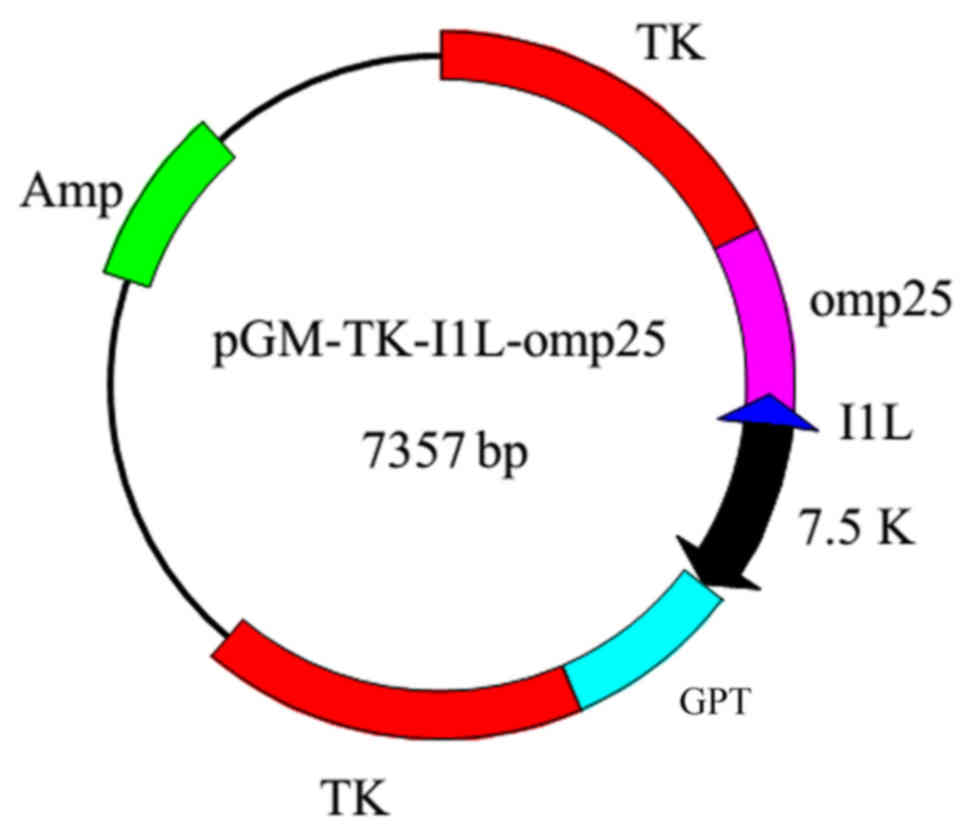
The Brucella OMP25 gene was amplified by PCR;
yielding fragments had sizes of ~642 bp. PCR fragments derived from
the genomic DNA of Brucella were digested and ligated to the
XhoI restriction sites of the transfer plasmid
pGM-TK-I1L-GPT, to construct the recombinant vector
pGM-TK-I1L-GPT-OMP25 (Fig. 1).
Construction of rGTPV expressing OMP25
of Brucella
Recombinant viruses were screened in LT and SFF
cells until a CPE was observed. Viruses were tested for genetic
stability at defined intervals after repeated passages. In the
present study, in a total of 10 generations, viral genomes were
extracted every 2–3 generations, and PCR was performed using
customized primers to yield amplified fragments with sizes of 642
bp.
The transfection product rGTPV-OMP25 was inoculated
into confluent SFF and LT cells that were cultured in screening
solution for 24 h, and then transfected cells were observed daily
for 14 days by microscopy. With the extension of the infection time
by rGTPV, the cytopathic phenomenon gradually increased (Fig. 2). The electron microscope
examination demonstrated that the viral particles had an oval or
round appearance, and brick-shaped virion were identified in
cytoplasm (Fig. 2).
The recombinant virus was passaged for 15
generations, and the viral genome was extracted every 2–3
generations; the genetic stability of rGTPV was confirmed by PCR
(Fig. 3).
 | Figure 3.Stability of rGTPV-outer membrane
protein 25. The recombinant vector was passaged for 10 generations,
and the viral genome was extracted every 2–3 generations.
Polymerase chain reaction was performed using designed primers for
amplification of the attachment gene of rGTPV. Lane 1, negative
control; lane 2, sample from the third generation; lane 3, sample
from the fifth generation; lane 4, sample from the seventh
generation; lane 5, sample from the eighth generation; lane 6,
sample from the tenth generation; lane 7, sample from the fifteenth
generation; lane M, DNA marker; rGTPV, recombinant goat pox
virus. |
Analysis of recombinant expression
vectors by western blotting
To confirm that the OMP25 was expressed in the
recombinant vector, whole cell extracts of LT/SFF cells transfected
with the GTPV or rGTPV strains were separated by electrophoresis. A
band with a molecular mass of 25 kDa was present in rGTPV
G14-STV44-55-OMP25 and GTPV G14-STV44-55; however, was missing in
untransfected SFF cells. Furthermore, when western blot analysis of
the expressed protein was performed using an anti-native
Brucella serum, the absence of OMP25 in rGTPV
G14-STV44-55-OMP25 was confirmed (Fig.
4).
Immunogenicity of rGTPV expressing
OMP25
The levels of OMP25 and GTPV in the sera of mice was
measured at 450 nm using a microtiter plate reader. Compared with
the control group, OMP25 levels were significantly upregulated in
the OMP25 group. However, the rGTPV-OMP25 group exhibited reduced
OMP25 levels compared with the OMP25 group (Fig. 5A). GTPV levels were significantly
upregulated in the GTPV group compared with the control group;
however, there were no significant differences between the GTPV and
rGTPV-OMP25 groups (Fig. 5B).
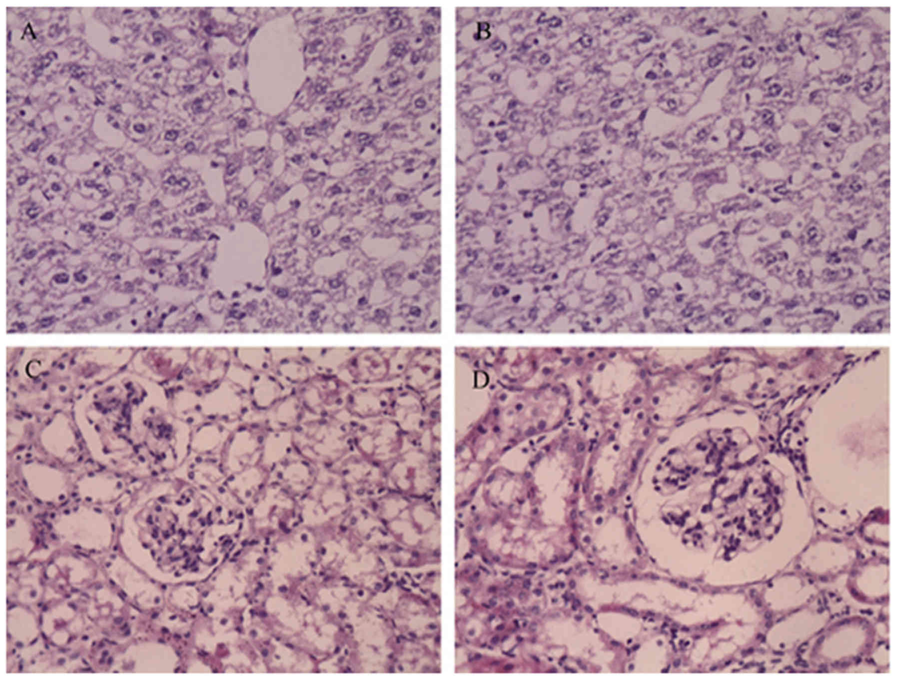
rGTPV-OMP25 is attenuated in BALB/c
mice
The liver and kidney are important organs that
harbor rGTPV during infection. To assess whether the increased
bacterial burden in mice altered liver and kidney pathology, the
number and area of granulomas in these organs were determined. As
presented in Fig. 6, the mice did
not exhibit alterations in liver granulomas after rGTPV infection,
indicating that the recombinant vaccine did not cause visible
damage to the liver or kidney.
Discussion
Brucellosis is a zoonotic disease caused by bacteria
of the genus Brucella (1),
which may lead to substantial economic loss and serious public
health problems. Brucellosis will continue to be an important
public health concern as long as natural reservoirs exist.
Traditional Brucella vaccines, particularly the attenuated
vaccine, serve important roles in regulating the disease; however,
limitations, including potential problems with safety and other
side effects, remain a principal concern. Nevertheless,
Brucella remains to be eradicated, and vaccines remain the
primary means of treatment, in addition to the most economical
means of prevention and control.
OMPs are types of proteins that are common to
bacteria. They maintain the stability of the bacterial outer
membrane and serve an important role in interactions with the host.
OMP25 is the most important Brucella OMP that serves a
critical role in maintaining the structural stability of the
bacteria. Additionally, OMP25 is a Brucella
virulence-associated gene (15).
GTPV vaccine vectors have a large genome structure,
which may accommodate large exogenous genes and have a good safety
profile. Importantly, it may replicate by itself and express
exogenous genes following entry into the body (16). Previously, the use of GTPV as a
vaccine vector has been reported in China and abroad, and numerous
ruminant mammal protective antigens have been expressed in the GTPV
vaccine vector expression system, which have demonstrated promising
results (17,18). The present study selected VVI1L
promoter-expressed exogenous genes as VVI1L is a strong promoter,
as its activity is 10-fold greater than that of VVP7.5 (10,12).
The present study demonstrated that the
immunogenicity of rGTPV-OMP25 is weak. A reason for this may be
that the immunological effect of rGTPV-OMP25 in non-ruminants is
weaker compared with ruminants. Therefore, rGTPV-OMP25 will be
further evaluated in goats and sheep. Furthermore,
Brucella-associated genes expressed by rGTPV may
additionally contribute to reduced immunogenicity.
In conclusion, in the present study, an
overexpression vector for the Brucella OMP OMP25 was
constructed and identified a robust. The construct was recombined
with GTPV, and the immunostimulatory activity was confirmed. This
novel vaccine may aid efforts to prevent brucellosis and goat pox
infections, and may yield novel insights for research efforts aimed
at generating Brucella and goat pox bivalent vaccines.
Acknowledgements
Not applicable.
Funding
The present study was supported by the National key
Research and Development Program of China (grant no.
2017YFD0500304), the Training Program for Excellent Young Teachers
Colleges and Universities of Corps (grant no. CZ027202), the
National Natural Science Foundation of China (grant nos. 31860691
and 31602080), the Youth Science and technology innovation leading
talent program of Corps (grant no. 2017CB002), the Foundation of
the Technology Department of Henan Province (grant no.
172102310335), and the Shihezi University international science and
technology cooperation promotion plan (grant no. GJHZ201709).
Availability of data and materials
The datasets used and/or analyzed during the current
study are available from the corresponding author on reasonable
request.
Authors' contributions
HZ, ZG and CC designed the experiments. ZS, LL, YL,
FW and QF performed the experiments. ZL, PW, YR and YZ analyzed the
data. ZS, LL and ZL wrote the manuscript. HZ and ZL revised the
manuscript. All authors read and approved the final manuscript.
Ethics approval and consent to
participate
The present study was approved by the Institutional
Committee of Post-Graduate Studies and Research at Shihezi
University.
Patient consent for publication
Not applicable.
Competing interests
The authors declare they have no competing
interests.
References
|
1
|
Bercovich Z: The use of skin delayed-type
hypersensitivity as an adjunct test to diagnose brucellosis in
cattle: A review. Vet Q. 22:123–130. 2000. View Article : Google Scholar : PubMed/NCBI
|
|
2
|
Wang Z and Wu Q: Research progress in live
attenuated Brucella vaccine development. Curr Pharm
Biotechnol. 14:887–896. 2013. View Article : Google Scholar : PubMed/NCBI
|
|
3
|
Cloeckaert A, de Wergifosse P, Dubray G
and Limet J: Identification of seven surface-exposed
Brucella outer membrane proteins by use of monoclonal
antibodies: Immunogold labeling for electron microscopy and
enzyme-linked immunosorbent assay. Infect Immun. 58:3980–3987.
1990.PubMed/NCBI
|
|
4
|
Shevtsov A, Tarlykov P, Zholdybayeva E,
Shevtsova E, Momynkulov D, Sytnik I, Karibaev T, Chsherbakov A and
Momynaliev K: Draft genome sequence of the live vaccine strain
Brucella abortus 82. Genome Announc. 1:e01101–01113. 2013.
View Article : Google Scholar : PubMed/NCBI
|
|
5
|
Schurig GG, Sriranganathan N and Corbel
MJ: Brucellosis vaccines: Past, present and future. Vet Microbiol.
90:479–496. 2002. View Article : Google Scholar : PubMed/NCBI
|
|
6
|
Diallo A, Minet C, Berhe G, Le Goff C,
Black DN, Fleming M, Barrett T, Grillet C and Libeau G: Goat immune
response to capripox vaccine expressing the hemagglutinin protein
of peste des petits ruminants. Ann N Y Acad Sci. 969:88–91. 2002.
View Article : Google Scholar : PubMed/NCBI
|
|
7
|
Venkatesan G, Balamurugan V, Yogisharadhya
R, Kumar A and Bhanuprakash V: Differentiation of sheeppox and
goatpox viruses by polymerase Chain reaction-restriction fragment
length polymorphism. Virol Sin. 27:353–359. 2012. View Article : Google Scholar : PubMed/NCBI
|
|
8
|
Zhao Z, Wu G, Zhu X, Yan X, Dou Y, Li J,
Zhu H, Zhang Q and Cai X: RNA interference targeting virion core
protein ORF095 inhibits Goatpox virus replication in Vero cells.
Virol J. 9:482012. View Article : Google Scholar : PubMed/NCBI
|
|
9
|
Mackett M, Smith GL and Moss B: Vaccinia
virus: A selectable eukaryotic cloning and expression vector. Proc
Natl Acad Sci USA. 79:7415–7419. 1982. View Article : Google Scholar : PubMed/NCBI
|
|
10
|
Liu X, Kremer M and Broyles SS: A natural
vaccinia virus promoter with exceptional capacity to direct protein
synthesis. J Virol Methods. 122:141–145. 2004. View Article : Google Scholar : PubMed/NCBI
|
|
11
|
Falkner FG and Moss B: Escherichia coli
gpt gene provides dominant selection for vaccinia virus open
reading frame expression vectors. J Virol. 62:1849–1854.
1988.PubMed/NCBI
|
|
12
|
Fick WC and Viljoen GJ: Identification and
characterisation of an early/late bi-directional promoter of the
capripoxvirus, lumpy skin disease virus. Arch Virol. 144:1229–1239.
1999. View Article : Google Scholar : PubMed/NCBI
|
|
13
|
Chen H, Li Y, Li Z, Shi J, Shinya K, Deng
G, Qi Q, Tian G, Fan S, Zhao H, et al: Properties and dissemination
of H5N1 viruses isolated during an influenza outbreak in migratory
waterfowl in western China. J Virol. 80:5976–5983. 2006. View Article : Google Scholar : PubMed/NCBI
|
|
14
|
Feng W, Zhiru G, Chuangfu C and Jun Q:
Construction of the recombinant goat pox virus expressing OMP 25
gene of B. melitensis. Prog Veterinary Med. 29:6–9. 2008.
|
|
15
|
Goel D, Rajendran V, Ghosh PC and
Bhatnagar R: Cell mediated immune response after challenge in Omp25
liposome immunized mice contributes to protection against virulent
Brucella abortus 544. Vaccine. 31:1231–1237. 2013. View Article : Google Scholar : PubMed/NCBI
|
|
16
|
Gershon PD and Black DN: A comparison of
the genomes of capripoxvirus isolates of sheep, goats, and cattle.
Virology. 164:341–349. 1988. View Article : Google Scholar : PubMed/NCBI
|
|
17
|
Tulman ER, Afonso CL, Lu Z, Zsak L, Sur
JH, Sandybaev NT, Kerembekova UZ, Zaitsev VL, Kutish GF and Rock
DL: The genomes of sheeppox and goatpox viruses. J Virol.
76:6054–6061. 2002. View Article : Google Scholar : PubMed/NCBI
|
|
18
|
Berhe G, Minet C, Le Goff C, Barrett T,
Ngangnou A, Grillet C, Libeau G, Fleming M, Black DN and Diallo A:
Development of a dual recombinant vaccine to protect small
ruminants against peste-des-petits-ruminants virus and
capripoxvirus infections. J Virol. 77:1571–1577. 2003. View Article : Google Scholar : PubMed/NCBI
|