Introduction
Obesity has reached epidemic proportions worldwide
and is associated with insulin resistance, dyslipidemias, type 2
diabetes and neurodegenerative disorders (1). Emerging evidence has revealed a link
between obesity and increased sensitivity to pain. For example,
obese Zucker rats were found to be more sensitive to thermal
stimulation of the tail (2), and
diet-induced obese rats exhibited increased paw edema following
Freund's complete adjuvant-induced arthritis (3). Also, diet-induced obese rats and
obese Zucker rats exhibited augmented peripheral inflammation and
inflammatory hyperalgesia in response to an intradermal injection
of carrageenan (4–6). In addition, obesity has been
associated with enhanced neuropathic pain independent of diabetes
(7), though the underlying
mechanism remains unclear. There are currently no available
pharmacological treatments for the prevention or cure of
obesity-associated pain. Therefore, a greater understanding of the
precise mechanisms of this pathology, as well as the identification
of potential drug targets, is of considerable interest.
AMP-activated protein kinase (AMPK) is an important sensor of
cellular energy status that serves a key role in cellular energy
homeostasis (8). AMPK has been
implicated in a number of diseases related to energy metabolism,
including obesity. In addition to the regulation of cellular energy
metabolism, AMPK may mediate the pain response (as indicated in
preclinical pain models) (9,10),
and targeting AMPK is now considered to be a novel strategy for the
prevention and treatment of pain (11). A recent study demonstrated that the
activation of AMPK alleviated trigeminal neuralgia by
downregulating the expression level of neuropeptide calcitonin
gene-related peptide (CGRP; a principal neurotransmitter of
nociceptive sensory C-fibers), and reducing neuroinflammation in
the spinal trigeminal nucleus (12). The activation of AMPK has also been
reported to be impaired in the central nervous system (CNS) of
diet-induced obese animals (13).
The present study hypothesized that obesity may
result in increased sensitivity to neuropathic pain by
downregulating AMPK and upregulating CGRP-associated pathways in
the spinal cord (SC) and dorsal root ganglion (DRG). For this
purpose, a high-fat diet (HF)-induced obese rat model that mimics
human obesity syndrome was utilized, and mechanical allodynia in
rats with or without spared nerve injury (SNI) was assessed.
Materials and methods
Animals
A total of 100 male Sprague-Dawley rats weighing ~70
g (4 weeks old; Liaoning Changsheng Biological Center, Shenyang,
China) were fed a high-fat diet (HF; fat-containing 45% kcal;
Research Diets, Inc.) to induce obesity as previously described
(14). A further 100 male
Sprague-Dawley rats were given a low-fat diet (LF; contains 10%
kcal of fat; Research Diets, Inc), and 10 rats were allocated to
each group as previously described (14). All animals were housed at a
constant temperature of 23–25°C, with 50% relative humidity and a
12-h light/dark cycle. The rats had ad libitum access to
food and autoclaved water. All experiments were conducted in
accordance with the Guiding Principles for Research Involving
Animal and Human Beings, and the experimental procedures were
approved by the Animal Care and Use Committee of China Medical
University (IACUC no. 20180121).
Chemicals and reagents
Phosphorylated (p)-AMPKThr172 and
total-AMPK antibodies were purchased from Cell Signaling
Technology, Inc. 5-Aminoimidazole-4-carboxamide riboside (AICAR),
dorsomorphin (DOR) and CGRP8-37 were purchased from APExBIO. The
CGRP ELISA kit was purchased from Cusabio Technology LLC (cat. no.
CSB-E08211r). All other chemicals were purchased from
Sigma-Aldrich; Merck KGaA.
Experimental protocols
To examine neuropathic pain in obese animals, body
weight and mechanical allodynia were measured once a week during HF
or LF feeding. At 10 and 12 weeks after feeding, 20 rats from each
group were sacrificed to collect the SC and DRG for molecular
studies. The remaining animals were treated with an intrathecal
AMPK activator (AICAR; 0.2 µmol/kg), AMPK inhibitor (DOR; 0.2
µmol/kg), CGRP antagonist (CGRP8-37; 20 pmol/kg) or the saline
vehicle control (VEH) after 12 weeks, for a further 7 days. During
treatment, mechanical allodynia was recorded daily, and the SC and
DRG were collected at the end of the treatment course.
To investigate neuropathic pain following nerve
injury in obese animals, 12 weeks after HF or LF feeding, rats from
each group underwent SNI, and mechanical allodynia was measured
once a week for 14 days. At the 14-day time-point, these animals
were treated with AICAR, DOR or VEH for 7 consecutive days. During
treatment, mechanical allodynia was measured daily, and the SC and
DRG were collected for further studies following treatment.
Quantitative assessment of
allodynia
Mechanical allodynia was assessed using the Von Frey
test as previously described (15). Briefly, the rats were placed in a
cage with a mesh bottom, and calibrated cilia-Von Frey filaments
were applied perpendicularly to the hind paws until a positive
response was observed, which included withdrawal and licking or
shaking of the paws. The up-down method was used to determine the
mechanical force required to induce paw withdrawal in 50% of
animals. A filament that was estimated to be close to the 50% paw
withdrawal threshold (PWT) was first used to stimulate the animal.
If the animal showed a positive response, the next lowest-force
filament was tested; if the animal did not show a response, the
next highest-force filament was tested. A total of 4 readings were
acquired and the 50% threshold was calculated using the following
formula: 50% contraction threshold=10log(x) + kδ; × was the
strength of the last stimulus used; k was the coefficient of
different stimulation modes; and δ (δ=0.224) was the average value
of the adjacent spacing of each stimulation intensity (g).
SNI animal model
An SNI model that mimics nerve injury-associated
human neuropathic pain was induced as previously described
(16). Briefly, rats were
anesthetized by inhalation of 3% isoflurane, and the main sciatic
nerve and its branches in the hind limb were exposed. The phrenic
nerve and the common peroneal nerve were ligated or severed, and
small sural nerves were preserved. After 24 h, a significant pain
response was triggered in the hind paw and the lateral aspect of
the foot.
Lumbar catheterization of the spinal
subarachnoid space
For drug delivery to the lumbar subarachnoid space,
lumber catheterization was performed as described previously
(17). Briefly, the rat was
anesthetized by inhalation of 3% isoflurane. An incision lateral to
the midline was made and a guided cannula (20 G) was inserted into
the subarachnoid space. The correct localization was indicated by a
tail-flicking action and hind limb paralysis after administration
of 2% lidocaine (10 µl) through the catheter in wakened
animals.
Specimen collection and
processing
Rats were euthanized individually in a transparent
sealed box; 5 ml/kg sevoflurane liquid was introduced into the box,
and the rat was allowed to inhale the vapor until death (~2 min).
The L4-6 SC and DRG were isolated from the rat and
stored at −80°C for subsequent western blot and ELISA analyses.
Western blotting
The L4-6 SC and DRG samples were
homogenized in 400 µl homogenization buffer containing RIPA lysis
buffer (cat. no. P0013B; Beyotime Institute of Biotechnology). The
samples were incubated at 0°C for 30 min, followed by
centrifugation at 12,000 × g at 4°C for 10 min. The supernatant was
collected and denatured by boiling for 5 min. Total protein was
quantified using the BCA method and was separated using 12%
SDS-PAGE gels (20 µg of protein was loaded per lane), and then
transferred to a polyvinylidene fluoride membrane. The membrane was
blocked with blocking buffer containing 5% fat-free milk for 30 min
at room temperature and incubated with the following primary
antibodies at 4°C overnight: Rabbit anti-p-AMPKThr172
(1:1,000; cat. no. 50081s; Cell Signaling Technology, Inc.), rabbit
anti-total APMK (1:1,000; cat. no. 5832s; Cell Signaling
Technology, Inc.) and rabbit anti-GAPDH (1:1,000; cat. no. 5174s;
Cell Signaling Technology, Inc.). The membrane was then washed and
incubated with a horseradish peroxidase-conjugated goat anti-rabbit
secondary antibody (1:10,000; cat. no. zb-2301; ZSGB BIO) for 2 h
at room temperature, prior to developing with ECL reagent (EMD
Millipore). The results were acquired and analyzed using a
molecular imager (Gel Doc™ XR; 170-8170) and the associated
Quantity One 4.6.5 software (Bio-Rad Laboratories, Inc.).
Statistical analysis was performed using the optical density ratio
of the target protein bands to that of the GAPDH band, and the
expression levels of phosphorylated proteins were normalized
according to the following formula: (phosphorylated
protein/GAPDH)/(total protein/GAPDH).
ELISA
The concentrations of CGRP in the SC and DRG were
measured using an ELISA kit (Thermo Fisher Scientific, Inc.)
according to the manufacturer's instructions.
Statistical analysis
All data are presented as the mean ± standard error
of the mean. Statistical analyses were performed using GraphPad
Prism 7 (GraphPad Software, Inc.,). Two-way repeated measures ANOVA
was conducted on PWT measurements, followed by the Bonferroni
correction to identify the significant differences between groups
at different time points. ELISA and western blotting results
between groups were compared using one- or two-way ANOVA followed
by the Bonferroni correction. P<0.05 was considered to indicate
a statistically significant difference.
Results
Mechanical allodynia and CGRP
signaling in obesity
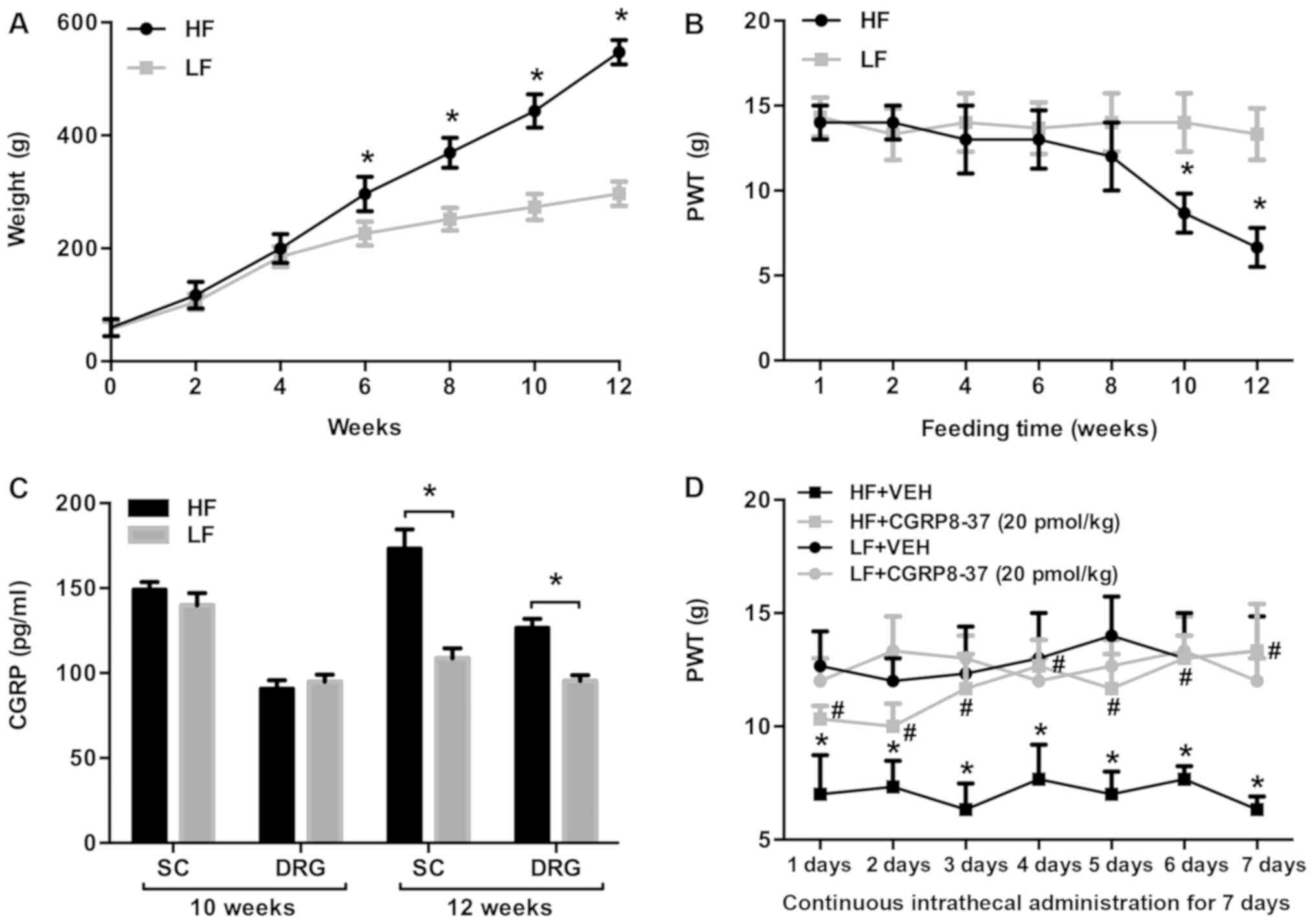
Prior to HF feeding, body weight was similar between
rats assigned to the HF and LF groups (Fig. 1A). After 6 weeks of feeding, the
HF-fed rats had gained significantly more body weight than the
LF-fed rats, and this trend continued throughout the 12-week
dietary period. After 10 weeks of feeding, the mean body weight in
HF-fed rats was approximately double that of the LF-fed rats,
indicating that a HF diet resulted in obesity in rats.
Mechanical allodynia was assessed with the Von-Frey
test and the 50% PWT was measured. The 50% PWT was significantly
lower in HF-fed rats compared with LF-fed rats at 10 weeks, and
this trend persisted until 12 weeks (Fig. 1B).
The CGRP pathway has been suggested to serve an
important role in mediating neuropathic pain (18,19).
To examine the possible molecular mechanisms responsible for
enhanced mechanical allodynia in HF-fed rats, the expression levels
of CGRP in the SC and DRG were measured and were comparable between
the groups at 10 weeks, but significantly higher in HF-fed rats
than in LF-fed rats at the 12-week point (Fig. 1C).
To further examine whether increased CGRP in the SC
and DRG was associated with enhanced mechanical allodynia in HF-fed
rats, HF- and LF-fed rats were treated with the intrathecal CGRP
antagonist CGRP8-37 or the saline VEH control for 7 consecutive
days, after 12 weeks of feeding. As shown in Fig. 1D, the 50% PWT was significantly
lower in HF-fed rats compared with LF-fed rats receiving
intrathecal VEH. Intrathecal CGRP8-37 significantly increased the
50% PWT in HF-fed rats throughout the 7 days when compared with VEH
treatment. Intrathecal CGRP8-37 did not alter the 50% PWT in LF-fed
rats.
Effects of modulating the AMPK pathway
on mechanical allodynia in obesity
Activation of AMPK has been shown to mediate
neuropathic pain via the CGRP pathway (11); therefore, the role of the AMPK
pathway in regulating neuropathic pain in obesity was examined. As
presented in Fig. 2, intrathecal
treatment with AICAR for 7 days significantly increased the 50% PWT
in HF-fed rats compared with VEH treatment (Fig. 2A). By contrast, intrathecal
treatment with the AMPK inhibitor DOR for 7 days further decreased
the 50% PWT in HF-fed rats compared with VEH treatment (Fig. 2B). Neither AICAR nor DOR had any
effect on the 50% PWT in LF-fed rats.
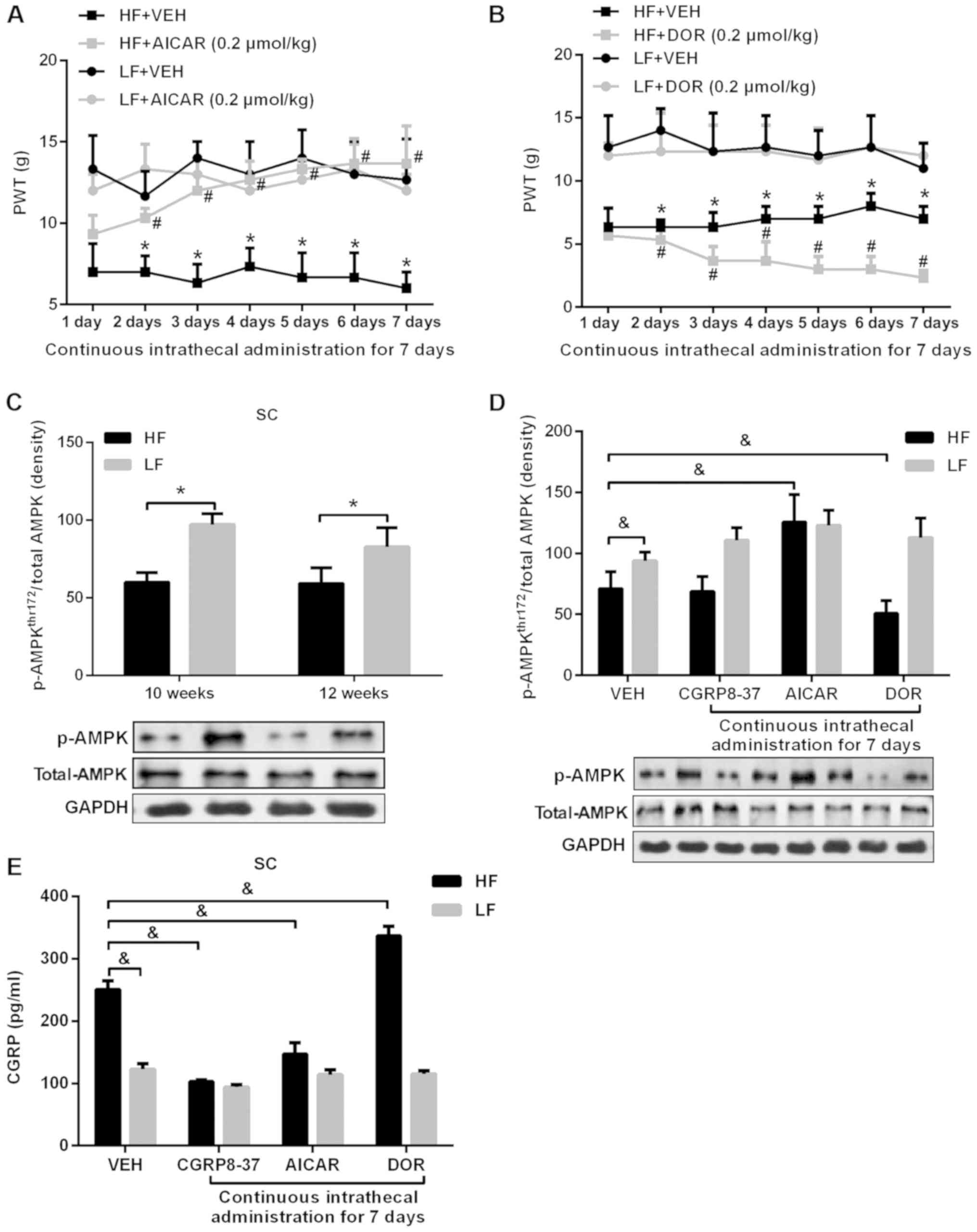 | Figure 2.PWT, expression of p-AMPK and CGRP in
rats given an AMPK agonist or inhibitor. (A) 50% PWT in HF-fed rats
and LF-fed rats treated with the intrathecal AMPK activator AICAR.
*P<0.05 vs. respective LF group; #P<0.05 vs.
respective HF+VEH group. (B) 50% PWT in HF-fed rats and LF-fed rats
treated with the intrathecal AMPK inhibitor DOR. *P<0.05 vs.
respective LF group; #P<0.05 vs. respective HF+DOR
group. (C) Expression levels of p-AMPKThr172 in the SC
at 10 and 12 weeks after HF or LF feeding. *P<0.05. (D) Effect
of intrathecal AICAR, DOR and CGRP8-37 on p-AMPKThr172
in HF- and LF-fed rats. (E) Effects of intrathecal AICAR, DOR and
CGRP8-37 on expression levels of CGRP in the SC of HF- and LF-fed
rats. &P<0.05. n=10 per group. PWT, paw
withdrawal threshold; HF, high fat; LF, low fat; AMPK,
AMP-activated protein kinase; AICAR, 5-aminoimidazole-4-carboxamide
riboside; DOR, dorsomorphin; p-AMPK, phosphorylated-AMPK; SC,
spinal cord; CGRP, calcitonin gene-related peptide; VEH,
vehicle. |
The expression of p-AMPKThr172 in the SC
was significantly lower in HF-fed rats at both 10 and 12 weeks,
compared with that in LF-fed rats (Fig. 2C). The level of
p-AMPKThr172 in the SC of HF-fed rats was increased by
intrathecal treatment with AICAR, decreased by DOR treatment, and
unchanged by CGRP8-37 treatment (Fig.
2D). Additionally, the levels of CGPR in the SC of HF-fed rats
were decreased by AICAR and CGRP8-37 treatment, but increased by
DOR treatment (Fig. 2E). None of
the treatments had any notable effect on p-AMPKThr172 or
CGPR expression levels in LF-fed rats.
Mechanical allodynia and the AMPK-CGRP
pathway in obesity after SNI
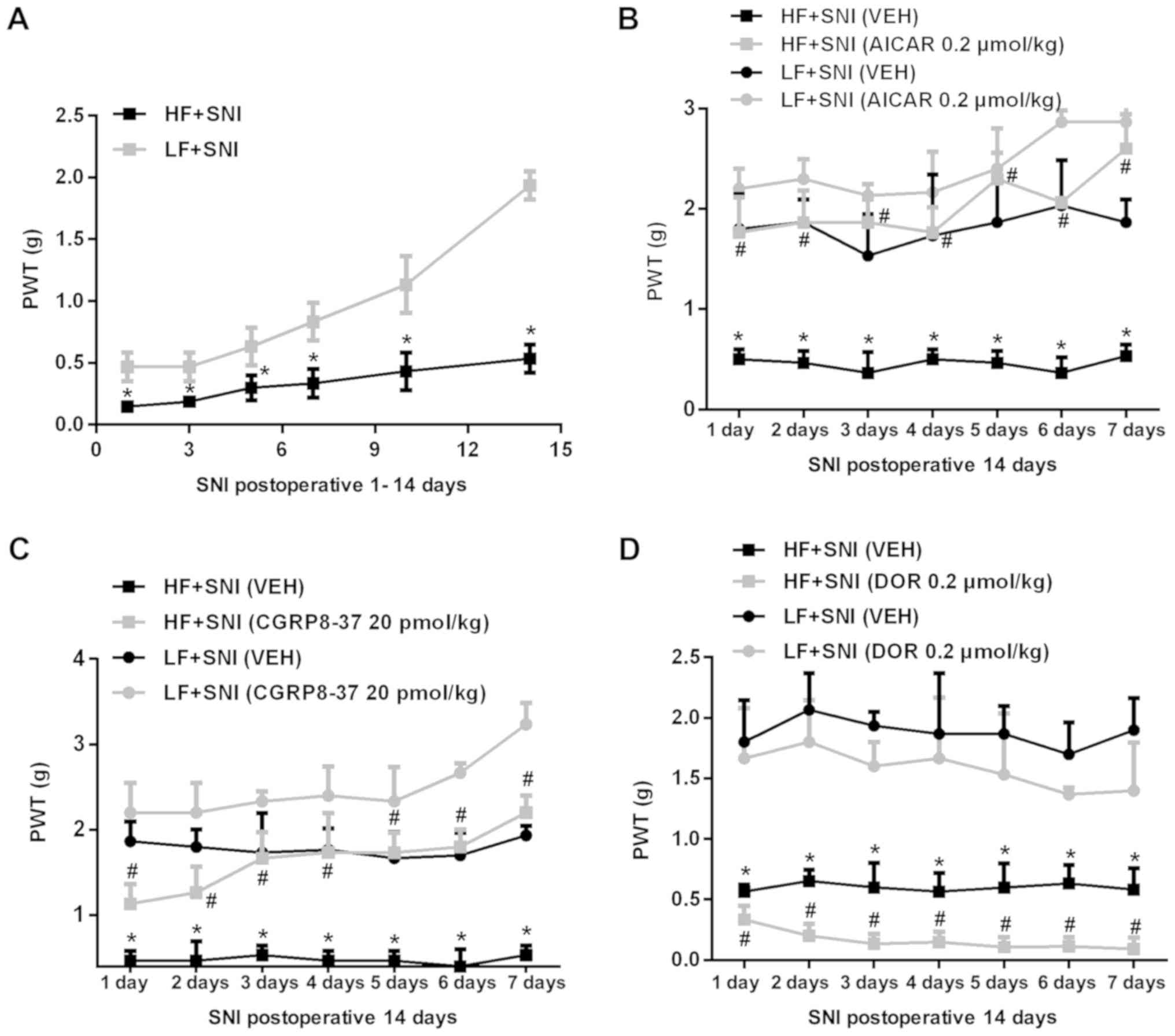
To investigate neuropathic pain following nerve
injury in obese animals, SNI was induced after 12 weeks of HF or LF
feeding, and mechanical allodynia was measured for 14 consecutive
days. At 2 weeks after SNI, rats were treated with intrathecal
AICAR, DOR, CGRP8-37 or VEH, and mechanical allodynia was measured
for another 7 consecutive days. As shown in Fig. 3, HF-fed rats exhibited a
significantly lower 50% PWT than LF-fed rats at all time points
following SNI (Fig. 3A).
Intrathecal treatment with AICAR (Fig.
3B) or CGRP8-37 (Fig. 3C)
increased the 50% PWT, whereas DOR (Fig. 3D) further reduced the 50% PWT in
HF-fed rats compared with LH-fed rats.
The expression levels of CGRP in the SC (Fig. 4A) and DRG (Fig. 4B) were significantly higher in
HF-fed rats than in LF-fed rats following SNI. Intrathecal
treatment with AICAR or CGRP8-37 reduced the levels of CGRP
expression in the SC and DRG in both study groups. By contrast,
intrathecal treatment with DOR augmented the expression levels of
CGRP in the SC and DRG of HF-fed rats, compared with those in
LF-fed rats. Moreover, p-AMPKThr172 (Fig. 4C) in the SC was lower in HF-fed
rats compared with LF-fed rats after SNI. Intrathecal treatment
with AICAR or CGRP8-37 upregulated p-AMPKThr172 in SC,
whereas intrathecal treatment with DOR decreased the expression of
p-AMPKThr172 in the SC of HF-fed rats compared with that
in LF-fed rats after SNI. As the present sudy focused on the effect
of neuropathic pain in rats fed a HF diet, there was no concern
about some changes in the rats fed a LF diet. A significant
decrease in the expression of p-AMPKThr172 was not
observed in the LF group after DOR. It is hypothesized that the SNI
model itself may have an effect on the expression of
p-AMPKThr172.
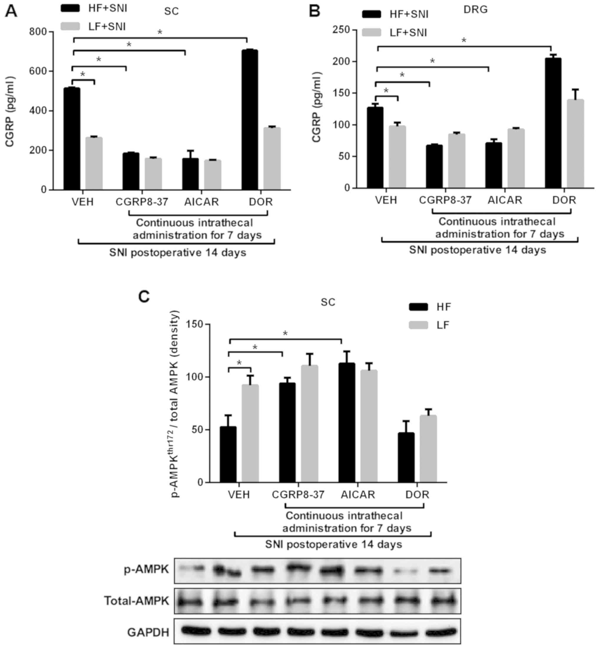 | Figure 4.Expression of p-AMPK and CGRP 14 days
after SNI and following administration of an AMPK agonist or
inhibitor for 7 days. Effects of intrathecal AICAR, DOR and
CGRP8-37 treatment on the expression levels of CGRP in the (A) SC
and (B) DRG of HF- and LF-fed rats with SNI. (C) Effect of
intrathecal AI, DOR or CGRP8-37 on p-AMPKThr172 in the
SC in HF- and LF-fed rats with SNI. *P<0.05. n=10 per group.
AICAR, 5-aminoimidazole-4-carboxamide riboside; DOR, dorsomorphin;
CGRP, calcitonin gene-related peptide; SC, spinal cord; DRG, dorsal
root ganglion; HF, high fat; LF, low fat; SNI, spared nerve injury;
p-AMPK, phosphorylated-AMPK; VEH, vehicle. |
Discussion
The primary findings of the present study were: i)
HF-induced obese rats with or without nerve injury exhibited
enhanced mechanical pain sensitivity, which was inhibited by the
AMPK activator AICAR or the CGRP antagonist CGRP8-37, and
exaggerated by the AMPK inhibitor DOR; ii) HF-induced obese rats
with or without nerve injury showed decreased levels of
p-AMPKThr172 and higher expression levels of CGRP in the
SC and DRG; iii) p-AMPKThr172 in the SC and DRG of
HF-induced obese rats was upregulated by AICAR, which also reduced
the expression levels of CGRP; and iv) p-AMPKThr172 in
the SC and DRG of HF-induced obese rats was downregulated by DOR,
which also augmented the expression levels of CGRP. Collectively,
these data suggested that the enhanced neuropathic pain in
HF-induced obese rats with or without nerve injury was due to
decreased AMPK activity in the SC and DRG, which resulted in
increased CGRP expression levels.
Neuropathic pain is defined as pain resulting from
injury or disease affecting the peripheral nervous system or CNS.
Injury to the peripheral nervous system due to trauma, metabolic
disease or exposure to drugs is the fundamental etiology of
neuropathic pain (20). The common
symptomology of neuropathic pain includes exquisite
hypersensitivity to mechanical stimulation (20). A recent study demonstrated that
HF-induced obese animals exhibited increased neuropathic pain
(21), but the mechanisms are not
fully understood. Additionally, it is unclear whether neuropathic
pain is enhanced in obesity following nerve injury. In the present
study, it was discovered that HF-induced obese rats possessed
enhanced mechanical pain sensitivity compared with LF-fed control
rats, which is consistent with a previous study (21). The present study also extended
previous research by demonstrating that mechanical pain sensitivity
remained higher in HF-induced obese rats than in LF-fed control
rats after nerve injury (22).
Emerging evidence has indicated that the AMPK
pathway in the SC and DRG serves a prominent role in the mediation
of neuropathic pain (9). The
activation of AMPK negatively regulates activity-dependent protein
synthesis and other signaling pathways that drive neuronal
plasticity in neuropathic pain, resulting in reduced nociceptor
excitability (23). Systemic
activation of AMPK with either metformin or A769662 in rodents,
following peripheral nerve injury, attenuates mechanical
hypersensitivity (24,25). Additionally, activation of AMPK in
the SC in a rat model of neuropathic pain reduced mechanical
hypersensitivity by inhibiting the expression of proinflammatory
cytokines, and upregulating that of the glutamate transporter after
nerve injury (26). In
vitro experiments using patch-clamp electrophysiology on DRG
neurons demonstrate that the activation of AMPK with metformin or
A769662 reduces the excitability of DRG neurons; these neurons are
widely believed to induce ectopic activity causing an ongoing
burning pain, a prominent feature of neuropathic pain following
injury (24). In the present
study, p-AMPKThr172 levels in the SC and DRG were
significantly lower in HF-induced obese rats compared with LF-fed
control rats after 10 weeks of HF feeding, indicating that AMPK
signaling was impaired in HF-induced obesity. This finding is
consistent with a previous study illustrating impaired
AMPK-signaling in the CNS of HF-induced obese rats (13). To understand whether impaired
AMPK-signaling in the SC and DRG accounts for enhanced mechanical
pain sensitivity in obesity, animals were treated with the
intrathecal AMPK activator AICAR or AMPK inhibitor DOR. This
revealed that AICAR increased AMPK activity in the SC and DRG of
HF-induced obese rats with or without nerve injury, leading to
decreased mechanical pain sensitivity. By contrast, intrathecal DOR
further reduced AMPK activity in the SC and DRG in HF-induced obese
rats (with or without nerve injury), resulting in exaggerated
mechanical pain sensitivity. These data clearly demonstrated that
impaired AMPK activity in the SC and DRG may be a primary
contributor to enhanced neuropathic pain in HF-induced obesity.
CGRP is a neuropeptide that is primarily generated
in the central and peripheral nervous systems, particularly in the
SC, DRG and trigeminal ganglion (27). CGRP in the SC serves an important
role in chronic pain by facilitating the introduction of synaptic
pain information via protein kinase A and protein kinase C second
messenger pathways, and participating in the generation and
maintenance of allodynia and hyperpathia (27–30).
CGRP expression levels were increased in animals after peripheral
nerve injury, and serve an important role in generating and
maintaining pain behavior (27,31).
In the present study, the 50% PWT in HF-fed rats was significantly
reduced compared with that of LF-fed rats, at both 10 and 12 weeks
of feeding. Additionally, the expression levels of CGRP in the SC
and DRG of HF-fed rats were significantly higher than in LF-fed
rats at 12 weeks. However, there was no significant difference
between the two groups at 10 weeks. Furthermore, the 50% PWT of the
HF group decreased significantly at 10 weeks, which may have been
associated with oxidative stress. Previously, increased levels of
oxidative stress biomarkers such as malondialdehyde (MDA), SOD and
reactive oxygen species (ROS). have been detected in the plasma of
obese patients (31,32). Animal models have also revealed
that weight loss, dietary restriction and exercise can also
decrease the expression levels of these biomarkers (33). There is increasing evidence that
the nervous system undergoes oxidative stress reactions following
noxious stimulation and inflammation, and that the production of
oxidation products increases; this results in damage to nerve
tissue, which is an important contributor to neuropathic pain
(34,35). Zhao et al (36) found that MDA concentrations
increased, and that superoxide dismutase activity was decreased in
a neuropathic pain rat model. The present study indicated that AMPK
phosphorylation was also significantly reduced in the HF group at
10 weeks; this suggests that AMPK may have an important regulatory
effect on intracellular oxidative stress, which may subsequently
inhibit the generation of ROS (37,38).
Therefore, it was hypothesized that the 50% PWT reduction may be
associated with reduced AMPK phosphorylation, resulting in
increased levels of oxidative stress; this will be the focus of
future investigations.
Inhibiting CGRP expression in the SC has been proven
to significantly ameliorate pain behavior after peripheral nerve
injury (27,29). A recent study revealed that AMPK
mediates neuropathic pain by regulating CGRP (12). Consistent with previous studies
(27,31), the present study observed a
significant increase in CGRP expression levels in the SC and DRG of
HF-induced obese rats (with or without nerve injury), and treatment
with CGRP8-37 prevented an increase in mechanical pain sensitivity
to a significant degree; this indicates that increased HF-induced
obesity may result in increased levels of CGRP expression in the SC
and DRG, contributing to enhanced neuropathic pain. Moreover, the
levels of CGRP expression in the SC and DRG of HF-induced obese
rats were decreased by AICAR, but further increased by the AMPK
inhibitor DOR, indicating that AMPK in the SC and DRG mediates
neuropathic pain in HF-induced obesity via the CGRP pathway.
The present study revealed that the administration
of an AMPK agonist (AICAR) or CGRP inhibitor (CGRP8-37)
significantly increased the 50% PWT compared with VEH
administration in HF-fed rats, but neither AICAR nor CGRP8-37 had
this effect in LF-rats. Obesity is associated with multiple chronic
conditions that lead to pain, such as osteoarthritis, peripheral
vascular disease, lower back pain and neuropathic pain (39–42).
In addition, overweight patients appear to experience more severe
paroxysmal pain, and their neuropathic-negative symptoms tend to be
aggravated (43). A large number
of studies have revealed that neuropathic pain is associated with
increased expression levels of proinflammatory factors (including
IL-1β, IL-6 and IL-8); adipose tissue is an endocrine organ,
generating various cytokines such as leptin, adiponectin, TNF-α,
interleukins (IL-1β, IL-6, IL-8, IL-10) and other biologically
active factors involved in metabolism and immunity (44,45).
Studies have shown that serum expression levels of TNF-α and IL-6
are significantly decreased in obese patients after strict dietary
intake (46). Therefore, it was
hypothesized that AMPK reduction, CGRP enhancement and increased
neuropathic pain may be reversed following weight loss in
HF-induced obese rats, though further experiments are needed to
verify this claim.
In conclusion, the present study demonstrated that
AMPK activity was reduced in the SC and DRG of HF-induced obese
rats. Reduced AMPK activity resulted in increased CGRP production
in the SC and DRG, contributing to enhanced neuropathic pain in
HF-induced obese rats, with or without nerve injury. These results
suggested that obesity may decrease AMPK activity in the CNS,
indicating that AMPK may be a novel therapeutic target for
obesity-related neuropathic pain.
Acknowledgements
Not applicable.
Funding
The present study was supported by the Medical
Health Joint Fund (grant no. 20180530063) and the Guidance Project
Fund (grant no. 20180550175) from the Liaoning Natural Science
Foundation of China.
Availability of data and materials
The datasets generated and analyzed during the
present study are available from the corresponding author on
reasonable request.
Authors' contributions
XG performed the majority of the experiments,
designed the study and acquired data, was involved in the analysis
and interpretation of data, participated in drafting the
manuscript, in critical revision of important content and gave
final approval of the version to be released. XT, TL and QT were
involved in drafting the manuscript and acquisition of data. DD,
XT, BZ and MZ contributed to data interpretation and analysis. TS
made a substantial contribution to the experimental design,
drafting and critically revising the manuscript. All of the authors
have read and approved the final manuscript.
Ethics approval and consent to
participate
All experiments were conducted in accordance with
the Guiding Principles for Research Involving Animal and Human
Beings, and the experimental procedures were approved by the Animal
Care and Use Committee of China Medical University (IACUC no.
20180121).
Patient consent for publication
Not applicable.
Competing interests
The authors declare that they have no competing
interests.
References
|
1
|
Schwartz MW, Seeley RJ, Zeltser LM,
Drewnowski A, Ravussin E, Redman LM and Leibel RL: Obesity
Pathogenesis: An endocrine society scientific statement. Endocr
Rev. 38:267–296. 2017. View Article : Google Scholar : PubMed/NCBI
|
|
2
|
Roane DS and Porter JR: Nociception and
opioid-induced analgesia in lean (Fa/-) and obese (fa/fa) Zucker
rats. Physiol Behav. 38:215–218. 1986. View Article : Google Scholar : PubMed/NCBI
|
|
3
|
Croci T and Zarini E: Effect of the
cannabinoid CB1 receptor antagonist rimonabant on nociceptive
responses and adjuvant-induced arthritis in obese and lean rats. Br
J Pharmacol. 150:559–566. 2007. View Article : Google Scholar : PubMed/NCBI
|
|
4
|
Iannitti T, Graham A and Dolan S:
Increased central and peripheral inflammation and inflammatory
hyperalgesia in Zucker rat model of leptin receptor deficiency and
genetic obesity. Exp Physiol. 97:1236–1245. 2012. View Article : Google Scholar : PubMed/NCBI
|
|
5
|
Wang J, Zhang Q, Zhao L, Li D, Fu Z and
Liang L: Down-regulation of PPARα in the spinal cord contributes to
augmented peripheral inflammation and inflammatory hyperalgesia in
diet-induced obese rats. Neuroscience. 278:165–178. 2014.
View Article : Google Scholar : PubMed/NCBI
|
|
6
|
Zhang Y, Song C, Li H, Hou J and Li D:
Ursolic acid prevents augmented peripheral inflammation and
inflammatory hyperalgesia in high-fat diet-induced obese rats by
restoring downregulated spinal PPARα. Mol Med Rep. 13:5309–5316.
2016. View Article : Google Scholar : PubMed/NCBI
|
|
7
|
Hozumi J, Sumitani M, Matsubayashi Y, Abe
H, Oshima Y, Chikuda H, Takeshita K and Yamada Y: Relationship
between neuropathic pain and obesity. Pain Res Manag.
2016:24879242016. View Article : Google Scholar : PubMed/NCBI
|
|
8
|
Kim M and Tian R: Targeting AMPK for
cardiac protection: Opportunities and challenges. J Mol Cell
Cardiol. 51:548–553. 2011. View Article : Google Scholar : PubMed/NCBI
|
|
9
|
Kialka M, Doroszewska K, Janeczko M and
Milewicz T: Metformin-new potential medicine in pain treatment?
Przegl Lek. 74:81–83. 2017.(In Polish). PubMed/NCBI
|
|
10
|
Price TJ, Das V and Dussor G: Adenosine
Monophosphate-activated Protein Kinase (AMPK) Activators For the
Prevention, Treatment and Potential Reversal of Pathological Pain.
Curr Drug Targets. 17:908–920. 2016. View Article : Google Scholar : PubMed/NCBI
|
|
11
|
Asiedu MN, Dussor G and Price TJ:
Targeting AMPK for the alleviation of pathological pain. Exp Suppl.
107:257–285. 2016.PubMed/NCBI
|
|
12
|
Yang YJ, Hu L, Xia YP, Jiang CY, Miao C,
Yang CQ, Yuan M and Wang L: Resveratrol suppresses glial activation
and alleviates trigeminal neuralgia via activation of AMPK. J
Neuroinflammation. 13:842016. View Article : Google Scholar : PubMed/NCBI
|
|
13
|
Fei W, Tian DR, Tso P and Han JS:
Diet-induced obese rats exhibit impaired LKB1-AMPK signaling in
hypothalamus and adipose tissue. Peptides. 35:23–30. 2012.
View Article : Google Scholar : PubMed/NCBI
|
|
14
|
Song T, Lv LY, Xu J, Tian ZY, Cui WY, Wang
QS, Qu G and Shi XM: Diet-induced obesity suppresses sevoflurane
preconditioning against myocardial ischemia-reperfusion injury:
Role of AMP-activated protein kinase pathway. Exp Biol Med
(Maywood). 236:1427–1436. 2011. View Article : Google Scholar : PubMed/NCBI
|
|
15
|
Chaplan SR, Bach FW, Pogrel JW, Chung JM
and Yaksh TL: Quantitative assessment of tactile allodynia in the
rat paw. J Neurosci Methods. 53:55–63. 1994. View Article : Google Scholar : PubMed/NCBI
|
|
16
|
Decosterd I and Woolf CJ: Spared nerve
injury: An animal model of persistent peripheral neuropathic pain.
Pain. 87:149–158. 2000. View Article : Google Scholar : PubMed/NCBI
|
|
17
|
Storkson RV, Kjorsvik A, Tjolsen A and
Hole K: Lumbar catheterization of the spinal subarachnoid space in
the rat. J Neurosci Methods. 65:167–172. 1996. View Article : Google Scholar : PubMed/NCBI
|
|
18
|
Kanai Y, Nakazato E, Fujiuchi A, Hara T
and Imai A: Involvement of an increased spinal TRPV1 sensitization
through its up-regulation in mechanical allodynia of CCI rats.
Neuropharmacology. 49:977–84. 2005. View Article : Google Scholar : PubMed/NCBI
|
|
19
|
Rossi HL, Luu AK, DeVilbiss JL and Recober
A: Obesity increases nociceptive activation of the trigeminal
system. Eur J Pain. 17:649–653. 2013. View Article : Google Scholar : PubMed/NCBI
|
|
20
|
Baron R, Binder A and Wasner G:
Neuropathic pain: Diagnosis, pathophysiological mechanisms, and
treatment. Lancet Neurol. 9:807–819. 2010. View Article : Google Scholar : PubMed/NCBI
|
|
21
|
Gavini CK, Bookout AL, Bonomo R, Gautron
L, Lee S and Mansuy-Aubert V: Liver X receptors protect dorsal root
ganglia from obesity-induced endoplasmic reticulum stress and
mechanical allodynia. Cell Rep. 25:271–277 e4. 2018. View Article : Google Scholar : PubMed/NCBI
|
|
22
|
Price TJ and Dussor G: AMPK: An emerging
target for modification of injury-induced pain plasticity. Neurosci
Lett 557 Pt A. 9–18. 2013. View Article : Google Scholar
|
|
23
|
Melemedjian OK, Asiedu MN, Tillu DV,
Sanoja R, Yan J, Lark A, Khoutorsky A, Johnson J, Peebles KA, Lepow
T, et al: Targeting adenosine monophosphate-activated protein
kinase (AMPK) in preclinical models reveals a potential mechanism
for the treatment of neuropathic pain. Mol Pain. 7:702011.
View Article : Google Scholar : PubMed/NCBI
|
|
24
|
Melemedjian OK, Tillu DV, Asiedu MN,
Mandell EK, Moy JK, Blute VM, Taylor CJ, Ghosh S and Price TJ: BDNF
regulates atypical PKC at spinal synapses to initiate and maintain
a centralized chronic pain state. Mol Pain. 9:122013. View Article : Google Scholar : PubMed/NCBI
|
|
25
|
Maixner DW, Yan X, Gao M, Yadav R and Weng
HR: Adenosine monophosphate-activated protein kinase regulates
interleukin-1beta expression and glial glutamate transporter
function in rodents with neuropathic pain. Anesthesiology.
122:1401–1413. 2015. View Article : Google Scholar : PubMed/NCBI
|
|
26
|
Ren H, Jin H, Jia Z, Ji N and Luo F:
Pulsed Radiofrequency Applied to the Sciatic Nerve Improves
Neuropathic Pain by Down-regulating The Expression of Calcitonin
Gene-related Peptide in the Dorsal Root Ganglion. Int J Med Sci.
15:153–160. 2018. View Article : Google Scholar : PubMed/NCBI
|
|
27
|
Pezet S and McMahon SB: Neurotrophins:
mediators and modulators of pain. Annu Rev Neurosci. 29:507–538.
2006. View Article : Google Scholar : PubMed/NCBI
|
|
28
|
Lee SE and Kim JH: Involvement of
substance P and calcitonin gene-related peptide in development and
maintenance of neuropathic pain from spinal nerve injury model of
rat. Neurosci Res. 58:245–249. 2007. View Article : Google Scholar : PubMed/NCBI
|
|
29
|
Sun RQ, Lawand NB and Willis WD: The role
of calcitonin gene-related peptide (CGRP) in the generation and
maintenance of mechanical allodynia and hyperalgesia in rats after
intradermal injection of capsaicin. Pain. 104:201–208. 2003.
View Article : Google Scholar : PubMed/NCBI
|
|
30
|
Hu P, Bembrick AL, Keay KA and McLachlan
EM: Immune cell involvement in dorsal root ganglia and spinal cord
after chronic constriction or transection of the rat sciatic nerve.
Brain Behav Immun. 21:599–616. 2007. View Article : Google Scholar : PubMed/NCBI
|
|
31
|
Vincent HK and Taylor AG: Biomarkers and
potential mechanisms of obesity-induced oxidant stress in humans.
Int J Obes. 30:400–418. 2006. View Article : Google Scholar
|
|
32
|
Natoli R, Fernando N, Dahlenburg T, Jiao
H, Aggio-Bruce R, Barnett NL, Chao de la Barca JM, Tcherkez G,
Reynier P, Fang J, Chu-Tan JA, et al: Obesity-induced metabolic
disturbance drives oxidative stress and complement activation in
the retinal environment. Mol Vis. 24:201–217. 2018.PubMed/NCBI
|
|
33
|
Dandona P, Mohanty P, Ghanim H, Aljada A,
Browne R, Hamouda W, Prabhala A, Afzal A and Garg R: The
suppressive effect of dietary restriction and weight loss in the
obese on the generation of reactive oxygen species by leukocytes,
lipid peroxidation, and protein carbonylation. J Clin Endocrinol
Metab. 86:355–362. 2001. View Article : Google Scholar : PubMed/NCBI
|
|
34
|
Kiasalari Z, Rahmani T, Mahmoudi N,
Baluchnejadmojarad T and Roghani M: Diosgenin ameliorates
development of neuropathic pain in diabetic rats: Involvement of
oxidative stress and inflammation. Biomed Pharmacothe. 86:654–661.
2017. View Article : Google Scholar
|
|
35
|
Siotto M, Aprile I, Simonelli I, Pazzaglia
C, Ventriglia M, Santoro M and Padua L: An exploratory study of
BDNF and oxidative stress marker alterations in subacute and
chronic stroke patients affected by neuropathic pain. J Neural
Transm (Vienna). 124:1557–1566. 2017. View Article : Google Scholar : PubMed/NCBI
|
|
36
|
Zhao B, Pan Y, Wang Z, Tan Y and Song X:
Intrathecal administration of tempol reduces chronic constriction
injury-induced neuropathic pain in rats by increasing SOD activity
and inhibiting NGF expression. Cell Mol Neurobiol. 36:893–906.
2015. View Article : Google Scholar : PubMed/NCBI
|
|
37
|
Lee SG, Wu HM, Lee CG, Oh CS, Chung SW and
Kim SG: Binge alcohol intake after hypergravity stress sustainably
decreases AMPK and transcription factors necessary for hepatocyte
survival. Alcohol Clin Exp Res. 41:76–86. 2017. View Article : Google Scholar : PubMed/NCBI
|
|
38
|
Shi L, Zhang T, Zhou Y, Zeng X, Ran L,
Zhang Q, Zhu J and Mi M: Dihydromyricetin improves skeletal muscle
insulin sensitivity by inducing autophagy via the AMPK-PGC-1α-Sirt3
signaling pathway. Endocrine. 50:378–389. 2015. View Article : Google Scholar : PubMed/NCBI
|
|
39
|
Reyes C, Leyland KM, Peat G, Cooper C,
Arden NK and Prieto-Alhambra D: Ass-ociation between overweight and
obesity and risk of clinically diagnosed knee, hip, and hand
osteoarthritis: A population-based cohort study. Arthritis
Rheumatol. 68:1869–1875. 2016. View Article : Google Scholar : PubMed/NCBI
|
|
40
|
Fischer K, PrzepieraBędzak H, Sawicki M,
Walecka A, Brzosko I and Brzosko M: Serum Interleukin-23 in polish
patients with systemic lupus erythematosus: Association with lupus
nephritis, obesity, and peripheral vascular disease. Mediators
Inflamm. 2017:94014322017. View Article : Google Scholar : PubMed/NCBI
|
|
41
|
Dario AB, Ferreira ML, Refshauge KM, Lima
TS, Ordoñana JR and Ferreira PH: The relationship between obesity,
low back pain, and lumbar disc degeneration when genetics and the
environment are considered: A systematic review of twin studies.
Spine J. 15:1106–1117. 2015. View Article : Google Scholar : PubMed/NCBI
|
|
42
|
Gallagher HC, Gallagher RM, Butler M,
Buggy DJ and Henman MC: Venlafaxine for neuropathic pain in adults.
Cochrane Database Syst Rev. 8:CD0110912015.
|
|
43
|
Hozumi J, Sumitani M, Matsubayashi Y, Abe
H, Oshima Y, Chikuda H, Takeshita K and Yamada Y: Relationship
between neuropathic pain and obesity. Pain Res Manag.
2016:24879242016. View Article : Google Scholar : PubMed/NCBI
|
|
44
|
Bruun JM, Stallknecht B, Helge JW and
Richelsen B: Interleukin-18 in plasma and adipose tissue: Effects
of obesity, insulin resistance and weght loss. EurJ Endocrinl.
157:465–471. 2007. View Article : Google Scholar
|
|
45
|
Wisse BE: The inflammatory syndrome: The
role of adipose tissue cytokines in metabolic disorders linked to
obesity. J Am Soc Nephrol. 15:2792–2800. 2004. View Article : Google Scholar : PubMed/NCBI
|
|
46
|
Barisione M, Carlini F, Gradaschi R,
Camerini G and Adami GF: Body weight at developmental age in
siblings born to mothers before and after surgically induced weight
loss. Surg Obes Relat Dis. 8:387–391. 2012. View Article : Google Scholar : PubMed/NCBI
|