Introduction
Candidiasis is a disease caused by Candida sp
which are part of the normal flora found in the upper respiratory,
gastrointestinal and female genital tract of the human body. Most
cases of Candida infection result from Candida
albicans, which is an opportunistic infection as it does not
induce disease in immunocompetent individuals but can only do so in
those with an impaired host immune defence system. The
Candida infection is generally classified into superficial
and deep. It commonly infects the nails, skin and mucous membranes,
especially the oropharynx, vagina, oesophagus and gastrointestinal
(GI) tract. Occasionally, Candida sp invade the bloodstream
and spread to other deep structure organs in the body such as
kidneys, lungs and brain, causing systemic candidiasis (1).
Although a decrease in bloodstream infection has
been noted, the number of risk factors which may eventually lead to
systemic candidiasis have been on the increase (2). Risk factors for systemic candidiasis
include immunosuppression due to chemotherapy or corticosteroid
therapy, diabetes mellitus, low birth weight in neonates, broad
spectrum antibiotics, long-term catheterization, haemodialysis and
parenteral nutrition. However, a significant observation was that
the three main groups of patients associated with systemic
candidiasis are those with neutropenic cancer, organ or stem cell
transplant patients and those undergoing intensive care
procedures.
Breast cancer is the most common cancer among
Malaysian women, with approximately 1 in 20 developing breast
cancer in their lifetime (3). There
is a marked geographical difference in the worldwide incidence of
breast cancer, with a higher incidence in developed countries
compared to developing countries. In a survey conducted in two
prominent hospitals in Malaysia, the age incidence was similar.
Moreover, it was discovered that on average, half of the cases were
delayed in presentation. This delay was possibly attributed to a
strong belief in traditional medicine, the negative perception of
the disease, poverty and poor education, coupled with fear and
denial (3,4).
While the exact mechanism leading to systemic
candidiasis is not known, the initiation and progression of
systemic candidiasis can be viewed as an imbalance in the
host-pathogen relationship in favour of Candida albicans.
Previous studies showed that invasive candidiasis is a common and
serious complication of cancer and its therapy (5). In cancer patients, it has been
hypothesized that it develops from initial GI colonization with
subsequent translocation into the bloodstream. It is unclear what
components of the innate immune system are necessary for the
prevention of Candida albicans dissemination from the GI
tract, but it is hypothesized that neutropenia and GI mucosal
damage are critical in allowing widespread invasive Candida
albicans disease (6).
Few studies have documented the co-existence and
plausible relationship between breast cancer and systemic
candidiasis (7–10). However, there have been no authentic
studies on systemic candidiasis and its relationship with breast
cancer in experimentally induced mice. This study aimed to
establish a hypothetical relationship between the most common type
of cancer in women in Malaysia and systemic candidiasis by using a
mouse breast cancer model with Candida inoculation. Results
from this study will provide the groundwork from which further
studies, such as immunology, can be carried out to better
understand the pathogenesis of Candida in cancer patients.
It may also help bring better insight into the current treatment
and pathophysiology of cancer which has itself been shown to be a
risk factor to the predisposition of candidiasis.
Materials and methods
Experimental animals
Female Balb/c mice were used for the investigation,
after prior approval from the Ethics committee. The mice were
divided into five groups (Table I).
Dosing began when the mice were 10 weeks old and weighed 15–25 g.
They were housed in groups of 6 mice for each metal cage located
within the Animal Housing Facility in the International Medical
University. The mice were fed with standard mice chow and were
given free access to water. The weight of the mice was recorded at
the start, once every week thereafter and finally at the end of the
experiment.
 | Table ICharacteristics of the five
groups. |
Table I
Characteristics of the five
groups.
| Group no. | Group
description | Concentration per
dose of 0.1 ml (cells/ml) | Duration before
dissection (weeks) |
|---|
| 1 | Control group
(injected with PBS only) | - | 2 |
| 2 | Mice inoculated with
Candida albicans | 5×10⁶ | 2 |
| 3 | Mice induced with
breast cancer | 1×10⁵ | 4 |
| 4 | Mice induced with
breast cancer and subsequently inoculated with Candida
albicans | 1×10⁵ of
4T1 breast cancer cells and 5×10⁶ for Candida
albicans | 3 + 1 |
| 5 | Mice induced with
breast cancer and subsequently inoculated and with Candida
albicans | 1×10⁵ of
4T1 breast cancer cells 5×10⁸ for Candida
albicans | 3 + 1 |
Culture of Candida yeast cells
The Candida yeast cells were obtained from
patient clinical isolates (International Medical University
Research Lab, Wong et al). Samples were used after
permission was obtained from the investigator. The cells were then
subcultured onto a solid media of Sabouraud agar by streaking
methods and stored in an incubator at 37°C. Before harvesting the
colonies for inoculation, one of the Candida colonies was
subcultured in the YPD broth and left for 72 h in a shaking
incubator (Certomat S11) fixed at 100 rpm at a controlled
temperature of 37°C. After 3 days or on the stipulated day of
inoculation, serum was added into the broth to allow for germ tube
formation to occur and left in the shaking incubator for an
additional 3 h with similar settings. The colonies were then
harvested by means of centrifugation. The volume and concentration
needed for inoculation were prepared by dilutions and calculated
using a haemocytometer.
Inoculation of mice with systemic
candidiasis
The mice were first placed inside a retainer. A 27G
needle syringe was then used to inject 0.1 ml of Candida
blastospores suspended in phosphate buffer solution (PBS) at a
concentration of 5×10⁶ cells/ml via the tail vein using
ethanol swap. This step was repeated with another group of mice at
a concentration of 5×10⁸ cells/ml.
Culture of 4T1 breast cancer cells
The breast cancer cells (4T1 cell line;
International Medical University Research Lab, Radhakrishnan et
al) were maintained and subcultured into a 25-cm3
culture flask until the cells were healthy and had achieved a
steady replicative rate. They were then harvested by means of
centrifugation and kept suspended in the culture medium. The volume
and concentration needed for inoculation was prepared by dilutions
and calculated using a haemocytometer.
Inducing mice with 4T1 cancer cells
The mice were first anesthesized with diethyl ether
before an injection of 0.1 ml of 1×10⁵ cells/ml was
administered subcutaneously into the mammary fatpad at the axilla
of the right arm.
Sample collection
The mice were weighed at the end of the experiment
before being sacrificed with diethyl ether in a desiccator. The
organs harvested were spleen, kidneys, lungs, heart and brain. The
tumours from groups 3, 4 and 5 were also harvested. They were
subsequently fixed in 10% formalin for at least 2 days.
Tissue processing
The fixed organs were then sectioned and processed
to paraffin blocks. Sections of 4 μm were taken on glass slides and
were stained with hematoxylin and eosin, periodic acid schiff (PAS)
and gomori methenamine silver (GMS). The sections were then
dehydrated, cleared and mounted with cover slips using DPX mountant
media.
Results
The slides were observed under a light microscope in
order to grade the primary tumour, presence of metastatic deposits,
and extent of candidiasis in all the organs by comparatively
examining both the hematoxylin and eosin-stained slides, PAS and
GMS, and the extent of organ inflammation and congestion. A
correlation was then made between the pathological lesions observed
in the groups and the mean gross weight changes.
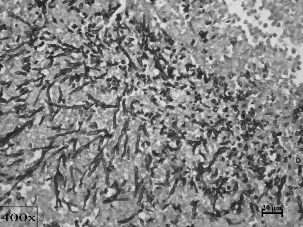
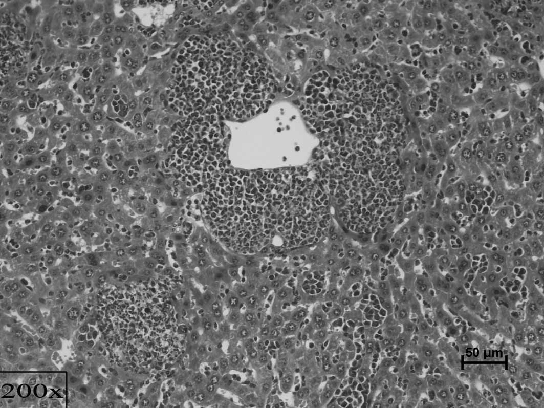
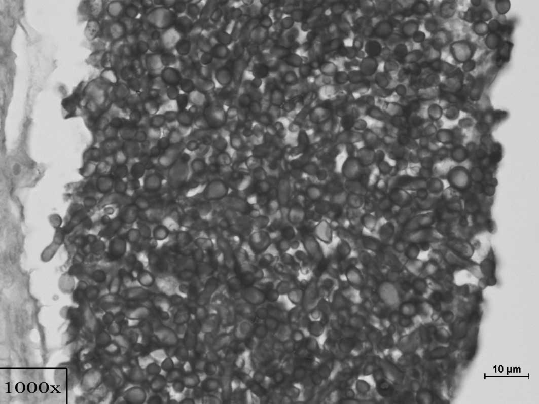
The histopathological scoring of inflammation and
congestion changes in these organs was based on the technique
employed by Kah Heng et al and Black et al (11,12)
(Figs. 3 and 4). Scoring of candidiasis was with
reference to Black et al and Balish et al (12,13)
(Figs. 2 and 3). Grading of the primary tumour was
carried out using the conventional method of analyzing the
similarity of the cells to its tissue of origin as poorly,
moderately and severely differentiated (14) (Figs.
1 and 4).
Statistical analysis
In this study, 60 samples were studied and analyzed.
Analytical data were expressed as a mean with standard deviation
and a 95% confidence interval. The level of significance was set at
0.05. Statistical tests used were: The i) paired t-test for
comparison of initial and final mean weight of mice in each group,
ii) Kruskal-Wallis test for global comparison of groups for all the
parameters, iii) non-parametric Mann-Whitney U test for comparison
between two groups for each parameter and iv) Spearman’s rho test
for the correlation of candidiasis, cancer metastases, inflammation
and congestion.
The statistical tests were conducted with the aid of
SPSS Statistical software version 16. For the individual tests,
p<0.05 was considered to be significant. The paired t-test is a
parametric method employed to test for any significant difference
between the means on the same or related subject over time or in
differing circumstances. From the test conducted, it was found that
p<0.05 in all the groups, with groups 1, 2 and 3 showing
p<0.01. Thus, a significant difference in the weight of the mice
in all the groups at the beginning and the end of the experiment
was noted (Table II).
| Table IIResults of the paired t-test for gross
weight of mice at the beginning and end of the experiment. |
Table II
Results of the paired t-test for gross
weight of mice at the beginning and end of the experiment.
| Group | Mean initial weight
(g) | Mean final weight
(g) | Asymptote
significance (p<0.05) |
|---|
| 1 | 17.71 | 19.09 | 0.001b |
| 2 | 18.36 | 16.85 | 0.009b |
| 3 | 19.00 | 20.00 | 0.000b |
| 4 | 19.40 | 18.01 | 0.039a |
| 5 | 20.25 | 18.04 | 0.032a |
Based on a global comparison for metastasis in each
of the organs for all groups, the Mann-Whitney U test for
comparison between groups 3 and 4 showed a significant difference
in all the organs except the brain (p<0.01; Table III). The kidneys showed a greater
level of significance (p<0.01) as compared to the other organs.
This shows that the presence of candidiasis, as in the case of
group 4, has an effect on the extent of the metastatic growth in
these organs.
| Table IIIResults obtained from the
Kruskal-Wallis test for global comparison of organ metastases among
the groups and the Mann-Whitney U test for comparison between
groups 3 and 4 for the extent of organ metastases. |
Table III
Results obtained from the
Kruskal-Wallis test for global comparison of organ metastases among
the groups and the Mann-Whitney U test for comparison between
groups 3 and 4 for the extent of organ metastases.
| Organs | Asymptote
significance (p<0.05) |
|---|
|
|
|---|
| Kruskal-Wallis test
of global comparison | Mann-Whitney U test
for comparison between groups 3 and 4 |
|---|
| Brain | 1.000 | - |
| Kidneys | 0.000b | 0.000b |
| Lungs | 0.000b | 0.016a |
| Liver | 0.000b | 0.015a |
| Spleen | 0.000b | 0.016a |
By comparing the median severity of candidiasis
between groups 2 and 4, a significant difference in severity was
observed in the kidneys and liver. In the kidneys of group 2, the
severity of candidiasis was mild, while that in group 4 was
moderate (Table IV). These
observations were also observed in slides stained in PAS and
GMS.
| Table IVHistopathological scoring of
candidiasis in hematoxylin and eosin. |
Table IV
Histopathological scoring of
candidiasis in hematoxylin and eosin.
| Experimental
group | Median of severity
of candidiasis |
|---|
|
|
|---|
| Brain | Kidneys | Lungs | Liver |
|---|
| Group 2- |
| Mice with
Candida (5×10⁶ cells/ml) | − | + | − | − |
| Group 4- |
| Mice with breast
cancer and Candida (5×10⁶ cells/ml) | − | ++ | − | ++ |
The Kruskal-Wallis test used for global comparison
between groups 2 and 4, and 4 and 5 for inflammation and congestion
showed a significant difference of p<0.01 in all the organs
(Tables V and VI). The Mann-Whitney U test used for
comparison between groups 2 and 4 for inflammation response showed
a significant difference in all the organs. This shows that the
co-existence of both candidiasis and cancer in the mice had a
heightened effect on the severity of inflammation as compared to
mice with candidiasis alone.
| Table VResults of Kruskal-Wallis for global
comparison among groups and Mann-Whitney U test for comparison
between groups 2 and 4 for organ inflammation response. |
Table V
Results of Kruskal-Wallis for global
comparison among groups and Mann-Whitney U test for comparison
between groups 2 and 4 for organ inflammation response.
| Organs | Asymptote
significance (p<0.05) |
|---|
|
|
|---|
| Kruskal-Wallis test
of global comparison | Mann-Whitney U test
for comparison between groups 2 and 4 |
|---|
| Brain | 0.000b | 0.000b |
| Kidneys | 0.000b | 0.005b |
| Lungs | 0.000b | 0.000b |
| Liver | 0.000b | 0.000b |
| Table VIResults of Kruskal-Wallis for global
comparison among groups and Mann-Whitney U test for comparison
between groups 4 and 5 for organ congestion. |
Table VI
Results of Kruskal-Wallis for global
comparison among groups and Mann-Whitney U test for comparison
between groups 4 and 5 for organ congestion.
| Organs | Asymptote
significance (p<0.05) |
|---|
|
|
|---|
| Kruskal-Wallis test
of global comparison | Mann-Whitney U test
for comparison between groups 4 and 5 |
|---|
| Brain | 0.000b | 0.000b |
| Kidneys | 0.000b | 0.000b |
| Lungs | 0.000b | 0.006b |
| Liver | 0.000b | 0.000b |
The Mann-Whitney test was used for comparison
between groups 4 and 5 to examine the extent of candidiasis. The
findings showed that the increase in the Candida dosage
injected into group 5 at a concentration of 5×10⁸
cells/ml compared to 5×10⁶ cells/ml in group 4, showed a
statistically significant difference in the brain and lungs
(p<0.01; Table VII). The
Mann-Whitney U test for comparison between groups 4 and 5 for
inflammation and congestion showed a significant difference in the
inflammatory response at the liver with p<0.01. This means that
the increased dose in group 5 showed a statistically significant
effect on the inflammatory response noted in the liver and
congestion observed in the brain and the lungs.
| Table VIIResults of hte Mann-Whitney U test
for comparison between groups 4 and 5 for the extent of
candidiasis, organ inflammation and organ congestion. |
Table VII
Results of hte Mann-Whitney U test
for comparison between groups 4 and 5 for the extent of
candidiasis, organ inflammation and organ congestion.
| Organs | Asymptote
significance (p<0.05) |
|---|
|
|
|---|
| Extent of
candidiasis | Organ
inflammation | Organ
congestion |
|---|
| Brain | 0.000b | 0.070 | 0.004b |
| Kidneys | 0.308 | 0.265 | 0.229 |
| Lungs | 0.000b | 0.314 | 0.013a |
| Liver | 0.808 | 0.000b | 1.000 |
The correlation between candidiasis and cancer
metastases was positive in the kidneys and liver for a significance
level of 0.01 (Table VIII).
However, no correlation was found in the brain, while in the lungs
the correlation was not significant. The correlation between
candidiasis and inflammation was shown to be positively correlated
with significance at a level of 0.01 in the kidneys, lungs and
liver, while the brain only showed significance at a level of 0.05.
The correlation between candidiasis and congestion was positive at
a significance level of 0.01 in all of the organs.
| Table VIIICorrelation coefficient between
candidiasis with cancer metastases, organ inflammation and organ
congestion. |
Table VIII
Correlation coefficient between
candidiasis with cancer metastases, organ inflammation and organ
congestion.
| Comparison
organs | Spearman rho
correlation coefficient between candidiasis and cancer
metastases | Spearman rho
correlation coefficient between candidiasis and organ
inflammation | Spearman rho
correlation coefficient between candidiasis and organ
congestion |
|---|
| Brain | 0 (no
correlation) | 0.272a | 0.553b |
| Kidneys | 0.347b | 0.619b | 0.537b |
| Lungs | 0.212 | 0.587b | 0.544b |
| Liver | 0.485b | 0.614b | 0.616b |
Discussion
Few studies have been conducted on experimental
systemic candidiasis in mice. Information obtained from these
studies on the necessary dosages, as well as previous observations
were used to make comparisons with this study (15–17).
Some of these studies have been dedicated to the observations of
the correlation between systemic candidiasis and other forms of
immunosuppression such as chemotherapy, steroid therapy, antibiotic
therapy, as well as other types of malignancies such as leukemia
and esophageal cancer.
However, an exclusive study on systemic candidiasis
and its relationship with breast cancer has yet to be conducted,
even though there are a few epidemiological studies that have shown
a co-existence of breast cancer and systemic candidiasis in humans
(7,10,18).
This study aimed to focus on the relationship between systemic
candidiasis and breast cancer by examining the behaviour of
candidiasis when the body is subjected to a chronic disease state.
Therefore, breast cancer was not only selected as an ideal
representation of a chronic illness, but also one that is capable
of suppressing the host immune system (19–22).
To study how the presence of a chronic illness such
as breast cancer can, by itself, be attributed to the increased
severity of candidiasis, this novel study focused on the growth of
systemic candidiasis following the induction of mice with breast
cancer. Scores were calculated based on the severity of candidiasis
and grading of the primary tumour, and identification of their
metastatic deposits was conducted. Other parameters taken into
consideration included gross weight of the mice at the beginning
and end of the study, as well as inflammation and congestion in the
respective organs which were studied by scoring on a
semi-quantitative scale using an established technique as mentioned
earlier.
Systemic candidiasis
In group 2, the mice were solely inoculated with
systemic candidiasis by intravenous injection via the tail vein for
a duration of two weeks. During the course of the experiment, signs
of the disease were noted in these mice. Their eyeballs protruded,
their fur roughened and they were generally less active as compared
to the normal group. Moreover, increased group huddle and sleep
were noted. The mice also appeared very weak and thin with the
curvatures of the bony structures beneath the mice visible to the
naked eye. In addition, the weight taken at the beginning and end
of the experiment showed that there was a statistically significant
reduction in their mean weight. This was attributed to the possible
loss of appetite and general cachexic state of the mice.
Histopathologically, favourable growth of the Candida
colonies in the form of hyphae, yeast cells and pseudohyphae were
discovered in the kidneys, pelvis and tubule region, but not in
many of the other organs. This was attributed to the mild dose of
5×10⁶ cells/ml Candida cells injected and the
short duration of the experiment as also shown by Wong et al
(16).
Breast cancer study
In group 3, the mice were injected at the mammary
fatpad with 4T1 cancer cells in the right axilla region at a
concentration of 1×10⁶ cells/ml (23). After four weeks of growth and
metastases, the mice were sacrificed for analysis. During the
course of the investigation, the weights of the mice were reduced
for the first week before gradually increasing in the 3rd week. The
growth of the primary tumour was detected as a palpable mass as
early as the 10th day and as late as the 14th day. The mice were
generally active for the first two weeks with no apparent
deviations from those usually observed in the normal control group.
However, by the 3rd week, the mice began to exhibit signs of
lethargy and did not move as often. Furthermore, the mass of tumour
began to appear significantly enlarged to the naked eye by the
middle of 3rd week. General appetite was good. No distinct changes
to the fur, eyes or prominent curvatures of the bony structures
were noted.
Grading for the primary tumour showed it to be
moderate to poorly differentiated with the majority of the tumours
being poorly differentiated. Metastatic deposits were discovered in
the lungs, liver and spleen with varying frequencies among the
mice. The gross morphology of the other organs did not exhibit any
crude changes, with the exception of the spleen which was markedly
enlarged as compared to that found in the normal group. The tumour
appeared hard and smooth in texture with a glistening surface.
Scoring for inflammation showed that the median of
severity of the entire group was mild in both the lungs and liver.
The median severity of congestion found in the kidneys, lungs and
liver were mild, while congestion found in the brain was mild to
moderate. In the liver, however, the micro-abscesses that were
observable in group 2, were not noted in the group as a whole.
Therefore, in the group with breast cancer, the severity of
inflammation and congestion observed in the organs were mostly mild
in severity with metastatic deposits found in the lungs, liver and
spleen.
Correlation between systemic candidiasis
and breast cancer
In group 4, the mice were first induced with breast
cancer for three weeks and subsequently inoculated with
Candida at a concentration of 5×10⁶ cells/ml for
one week. The time of induction with breast cancer was set at three
weeks, based on studies showing that by this period, adequate
metastases have occurred in all these organs (23,24).
The initial stages of tumour growth and changes in the mice were
similar to those found in group 3. However, when Candida was
injected, changes in group 2 were noted within days instead of the
2nd week as in the case of group 2. These changes included
protruding eyes, roughened fur and general inertia, with increased
huddle and sleep. In the final stages of the investigation, a surge
in the growth of tumour size was observed.
Grading carried out for the primary tumour showed
poorly differentiated tissue with atypical cells and a high number
of mitotic figures. Metastatic deposits were also found in the
lungs, liver, spleen and even in the kidneys at a higher frequency
as compared to that of group 3. These differences were
statistically significant (p<0.05). Thus, an increased frequency
of metastatic deposits occurred in these organs in group 4 as
compared to that in group 3. This suggests a possible role of
Candida causing immunosuppression which, by itself,
attributed to the increased metastatic deposits of the cancer found
in these organs. It also explains the late surge in tumour growth
noted late in the investigation.
Notable changes in the kidneys include candidiasis
involvement in the renal parenchyma, renal tubules and pelvis.
Within the liver parenchyma and vasculature, distinct changes such
as micro-abcesses, chronic inflammation and congestion were
observed at a greater level in this group as compared to that noted
in group 2. This group also showed an increased group median of
severity in Candida infection in the kidneys and liver. The
kidneys showed a moderate severity as compared to a mild one in
group 2, while the liver showed a moderate severity of candidiasis
as compared to the absence of candidiasis noted in group 2. Thus,
this group showed extra involvement of the liver compared to only
the kidneys as was the case in group 2. This observation holds true
in scoring performed for both PAS and GMS.
Scoring of inflammation showed a moderate severity
as noted in the brain, kidneys and lungs, while the liver showed
severe changes as compared to only mild ones found in all the
organs in group 2. Comparison of inflammation severity between the
two groups was statistically significant (p<0.01).
As for congestion, group 4 showed a moderate
congestion in the brain and kidneys as compared to a mild one in
group 2 and while congestion in the lungs was not observed in group
2, group 4 showed mild congestion. Furthermore, the liver showed
severe congestion as compared to just moderate congestion found in
group 2. A comparison between groups 2 and 4 for congestion was
statistically significant (p<0.05). In conclusion, severity of
candidiasis, inflammation and congestion were found at greater
levels in breast cancer induced mice with candidiasis as compared
to mice with only candidiasis.
Dose-dependent study
In group 5, the mice were first induced with breast
cancer and, subsequently, with candidiasis at a higher dose of
5×10⁸ cells/ml. These mice were similar to group 3 at
the initial stages of cancer growth. However, when candidiasis was
injected, the mice died within the first week of inoculation at
varied periods as compared to group 4 where the time of inoculation
with candidiasis was one week and mice survived till the end of
investigation. The sudden immediate death was attributed to
septicaemia.
Grading on the primary tumour showed the tumours to
be poorly differentiated. Metastatic deposits were found in the
kidneys, lungs, liver and spleen. Scoring of candidiasis showed
mild severity in the brain while the kidneys showed moderate and
the lungs severe candidiasis. The liver, on the other hand, showed
mild to moderate severity. Statistical tests comparing differences
in candidiasis severity between groups 4 and 5 found a significant
difference in the brain and lungs (p<0.01). This means that with
an increased dose, the brain and lungs exhibit candidiasis with
increased levels of severity. It is possible that with a higher
dose, the higher reaches of the body are more easily accessed as
the amount of dose eliminated by the liver or spleen is less.
In the scoring for inflammation, the brain showed
mild severity while the kidneys, lungs and liver showed moderate
severity. However, only the liver showed a statistically
significant difference when compared to group 4. Inflammation was
much less in severity compared to that in group 4, which may be
attributed to the short period of inoculation time before the
demise of the mice resulting in inadequate time for chronic
inflammation to occur.
In the scoring for congestion, group 5 showed severe
congestion in all the organs. However, only the brain and the lungs
showed a statistically significant difference with group 4, which
was shown to have a greater level of congestion. This may be
attributed to the acute changes noted in the host response to a
foreign pathogen.
Correlation of candidiasis, cancer
metastases, organ inflammation and organ congestion
The correlation between candidiasis and cancer
metastases was found to be significant in the kidneys and liver
(p<0.01). This shows that in the kidneys and liver, an increase
in cancer metastatic deposits was accompanied by an increase in
candidiasis severity. When correlating candidiasis with
inflammation and congestion, it was found to be statistically
significant (p<0.05) in all the organs. Thus, increased levels
of candidiasis are accompanied by increased levels of inflammation
and congestion in the respective organs studied.
In conclusion, the mouse model of inducing breast
cancer was successful, as well as the method and technique of
inducing candidiasis was effective. The mouse model and method and
technique were attributed to the efficient culture methods.
Moreover, growth of breast cancer and candidiasis were observable
in all the relevant groups. The weight of the mice was also
correlated with the pathology suffered by the mice. All the
objectives were carried out with precision and successfully
achieved.
An analysis was performed based on the scoring of
candidiasis, grading of metastatic deposits, inflammation and
congestion in the brain, kidneys, lungs and liver of the mice in
all the groups. The inflammation and congestion parameters showed a
statistically significant increase in severity in all the organs as
compared to the group of mice with systemic candidiasis and breast
cancer, and that of systemic candidiasis alone. The median severity
of the entire group for candidiasis scoring in the kidneys and
liver also increased for the group of mice with systemic
candidiasis and breast cancer.
Therefore, based on these evidences, systemic
candidiasis appears to be more severe in experimentally induced
mice with breast cancer than in mice without.
Acknowledgements
This investigation was funded by research grant no.
BMS I-02/2008 (12) from the
International Medical University, Kuala Lumpur, Malaysia.
References
|
1
|
Levinson W: Review of Medical Microbiology
and Immunology. 9th edition. The McGraw-Hill Companies; pp.
134–136. 2006
|
|
2
|
Richardson MD: Changing patterns and
trends in systemic fungal infections. J Antimicrob Chemother.
56(Suppl 1): 5–11. 2005. View Article : Google Scholar
|
|
3
|
Yip CH, Taib NA and Mohamed I:
Epidemiology of breast cancer in Malaysia. Asian Pac J Cancer Prev.
7:369–374. 2006.PubMed/NCBI
|
|
4
|
Hisham AN and Yip CH: Overview of breast
cancer in Malaysian women: a problem with late diagnosis. Asian J
Surg. 27:130–133. 2004. View Article : Google Scholar : PubMed/NCBI
|
|
5
|
DiNubile MJ, Hille D, Sable CA and
Kartsonis NA: Invasive candidiasis in cancer patients: observations
from a randomized clinical trial. J Infect. 50:443–449. 2005.
View Article : Google Scholar : PubMed/NCBI
|
|
6
|
Koh AY, Kohler JR, Coggshall KT, van
Rooijen N and Pier GB: Mucosal damage and neutropenia are required
for candida albicans dissemination. PLoS Pathogens. 4:35–38. 2008.
View Article : Google Scholar : PubMed/NCBI
|
|
7
|
Gottfredsson M, Vredenburgh JJ, Xu J,
Schell WA and Perfect JR: Candidemia in women with breast carcinoma
treated with high-dose chemotherapy and autologous bone marrow
transplantation. Cancer. 98:24–30. 2003. View Article : Google Scholar : PubMed/NCBI
|
|
8
|
Ghoneum M and Gollapudi S: Phagocytosis of
Candida albicans by metastatic and non metastatic human breast
cancer cell lines in vitro. Cancer Detect Prev. 28:17–26. 2004.
View Article : Google Scholar : PubMed/NCBI
|
|
9
|
Anderson LM, Krotz S, Weitzman SA and
Thimmapaya B: Breast cancer-specific expression of the Candida
albicans cytosine deaminase gene using a transcriptional targeting
approach. Cancer Gene Ther. 7:845–852. 2000. View Article : Google Scholar
|
|
10
|
Safdar A, Chaturvedi V, Cross EW, Park S,
Bernard EM and Armstrong D: Prospective study of Candida species in
patients at a comprehensive cancer center. Antimicrob Agents
Chemother. 45:2129–2133. 2001. View Article : Google Scholar : PubMed/NCBI
|
|
11
|
Lee KH, Chen YS, Judson JP, Chakravarthi
S, Sim YM and Er HM: The effect of water extracts of Euphorbia
hirta on cartilage degeneration in arthritic rats. Malays J Pathol.
30:95–102. 2008.PubMed/NCBI
|
|
12
|
Black CA, Eyers FM, Russell A, Dunkley ML,
Clancy RL and Beagley KW: Increased severity of Candida vaginitis
in BALB/c nu/nu mice versus the parent strain is not abrogated by
adoptive transfer of T cell enriched lymphocytes. J Reprod Immunol.
45:1–18. 1999. View Article : Google Scholar : PubMed/NCBI
|
|
13
|
Balish E: A URA3 null mutant of Candida
albicans (CAI-4) causes oro-oesophageal and gastric candidiasis and
is lethal for gnotobiotic, transgenic mice (Tgepsilon26) that are
deficient in both natural killer and T cells. J Med Microbiol.
58:290–295. 2009. View Article : Google Scholar : PubMed/NCBI
|
|
14
|
Kumar VRSC and Robbins SL: Robbins Basic
Pathology. 7th edition. Saunders; pp. 436–438. 2003
|
|
15
|
De Repentigny L: Animal models in the
analysis of Candida host-pathogen interactions. Curr Opin
Microbiol. 7:324–329. 2004.PubMed/NCBI
|
|
16
|
Wong SF, Mak JW and Pook CK: Potential use
of a monoclonal antibody for the detection of Candida antigens in
an experimental systemic candidiasis model. Hybridoma. 27:361–373.
2008. View Article : Google Scholar : PubMed/NCBI
|
|
17
|
Ashman RB and Papadimitriou JM: Murine
candidiasis. Pathogenesis and host responses in genetically
distinct inbred mice. Immunol Cell Biol. 65:163–171. 1987.
View Article : Google Scholar : PubMed/NCBI
|
|
18
|
Talarmin JP, Boutoille D, Tattevin P, et
al: Epidemiology of candidemia: a one-year prospective
observational study in the west of France. Med Mal Infect.
4:106–112. 2009.PubMed/NCBI
|
|
19
|
Mandeville R, Lamoureux G, Legault-Poisson
S and Poisson R: Biological markers and breast cancer. A
multiparametric study II Depressed immune competence. Cancer.
50:1280–1288. 1982. View Article : Google Scholar : PubMed/NCBI
|
|
20
|
Semiglazov VF, Kondrat’ev VB, Mar’enko AI,
L’Vovich EG and Sofronov BN: Immunologic reactivity of breast
cancer patients. Vopr Onkol. 24:74–79. 1978.PubMed/NCBI
|
|
21
|
Contreras Ortiz O and Stoliar A:
Immunological changes in human breast cancer. Eur J Gynaecol Oncol.
9:502–514. 1988.PubMed/NCBI
|
|
22
|
Das SN, Khanna NN and Khanna S: A
multiparametric observation of immune competence in breast cancer
and its correlation with tumour load and prognosis. Ann Acad Med
Singapore. 14:374–381. 1985.PubMed/NCBI
|
|
23
|
Pulaski BA and Ostrand-Rosenberg S: Mouse
4T1 breast tumor model. Curr Protoc Immunol. 20:2–11. 2001.
|
|
24
|
Tao K, Fang M, Alroy J and Sahagian GG:
Imagable 4T1 model for the study of late stage breast cancer. BMC
Cancer. 8:228–236. 2008. View Article : Google Scholar : PubMed/NCBI
|