Introduction
Compared with the general population, recipients of
a kidney transplant have an increased risk of developing various
types of cancer (associated and non-associated with infections),
due to the immunosuppressant treatment that must be maintained in
order to prevent and treat acute rejection of the organ (1). The incidence of secondary cancer
increases with the time from the transplant (2). Post-transplant lymphoproliferative
disorders (PTLDs) are frequent in patients who receive
immunosuppressants, including anti-thymocyte and -lymphocyte
globulins or muromonab-CD3, or those who are infected de
novo by Epstein-Barr virus (EBV) through the transplanted
kidney (3). PTLDs may present early
or late; be nodal, extranodal, polymorphic or monomorphic; and
follow an indolent or aggressive clinical course (3). In contrast to classical PTLDs, there are
limited studies on plasma cell malignancies that occur following
solid organ transplantation. Multiple myeloma (MM) represents ≤4%
of all PTLDs, and is associated with a poor response to
discontinuation of immunosuppression and conventional therapy, and
a short median survival rate (4,5).
Generally, the presence of plasma cells in the liver is associated
with aggressive forms of MM (6,7). In the
present study, the case of a patient who developed post-transplant
MM with extramedullary liver plasmacytoma 11 years following renal
transplantation, and was successfully treated with lenalidomide, is
reported.
Case report
In February 2012, a 45-year-old female was admitted
to The First People's Hospital of Changzhou (Changzhou, China) with
complaints of pain in the left shoulder. Due to chronic renal
failure, the patient had received a cadaveric kidney
transplantation at the same hospital on November 2nd
2001, and was subsequently administered immunosuppression composed
of 250 mg/day cyclosporine, 50 mg/day azathioprine and 25 mg/day
prednisone, in a dose-tapering manner. From June 2002, the patient
received 200 mg/day cyclosporine for maintenance therapy to prevent
renal rejection. The patient exhibited normal renal function during
the follow-up period.
In March 2008, the patient developed a left nasal
obstruction. Computed tomography (CT) scan revealed a soft tissue
mass in the left nasal cavity, which was surgically excised. The
post-operative histological study confirmed the presence of
extramedullary plasmacytoma, and immunochemical examination
demonstrated the specimen to be CD79α+, partially
CD138+, CD20−, CD3−,
CD45RO−, vimentin+, epithelial membrane
antigen (EMA)−, cytokeratin AE1/3− and 50%
Ki-67+. Treatment with cyclosporine and azathioprine was
discontinued, and the patient was administered instead rapamycin (2
mg/day) and mycophenolate mofetil (MMF; 1 mg twice daily) to
prevent renal rejection.
From March 26th to May 14th
2008, the patient received local radiotherapy in the bilateral
nasal cavity, ethmoid sinus and maxillary sinus at a total dose of
48 Gy, and continued receiving immunosuppressants (rapamycin and
MMF). During the follow-up period, the patient displayed normal
renal function.
In February 2012, the patient reported idiopathic
pain in the left shoulder. CT scan revealed marked bone destruction
of the left scapula and reactive bone formation at the right tenth
rib. Positron emission tomography-CT scan revealed high metabolism
of 18fluorodeoxyglucose at the left scapula, with a
standardized uptake value of 5.5, in addition to bone destruction.
Consequently, the patient was subjected to surgery, and
postoperative pathological examination suggested plasma cell
myeloma, while immunohistochemical staining indicated the specimen
to be CD20−, CD3−, CD38+,
CD138+, partially CD79α+, EMA−,
melanoma associated antigen mutated 1 (MUM1)− and <5%
Ki-67+. Bone marrow smear identified 2% mature plasma
cells with normal female chromosome karyotype. Interphase
fluorescence chromosomal in situ hybridization (FISH) of the
bone marrow cells revealed no gene abnormalities in 1q21, RB1, P53,
D13S319 and IgH. The serum concentrations of IgG and λ-light chain
were 43.8 g/l and 4,930 mg/dl, respectively. Serum protein
electrophoresis disclosed a monoclonal spike in the γ-globulin
region, whereas urine electrophoresis revealed no monoclonal spike.
Serum immunofixation electrophoresis confirmed the presence of an
IgG-λ chain monoclonal M component. The renal function and the
levels of calcium, hemoglobin, serum albumin, β2-microglobulin and
lactate dehydrogenase were normal. No significant alteration was
detected in the titers of anti-cytomegalovirus, -EBV or -hepatitis
B virus (HBV) surface antigen (HBsAg) antibodies. Thus, the patient
was diagnosed with post-renal-transplantation secondary MM IgG-λ
chain-type, group A and stage I, according to the International
Staging System (8). The patient
refused treatment with the novel agent bortezomib, due to the high
cost, and instead, the patient received modified VADT regimen
(vincristine, 0.4 mg, days 1–4; doxorubicin, 10 mg, days 1–4;
dexamethasone, 40 mg, days 1–4; and thalidomide, 50–150 mg, days
1–28) for a total of 3 cycles (28 days/cycle).
The patient experienced clinical complete remission
in June 2012, and exhibited negative serum immunofixation
electrophoresis, 2% mature plasma cells in the bone marrow smear
and no hypercalcemia, renal failure or anemia. Next, the patient
received a fourth cycle of VADT regimen, followed by 3 cycles (28
days/cycle) of TD regimen (thalidomide, 100 mg, days 1–28; and
dexamethasone, 40 mg, days 1–4), as maintenance therapy. During the
chemotherapy treatment, the patient was administered a reduced dose
of immunosuppressive therapy (rapamycin, 2 mg/day; and MMF, 1.0 g
twice daily, tapered to 0.5 g twice daily). During the follow-up,
the patient underwent monthly urine analysis, and the measurements
of blood urea nitrogen, creatinine, serum Igs and light chain
remained unaltered.
In March 2013, during a routine follow-up, increased
levels of serum IgG (28 g/l) were detected, which were confirmed to
be M protein by serum immunofixation electrophoresis, suggesting
asymptomatic relapse of MM. In consequence, the patient was again
advised to receive novel therapeutic agents such as bortezomib or
lenalidomide, but the patient selected to receive 3 cycles of the
previous VADT regimen. However, the patient's condition gradually
deteriorated, and in June 2013, levels of serum IgG increased to 57
g/l, with positive serum immunofixation electrophoresis, indicating
that the VADT regimen was no longer effective to treat the relapse.
Therefore, the patient was administered novel agents for rescue
therapy. On July 26th, August 23rd, September
19th and October 25th 2013, the patient
received a CyBorDT regimen (cyclophosphamide, 300 mg/m2,
days 1,8; bortezomib, 1.3 mg/m2, days 1,4,8,11;
dexamethasone, 20 mg, days 1, 2, 4, 5, 8, 9, 11 and 12; and
thalidomide, 100 mg, days 1–21) for 4 cycles (21 days/cycle). The
subsequent evaluation demonstrated negative serum immunofixation
electrophoresis with normal IgG levels and absence of abnormal
plasma cells in bone marrow smear. Thus, the patient experienced a
complete clinical remission for the second time in November 2013.
The patient then accepted TD regimen instead of bortezomib for
maintenance therapy, followed by monthly routine laboratory
investigations, including ultrasonography of the abdomen, which
remained unaltered.
However, 3 months later, in early February 2014, the
patient suddenly experienced fever with a temperature of 38.7°C and
onset of fatigue, progressive lumbago and non-tender skin mass of
the submaxilla. The subsequent abdominal ultrasound identified a
diffused hypoechoic lesion in the liver, with a maximum diameter of
8.0×6.5 cm. Full blood count examination demonstrated levels of
hemoglobin (84 g/l), hematocrit (35.6%), mean corpuscular volume
(86 fl), white blood cells (8.79) and platelets (134×109
cells/l). The results of the liver function tests were in the
normal range. The levels of serum total protein were 28 g/l, and
protein electrophoresis detected the presence of a monoclonal band
in the γ region, which was identified as IgG-λ by immunofixation.
Markers for hepatitis C and B virus and serum anti-EBV antibody
were negative. Upper abdominal non-enhanced CT scan indicated the
presence in the hepatic parenchyma of multiple round-shaped and
well-defined lesions of variable sizes with low attenuations.
Following administration of contrast agents, the enhancements of
the lesions appeared mild and heterogeneous. Based on the clinical
information available, these observations were considered to be due
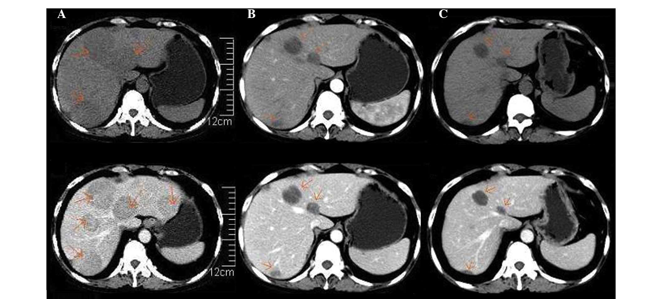
to myeloma (Fig. 1A). Magnetic
resonance imaging (MRI) of the lumbar vertebrae detected the
presence of a soft mass and multiple patchy lesions with high
attenuations in the T12 thoracic and L1-L4 lumbar vertebrae, and
swelling of the spine, spinal accessory, S1, S2 and soft tissue
around S1 and S2 (Fig. 2A).
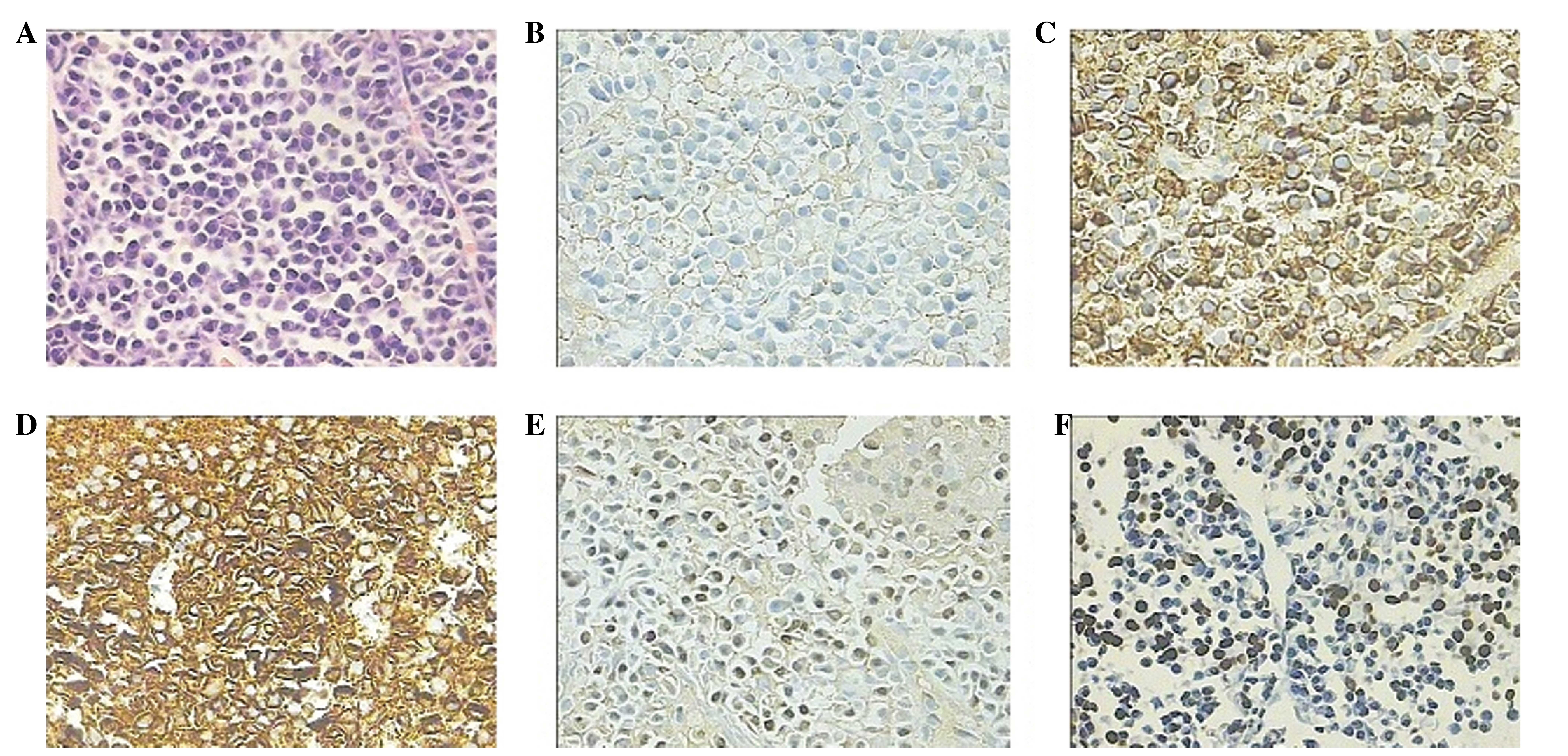
Therefore, a CT-guided percutaneous needle biopsy of the hepatic
lesion was performed, and the histological study demonstrated
diffused proliferation of plasma cells by hematoxylin and eosin
staining. The immunochemical examination demonstrated the specimen
to be CD20−, CD3−, CD38+,
CD138+, κ+<λ+, EBV+,
MUM1+ and ~30–40% Ki67+, which was indicative
of extramedullary liver plasmacytoma (Fig. 3). The bone marrow aspirate revealed 6%
plasma cell infiltration, and CD138 sorting interphase FISH of bone
marrow cells detected 1q21 amplification. Furthermore, increased
levels of serum IgG (62 g/l) were measured, which were confirmed to
correspond to M protein by serum immunofixation
electrophoresis.
 | Figure 1.Differences observed in the upper
abdominal CT scan images performed prior and subsequent to
lenalidomide therapy. (A) non-enhanced (top) and enhanced (bottom)
CT preceding lenalidomide therapy, performed February
25th 2014. Multiple round-shaped and well-defined
lesions of different sizes with low attenuations were present in
the hepatic parenchyma on non-enhanced CT. Following the
administration of contrast reagents, the enhancements of the
lesions were mild and heterogeneous. (B) Non-enhanced (top) and
enhanced (bottom) CT subsequent to 4 cycles of RCD regimen therapy
(July 1st 2014). A reduction in the number and size of
the round-shaped lesions in the hepatic parenchyma was observed,
compared with prior to treatment. (C) Non-enhanced (top) and
enhanced (bottom) CT following 5 cycles of RCD regimen (August
25th 2014). Further reduction in the size of the
round-shaped lesions present in the hepatic parenchyma was
observed, although the lesions did not disappear completely. CT,
computed tomography; RCD regimen, lenalidomide, 25 mg, days 1–21;
cyclophosphamide, 50 mg, days 1–21; and dexamethasone, 20 mg, days
1, 8, 15 and 22; 28 days/cycle. |
 | Figure 2.Differences in MRI images of the
lumbar vertebrae prior and subsequent to lenalidomide therapy. (A)
MRI preceding lenalidomide therapy (March 11th 2014). A
soft mass and multiple patchy lesions with high attenuations were
clearly observed in the T12 thoracic and L1-L4 lumbar vertebrae, in
addition to swelling of the spine, spinal accessory, S1, S2 and
soft tissue around S1 and S2. (B) MRI subsequent to 5 cycles of RCD
regimen, conducted August 26th 2014. Alleviated swelling
of the soft tissue around S1 and S2 was observed. MRI, magnetic
resonance imaging; RCD regimen, lenalidomide, 25 mg, days 1–21;
cyclophosphamide, 50 mg, days 1–21; and dexamethasone, 20 mg, days
1, 8, 15 and 22; 28 days/cycle. H, head. |
The patient experienced a rapid relapse 3 months
subsequent in February 2014 to the CyBorD regimen, thus, the
patient was initiated instead on an RCD regimen (lenalidomide, 25
mg, days 1–21; cyclophosphamide, 50 mg, days 1–21; dexamethasone,
20 mg, days 1, 8, 15 and 22) for 2 cycles (28 days/cycle), from
March 14th to May 10th 2014. The next
re-evaluation demonstrated that the levels of serum IgG had reduced
to 27 g/l, and ultrasonography indicated that the hepatic mass had
markedly reduced in size to 6.8×5.5 cm, indicating that the
extramedullary liver plasmacytoma was sensitive to the RCD
regimen.
Subsequently, the patient received third and fourth
cycles of RCD, and the following upper abdominal CT scan, performed
July 1st 2014, revealed that the extramedullary liver
plasmacytoma had markedly reduced (Fig.
1B), while the levels of hemoglobin and serum IgG were 114 and
13.6 g/l, respectively. During the fourth cycle of RCD, the patient
experienced transient agranulocytosis with pulmonary infection.
Once the infection had been controlled, the patient did not present
symptoms of fever or lumbago. Furthermore, the patient experienced
a recovery of myelosuppression, indicating that the patient had
achieved partial remission following 4 cycles of RCD regimen.
On August 25th 2014, the patient received
the fifth cycle of RCD, and the subsequent abdominal CT
demonstrated a further reduction in size, although no complete
disappearance, of the round-shaped lesions present in the hepatic
parenchyma (Fig. 1C). On August
26th 2014, lumbar vertebrae MRI disclosed a marked
reduction of the soft tissue mass in T12 and alleviated swelling of
the soft tissue around S1 and S2 (Fig.
2B). Currently, the patient is undergoing outpatient follow-up
and receiving RCD regimen for maintenance therapy.
The study was approved by the ethics committee of
the First People's Hospital of Changzhou, Third Affiliated Hospital
of Suzhou University (Changzhou, P.R. China).
Discussion
Secondary cancer is a major complication of renal
transplant, due to the immunosuppressant treatment required for
preventing renal rejection. Secondary cancer following renal
transplant results in significant rate of short- and long-term
mortality, accounting for 30% of mortalities among renal transplant
recipients with a follow-up >20 years (2,9–11). PTLD is a potentially fatal
complication of solid organ transplantation, and comprises various
lymphoid lesions, ranging from polymorphous reactive proliferations
to monomorphous malignant lymphomas (12,13). The
majority of these disorders are considered to be driven by EBV
infection and subsequent proliferation of B cells in a weakened
host. Previous studies have detected EBV-encoded RNA (EBER) in the
neoplastic cells of the host, suggesting the implication of the EBV
genome in the pathogenesis of PTLD (2). EBV-naïve patients who receive an organ
from an EBV-infected donor present the highest risk of developing
PTLD (10). There are several
treatments available for PTLD, including reduced immunosuppression,
surgical excision, chemoradiotherapy, anti-CD20 antibody therapy
and infusion of EBV-specific cytotoxic T cells, all with variable
results (13).
Post-transplantation plasma cell neoplasms (PCNs)
are a monomorphous type of PTLD derived from plasma cells, which
are the mature, terminally differentiated B cells responsible for
the production of antibodies (14).
Clinically, 2 diagnostic categories of PCN are recognized: MM,
which arises in the bone marrow; and plasmacytoma, which presents
as an isolated tumor of plasma cells. Patients with plasmacytoma
are commonly diagnosed with MM within months to a few years, which
indicates that these conditions are associated (2). Thus far, post-transplantation PCNs have
been observed in recipients of solid organ transplantation, but not
in recipients of hematopoietic stem cell transplantation (HSCT)
(4,5,15).
Compared with lymphoma, and in particular diffused large B cell
lymphoma, post-transplantation PCN is rarely observed, and it
accounts for the mortality of 0.24–0.69% of recipients of solid
organ transplantation (4,5,15). Unlike
non-post-transplantation PCN, post-transplantation PCN occurs in
recipients of different ages, including children (16–18).
Potential association factors that contribute to
post-transplantation PCN have been suggested as follows: i) Old age
of the recipient; ii) deceased donor; iii) onset of EBV infection
post-transplantation; iv) HCV infection in the recipient; and v)
administration of anti-thymocyte or -lymphocyte globulins following
solid organ transplantation (4,15).
The incidence of MM is rare in renal graft
recipients, despite the frequent findings of monoclonal
gammopathies following transplantation (19,20). Sun
et al (21) reported 3 cases
of plasma cell myeloma PTLD that exhibited clinical, radiological
and pathological features of conventional plasma cell myeloma, and
Tcheng et al (16) reported
the occurrence of EBV-associated post-transplant MM in a
16-year-old male. Plasmacytoma is a tumor of terminally
differentiated monoclonal plasma cells, and a frequent complication
of MM, appearing at diagnosis or during disease progression
(5,20–23).
Post-transplant plasmacytomas have been described at several sites,
including the allograft, skin, peritoneum, gastrointestinal tract
and gingiva. To the best of our knowledge, extramedullary
plasmacytoma of liver is associated with aggressive forms of MM,
and its presumptive ante mortem diagnosis is often based on
clinical findings, such as hepatomegaly and alterations of the liver
function detected by laboratory tests. Cases of myeloma with
involvement of the liver have been previously reported (24). In the present case report, the patient
developed nasal plasmacytoma 7 years following renal
transplantation, and experienced relapsed MM with extramedullary
liver plasmacytoma 4 years subsequent to this. The patient did not
exhibit a large number of abnormal plasma cells in the bone marrow
cells, in agreement with previous studies (5,20,22,23). In
this case, deceased donor and EBV infection are the 2 major
association factors for the post-transplantation PCN observed in
the patient.
Various treatment options currently exist for
eligible patients with MM, including induction therapy, which
usually involves novel biologic agents, followed by autologous stem
cell transplantation (25). CyBorD
regimen (cyclophosphamide, bortezomib and dexamethasone) has been
demonstrated to be effective in the treatment of MM, and produces a
rapid and complete hematological response in the majority of
patients (26). However, this
treatment is generally not considered curative, and relapses
usually occur. To the best of our knowledge, extramedullary relapse
is an uncommon presentation, and relapses that involve the liver
have rarely been described (24).
Extramedullary plasmacytoma confers a poor prognosis, since it is
resistant to conventional treatments, due to its distinct molecular
and histological features (27–29). There
are very limited data from randomized trials regarding the most
appropriate systemic treatment for extramedullary plasmacytoma.
Previous case reports suggested that lenalidomide is an effective
agent, in combination with dexamethasone, and previous cases of MM
relapse following autologous stem cell transplant presenting with
diffuse pulmonary nodules exhibited good response to the treatment
with bortezomib, dexamethasone and lenalidomide (27–31).
Au et al (32)
reported a case of PTLD presenting as an EBV-associated nasal
plasmacytoma in a renal allograft recipient 13 years following
transplantation. In order to treat the condition, the authors
conducted a non-myeloablative HSCT with peripheral blood
hematopoietic stem cells from the kidney donor. In their report, Au
et al (32) described the
occurrence, in a renal allograft recipient, of an MM with
extramedullary liver plasmacytoma. In the case of the patient in
the present study, the existence of nasal, scapula and liver
plasmacytoma was confirmed by post-operative histopathological
examination and CT-guided percutaneous needle biopsy of the hepatic
lesion, despite the clinical findings of multiple hepatic masses.
FISH analysis for EBER was positive in the hepatic lesion,
indicating that the PCN was associated with EBV infection.
Following the occurrence of a second relapse, CyBorDT regimen was
initiated, which resulted in a fast and positive clinical response,
in agreement with previous literature reports (26). However, the patient experienced a
third relapse with extramedullary liver plasmacytoma 3 months
later, and in consequence, the RCD regimen was initiated to avoid
bortezomib resistance. The RCD was combined with intermittent
ganciclovir for EBV infection. The patient experienced partial
remission, suggesting that the novel immunomodulator lenalidomide
is an effective agent to treat extramedullary liver plasmacytoma.
However, this treatment is generally not considered curative, and
relapses may occur. Therefore, auto-transplantation or
non-myeloablative HSCT should be considered in the future.
In summary, the unusual case reported in the present
study raises several notable questions with regards to the
pathogenesis, clinical behavior and treatment of post-transplant
extramedullary liver plasmacytoma. Additional studies are required
to establish the appropriate management of this condition.
Acknowledgements
The authors are grateful to Dr Changqing Lu and Dr
Tongbin Chen for their help with the pathological analyses. The
present study was supported by the 135th opening project
of Jiangsu, China (grant no. KF200947).
References
|
1
|
Penn I: Cancers complicating organ
transplantation. N Engl J Med. 323:1767–1769. 1990. View Article : Google Scholar : PubMed/NCBI
|
|
2
|
Andrés A: Cancer incidence after
immunosuppressive treatment following kidney transplantation. Crit
Rev Oncol Hematol. 56:71–85. 2005. View Article : Google Scholar : PubMed/NCBI
|
|
3
|
Caillard S, Dharnidharka V, Agodoa L,
Bohen E and Abbott K: Posttransplant lymphoproliferative disorders
after renal transplantation in the United States in era of modern
immunosuppression. Transplantation. 80:1233–1243. 2005. View Article : Google Scholar : PubMed/NCBI
|
|
4
|
Engels EA, Clarke CA, Pfeiffer RM, Lynch
CF, Weisenburger DD, Gibson TM, Landgren O and Morton LM: Plasma
cell neoplasms in US solid organ transplant recipients. Am J
Transplant. 13:1523–1532. 2013. View Article : Google Scholar : PubMed/NCBI
|
|
5
|
Karuturi M, Shah N, Frank D, Fasan O,
Reshef R, Ahya VN, Bromberg M, Faust T, Goral S, Schuster SJ, et
al: Plasmacytic post-transplant lymphoproliferative disorder: A
case series of nine patients. Transpl Int. 26:616–622. 2013.
View Article : Google Scholar : PubMed/NCBI
|
|
6
|
Ueda K, Matsui H, Watanabe T, Seki J,
Ichinohe T, Tsuji Y, Matsumura K, Sawai Y, Ida H, Ueda Y and Chiba
T: Spontaneous rupture of liver plasmacytoma mimicking
hepatocellular carcinoma. Intern Med. 49:653–657. 2010. View Article : Google Scholar : PubMed/NCBI
|
|
7
|
El Maaroufi H, Doghmi K, Rharrassi I and
Mikdame M: Extramedullary plasmacytoma of the liver. Hematol Oncol
Stem Cell Ther. 5:172–173. 2012. View Article : Google Scholar : PubMed/NCBI
|
|
8
|
Greipp PR, San Miguel J, Durie BG, Crowley
JJ, Barlogie B, Bladé J, Boccadoro M, Child JA, Avet-Loiseau H,
Kyle RA, et al: International staging system for multiple myeloma.
J Clin Oncol. 23:3412–3420. 2005. View Article : Google Scholar : PubMed/NCBI
|
|
9
|
Mahony JF, Caterson RJ, Coulshed S,
Stewart JH and Sheil AG: Twenty and 25 years survival after
cadaveric renal transplantation. Transplant Proc. 27:2154–2155.
1995.PubMed/NCBI
|
|
10
|
Harris NL, Swerdlow SH, Frizzera G, et al:
Post-transplant lymphoproliferative disorders. Pathology and
Genetics: Tumours of Haematopoietic and Lymphoid Tissues: World
Health Organization Classification of Tumours. Jaffe ES, Harris NL
and Stein H: (Lyon, France). IARC Press. 264–269. 2001.
|
|
11
|
Davis NF, McLoughlin LC, Dowling C, Power
R, Mohan P, Hickey D, Smyth G, Eng M and Little DM: Incidence and
long-term outcomes of squamous cell bladder cancer after deceased
donor renal transplantation. Clin Transplant. 27:E665–E668. 2013.
View Article : Google Scholar : PubMed/NCBI
|
|
12
|
Branco F, Cavadas V, Osório L, Carvalho F,
Martins L, Dias L, Castro-Henriques A and Lima E: The incidence of
cancer and potential role of sirolimus immunosuppression conversion
on mortality among a single-center renal transplantation cohort of
1,816 patients. Transplant Proc 2011. 43:137–141. 2011.
|
|
13
|
Knight JS, Tsodikov A, Cibrik DM, Ross CW,
Kaminski MS and Blayney DW: Lymphoma after solid organ
transplantation: Risk, response to therapy, and survival at a
transplantation center. J Clin Oncol. 27:3354–3362. 2009.
View Article : Google Scholar : PubMed/NCBI
|
|
14
|
McKenna RW, Kyle RA, Kuehl RA, Grogan TM,
Harris NL and Coupland RW: Plasma cell neoplasms. WHO
classification of tumours of haematopoietic and lymphoid tissues
(Lyon, France). IARC Press. 200–213. 2008.
|
|
15
|
Caillard S, Agodoa LY, Bohen EM and Abbott
KC: Myeloma, Hodgkin disease, and lymphoid leukemia after renal
transplantation: Characteristics, risk factors and prognosis.
Transplantation. 81:888–895. 2006. View Article : Google Scholar : PubMed/NCBI
|
|
16
|
Tcheng WY, Said J, Hall T, Al-Akash S,
Malogolowkin M and Feig SA: Post-transplant multiple myeloma in a
pediatric renal transplant patient. Pediatr Blood Cancer.
47:218–223. 2006. View Article : Google Scholar : PubMed/NCBI
|
|
17
|
Perry AM, Aoun P, Coulter DW, Sanger WG,
Grant WJ and Coccia PF: Early onset, EBV− PTLD in
pediatric liver-small bowel transplantation recipients: A spectrum
of plasma cell neoplasms with favorable prognosis. Blood.
121:1377–1383. 2013. View Article : Google Scholar : PubMed/NCBI
|
|
18
|
Plant AS, Venick RS, Farmer DG, Upadhyay
S, Said J and Kempert P: Plasmacytoma-like post-transplant
lymphoproliferative disorder seen in pediatric combined liver and
intestinal transplant recipients. Pediatr Blood Cancer.
60:E137–E139. 2013. View Article : Google Scholar : PubMed/NCBI
|
|
19
|
Renoult E, Bertrand F and Kessler M:
Monoclonal gammopathies in HBsAg-positive patients with renal
transplants. N Engl J Med. 318:12051988. View Article : Google Scholar : PubMed/NCBI
|
|
20
|
Radl J, Valentijn RM, Haaijman JJ and Paul
LC: Monoclonal gammapathies in patients undergoing
immunosuppressive treatment after renal transplantation. Clin
Immunol Immunopathol. 37:98–102. 1985. View Article : Google Scholar : PubMed/NCBI
|
|
21
|
Sun X, Peterson LC, Gong Y, Traynor AE and
Nelson BP: Post-transplant plasma cell myeloma and polymorphic
lymphoproliferative disorder with monoclonal serum protein
occurring in solid organ transplant recipients. Mod Pathol.
17:389–394. 2004. View Article : Google Scholar : PubMed/NCBI
|
|
22
|
Richendollar BG, Hsi ED and Cook JR:
Extramedullary plasmacytoma-like post-transplantation
lymphoproliferative disorders: Clinical and pathologic features. Am
J Clin Pathol. 132:581–588. 2009. View Article : Google Scholar : PubMed/NCBI
|
|
23
|
Trappe R, Zimmermann H, Fink S, Reinke P,
Dreyling M, Pascher A, Lehmkuhl H, Gärtner B, Anagnostopoulos I and
Riess H: Plasmacytoma-like post-transplant lympho-proliferative
disorder, a rare subtype of monomorphic B-cell post-transplant,
lympho-proliferation is associated with a favorable outcome in
localized as well as in advanced disease: A prospective analysis of
8 cases. Haematologica. 96:1067–1071. 2011. View Article : Google Scholar : PubMed/NCBI
|
|
24
|
Petrucci MT, Tirindelli MC, De Muro M,
Martini V, Levi A and Mandelli F: Extramedullary liver plasmacytoma
a rare presentation. Leuk Lymphoma. 44:1075–1076. 2003. View Article : Google Scholar : PubMed/NCBI
|
|
25
|
Gertz MA and Dingli D: How we manage
autologous stem cell transplantation for patients with multiple
myeloma. Blood. 124:882–890. 2014. View Article : Google Scholar : PubMed/NCBI
|
|
26
|
Mikhael JR, Schuster SR, Jimenez-Zepeda
VH, Bello N, Spong J, Reeder CB, Stewart AK, Bergsagel PL and
Fonseca R: Cyclophosphamide-bortezomib-dexamethasone (CyBorD)
produces rapid and complete hematologic response in patients with
AL amyloidosis. Blood. 119:4391–4394. 2012. View Article : Google Scholar : PubMed/NCBI
|
|
27
|
Nakazato T, Mihara A, Ito C, Sanada Y and
Aisa Y: Lenalidomide is active for extramedullary disease in
refractory multiple myeloma. Ann Hematol. 91:473–474. 2012.
View Article : Google Scholar : PubMed/NCBI
|
|
28
|
Ito C, Aisa Y, Mihara A and Nakazato T:
Lenalidomide is effective for the treatment of bortezomib-resistant
extramedullary disease in patients with multiple myeloma: Report of
2 cases. Clin Lymphoma Myeloma Leuk. 13:83–85. 2013. View Article : Google Scholar : PubMed/NCBI
|
|
29
|
Saboo SS, Fennessy F, Benajiba L, Laubach
J, Anderson KC and Richardson PG: Imaging features of
extramedullary, relapsed, and refractory multiple myeloma involving
the liver across treatment with cyclophosphamide, lenalidomide,
bortezomib, and dexamethasone. J Clin Oncol. 30:e175–e179. 2012.
View Article : Google Scholar : PubMed/NCBI
|
|
30
|
Calvo-Villas JM, Alegre A, Calle C,
Hernández MT, García-Sánchez R and Ramirez G: GEM-PETHEMA/Spanish
Myeloma Group, Spain: Lenalidomide is effective for extramedullary
disease in relapsed or refractory multiple myeloma. Eur J Haematol.
87:281–284. 2011. View Article : Google Scholar : PubMed/NCBI
|
|
31
|
Sumrall B, Diethelm L and Brown A Jr:
Multiple myeloma relapse following autologous stem cell transplant
presenting with diffuse pulmonary nodules. Ochsner J. 13:553–557.
2013.PubMed/NCBI
|
|
32
|
Au WY, Lie AK, Chan EC, Pang A, Ma SK,
Choy C and Kwong YL: Treatment of postrenal transplantation
lymphoproliferative disease manifesting as plasmacytoma with
nonmyeloablative hematopoietic stem cell transplantation from the
same kidney donor. Am J Hematol. 74:283–286. 2003. View Article : Google Scholar : PubMed/NCBI
|


