Introduction
Biliary papillomatosis, or intraductal papillary
neoplasm of the bile duct, is a rare disease of the biliary tract
characterized by the distinctive papillary proliferation of bile
duct epithelial cells (1). The
pathogenesis of biliary papillomatosis remains to be elucidated,
however bile stasis and recurrent infection induced by
hepatolithiasis or clonorchiasis likely contribute to the chronic
inflammation and subsequent mucosal changes that lead to biliary
papillomatosis (2). Although it is
histologically categorized as benign, biliary papillomatosis should
be considered to be a premalignant lesion, due to the fact that
malignant transformation has been observed in 35% of cases at the
time of presentation or at subsequent follow-up (3). As biliary papillomatosis is a relatively
rare tumor, initial and differential diagnosis is complex, and
management of the tumor is typically achieved by surgical
resection. In the current case report, a single case of biliary
papillomatosis with malignant transformation is presented. Informed
consent was obtained from the patient.
Case report
A 63-year-old female presented with right epigastric
pain, and was diagnosed with cholelithiasis at the People's
Hospital of Gaozhou (Maoming, China) on October 5, 2013 due to
existing medical history. The patient additionally complained of
fatigue, however did not exhibit any loss of weight or appetite.
Previously, imaging in another hospital had revealed a mass located
in the intra- and extrahepatic biliary tree, with diffuse bile duct
dilation and biliary sludge. The patient was admitted to the
Department of General Surgery, Guangdong General Hospital
(Guangzhou, China) for further examination and treatment on October
12, 2013. During the previous 19 years, the patient had undergone
three associated surgeries in other hospitals: In 1995, the patient
underwent cholecystectomy due to cholecystolithiasis. In 2002, a
choledocholithotomy was performed for cholangiolithiasis, and in
2007, another choledocholithotomy was performed to treat recurring
cholangiolithiasis. The patient did not exhibit any respiratory,
cardiovascular or constitutional symptoms. Physical examination
revealed normal vital signs and mild right epigastric tenderness to
palpation.
Hematological examination revealed that routine
blood test results were normal. Liver function tests revealed
elevated γ-glutamyl transferase (101 U/l; normal range, <32) and
slightly elevated alkaline phosphatase (170 U/l; normal range,
<150). Aspartate aminotransferase, alanine aminotransferase and
bilirubin levels were normal. Hypokalemia was observed in the
patient. With regard to tumor markers, carcinoembryonic antigen was
7.24 ng/ml (normal range, <5), and the serum levels of certain
tumor markers, including α-fetoprotein, carbohydrate antigen 19–9
and cancer antigen 125, were observed to be normal.
Abdominal ultrasonography revealed a hyperechoic
mass, with an irregular surface and uneven boundary, located in the
liver. Magnetic resonance imaging (MRI) and magnetic resonance
cholangiopancreatography demonstrated distinct intra- and
extrahepatic bile duct dilation and a T1 hypointense, T2
hyperintense, diffuse and complex solid neoplasm, extending from
the right intrahepatic bile duct into the common bile duct
(Fig. 1).
Preoperative diagnosis of the patient was not able
to be established, however biliary papillomatosis and
cholangiocarcinoma were considered during the differential
diagnosis.
Surgical resection was performed on October 23,
2013. A right hepatectomy was performed by means of half liver
vascular occlusion. The extrahepatic bile ducts were removed and
the branch of the left hepatic duct was attached to a jejuna loop
in a Roux-en-Y anastomosis. The resected specimen consisted of
liver segments V–VIII and extrahepatic bile ducts. During gross
inspection, the common hepatic duct appeared dilated and was
occluded with a 35×25×15 mm pedunculated polypoid mass. In
addition, the bile ducts of the liver were encased by a diffuse
villous tumor (Fig. 2). During
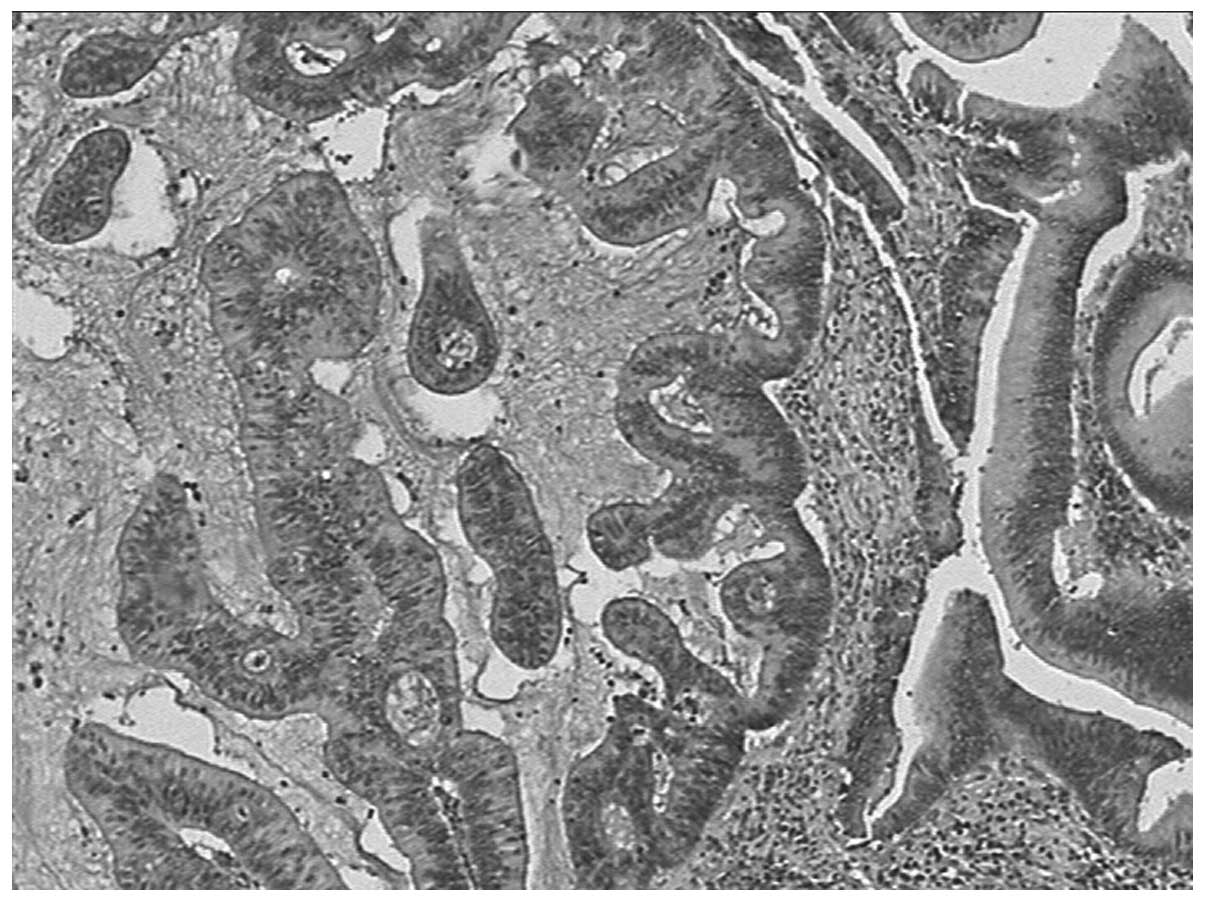
microscopic examination, papillary hyperplasia with the cellular
characteristics of high-grade dysplasia and marked nuclear
pleomorphism was observed in the dilated intra- and extrahepatic
bile ducts (Fig. 3). Thus, a
diagnosis of biliary papillomatosis with malignant transformation
was confirmed. Following surgical resection of the tumor, the
patient was followed up at seven months post surgery and no
recurrence was reported.
Discussion
Biliary papillomatosis was initially described by
Chappet in 1894 (4), and ~200 cases
have since been described (5).
Biliary papillomatosis characterized by intraductal papillary
growth of biliary epithelia has been sporadically reported. This is
a rare papillary or villous tumor that extends into the intra-
and/or extrahepatic biliary tree (2).
Biliary papillomatosis may demonstrate dysplastic changes, as well
as progression to carcinoma in situ and invasive
adenocarcinoma (6). In addition,
biliary papillomatosis is classified as one of two types,
mucin-hypersecreting or non mucin-producing, depending on the
presence or absence of mucin hypersecretion (7).
Biliary papillomatosis is most frequently observed
in middle-aged and elderly patients, possessing a male-to-female
ratio of 2:1 (7). The primary
clinical symptoms of biliary papillomatosis are obstructive
jaundice, repeated episodes of abdominal pain and cholangitis
(2,7,8). In the
present case, the patient presented with right epigastric pain
without jaundice and cholangitis. When the bile duct is not
completely obstructed by the tumor, the above significant principal
clinical symptoms may not be observed. Therefore, biliary
papillomatosis may also exist without inducing significant
symptoms.
In addition, laboratory findings did not aid
preoperative diagnosis, owing to the absence of specificity
(8). However, imaging modalities may
be capable of providing more direct information. In the present
case, ultrasound detection revealed dilated bile ducts and a
hyperechoic mass, which was nonspecific. In computed tomography
images, dilatation of intra- and extrahepatic bile ducts, as well
as hypoattenuating intraductal soft-tissue masses, may be observed
prior to and following administration of intravenous contrast
material (9). The lesions possess low
signal intensity on T1-weighted images, appear slightly
hyperintense on T2-weighted images under MRI and lack significant
enhancement following gadolinium administration (10).
Endoscopic retrograde cholangiopancreaticography
(ERCP) demonstrates multiple filling defects and a dilated biliary
tree with serrated irregularity of the bile duct wall (9,11). During
ERCP, a standard bile duct biopsy and/or a brush biopsy may be
performed, which may aid preoperative diagnosis. However, the
availability of ERCP is restricted by the associated complications,
including hemorrhage, cholangitis and pancreatitis (8).
Due to the high risk of malignant transformation and
the high rate of local recurrence of biliary papillomatosis,
radical excision is typically the recommended treatment. When
lesions are confined to a single hepatic lobe, as in the present
case, partial hepatectomy is advocated (12). For lesions dispersed over the entire
surface of the liver, the sole potentially curative treatment is
liver transplantation (13). Curative
surgical resection has been observed to result in a 5-year survival
rate of up to 81% (7). Increased
length of follow-up periods may be required following surgical
resection, as it is estimated that biliary papillomatosis may
involve the two lobes of the liver in a third of cases (14).
In conclusion, to the best of our knowledge, only a
small number of cases of biliary papillomatosis have previously
been reported. The present case supports the hypothesis that the
progression from benign to malignant disease may follow the
papilloma-carcinoma sequence (3).
Although biliary papillomatosis is a rare disease, it requires
increased attention due to its high malignant potential,
particularly in patients with a history of cholelithiasis.
References
|
1
|
Nakajima T, Kondo Y, Miyazaki M and Okui
K: A histopathologic study of 102 cases of intrahepatic
cholangiocarcinoma: Histologic classification and modes of
spreading. Hum Pathol. 19:1228–1234. 1988. View Article : Google Scholar : PubMed/NCBI
|
|
2
|
Nakanuma Y, Sasaki M, Ishikawa A, Tsui W,
Chen TC and Huang SF: Biliary papillary neoplasm of the liver.
Histol Histopathol. 17:851–861. 2002.PubMed/NCBI
|
|
3
|
Holtkamp W and Reis HE: Papillomatosis of
the bile ducts: Papilloma-carcinoma sequence. Am J Gastroenterol.
89:2253–2255. 1994.PubMed/NCBI
|
|
4
|
Chappet V: Cancer epithelial primitif du
canal cholédoque. Lyon Med. 76:145–157. 1894.
|
|
5
|
Sen I, Raju RS, Vyas FL, Eapen A and
Sitaram V: Benign biliary papillomatosis in a patient with a
choledochal cyst presenting as haemobilia: A case report. Ann R
Coll Surg Engl. 94:e20–e21. 2012. View Article : Google Scholar : PubMed/NCBI
|
|
6
|
Neumann RD, LiVolsi VA, Rosenthal NS,
Burrell M and Ball TJ: Adenocarcinoma in biliary papillomatosis.
Gastroenterology. 70:779–782. 1976.PubMed/NCBI
|
|
7
|
Lee SS, Kim MH, Lee SK, Jang SJ, Song MH,
Kim KP, Kim HJ, Seo DW, Song DE, Yu E, et al: Clinicopathologic
review of 58 patients with biliary papillomatosis. Cancer.
100:783–793. 2004. View Article : Google Scholar : PubMed/NCBI
|
|
8
|
Jiang L, Yan LN, Jiang LS, Li FY, Ye H, Li
N, Cheng NS and Zhou Y: Biliary papillomatosis: Analysis of 18
cases. Chin Med J (Engl). 121:2610–2612. 2008.PubMed/NCBI
|
|
9
|
Levy AD, Murakata LA, Abbott RM and
Rohrmann CA Jr: Armed Forces Institute of Pathology: From the
archives of the AFIP. Benign tumors and tumorlike lesions of the
gallbladder and extrahepatic bile ducts: Radiologic-pathologic
correlation. Radiographics. 22:387–413. 2002. View Article : Google Scholar : PubMed/NCBI
|
|
10
|
Hoang TV and Bluemke DA: Biliary
papillomatosis: CT and MR findings. J Comput Assist Tomogr.
22:671–672. 1998. View Article : Google Scholar : PubMed/NCBI
|
|
11
|
Kim YS, Myung SJ, Kim SY, Kim HJ, Kim JS,
Park ET, Lim BC, Seo DW, Lee SK, Kim MH, et al: Biliary
papillomatosis: Clinical, cholangiographic and cholangioscopic
findings. Endoscopy. 30:763–767. 1998. View Article : Google Scholar : PubMed/NCBI
|
|
12
|
Jiang L, Yan LN, Jiang LS, Li FY, Ye H, Li
N, Cheng NS and Zhou Y: Biliary papillomatosis: Analysis of 18
cases. Chin Med J (Engl). 121:2610–2612. 2008.PubMed/NCBI
|
|
13
|
Imvrios G, Papanikolaou V, Lalountas M,
Patsiaoura K, Giakoustidis D, Fouzas I, Anagnostara E, Antoniadis N
and Takoudas D: Papillomatosis of intra- and extrahepatic biliary
tree: Successful treatment with liver transplantation. Liver
Transpl. 13:1045–1048. 2007. View
Article : Google Scholar : PubMed/NCBI
|
|
14
|
Yeung YP, AhChong K, Chung CK and Chun AY:
Biliary papillomatosis: Report of seven cases and review of English
literature. J Hepatobiliary Pancreat Surg. 10:390–395. 2003.
View Article : Google Scholar : PubMed/NCBI
|


