Introduction
Epidermal growth factor receptor (EGFR/ErbB1/HER1)
overexpression is found in the majority of oral squamous cell
carcinoma (OSCC) tumors, and associations have been made between
increased expression levels and an aggressive phenotype, poor
prognosis and resistance to anticancer therapy (1). A number of anti-EGFR antibodies
(including cetuximab and panitumumab) and small molecule tyrosine
kinase inhibitors (including gefitinib and erlotinib), which target
EGFR, have been developed (2).
However, only the monoclonal antibody cetuximab (Erbitux®) is
currently approved for the treatment of advanced OSCC (3). Cetuximab therapy has been observed to be
effective in the treatment of recurrent and metastatic OSCC, as a
first-line treatment in combination with platinum-based
chemotherapy (4–7), and as a second-line therapy in patients
exhibiting platinum-resistant disease (8–10). In
clinical practice, EGFR expression may be evaluated by utilizing a
standardized immunohistochemistry (IHC) assay, which is designed to
assess cell membrane staining (11).
IHC studies have demonstrated EGFR overexpression in >75% of the
total analyzed OSCC cases, demonstrating values of 73.42% (12), 87.5% (13) and 72% (14). However, the cetuximab response rate is
typically not greater than 20% (15),
therefore the role of EGFR expression as a predictor of patient
response to EGFR-targeted therapeutic agents remains to be fully
elucidated.
The sensitivity of certain tumors to cetuximab may
be explained by the presence of mutations in the EGFR tyrosine
kinase domain (16,17). However, such mutations are rare in
OSCC (18). Thus, there is a
requirement to elucidate the mechanisms underlying the differential
drug response of cancer cells with wild-type EGFR. This may assist
with the identification of patients who may respond and clinically
benefit from cetuximab treatment, and furthermore may aid in the
development of novel therapeutic strategies, in order to circumvent
the de novo or acquired resistance of tumors to cetuximab.
Tumor resistance mechanisms to cetuximab may also be targetable,
and may thus present a potential opportunity to increase the
efficacy of cetuximab therapy. In general, tumors that are
frequently therapy-resistant may originate from tumor cells
exhibiting an epithelial-mesenchymal transition (EMT) phenotype
(19). During EMT, epithelial cells
lose their characteristic polarity and cell-cell contact, and
acquire a more migratory mesenchymal phenotype (20). EMT has additionally been demonstrated
to promote stem cell properties. Furthermore, the in vitro
sensitivity of head and neck cancer cell lines to anticancer agents
has been revealed to be affected by EMT-associated gene expression
(21,22).
In the present study, the expression of EGFR in
human OSCC tissues and cell lines was evaluated and the association
between EGFR expression and clinicopathological features in these
tissues and cells was investigated. Furthermore, the OSCC cells and
tissues were analyzed with regard to their EMT characteristics, and
the OSCC cell lines were also examined for their intrinsic
sensitivity to cetuximab treatments.
Materials and methods
Tissue samples
The present study was approved by the Ethics
Committee of the Kanazawa University Graduate School of Medical
Science (Kanazawa, Japan) and written informed consent was obtained
from each patient. The subjects consisted of 24 patients exhibiting
primary OSCC who underwent surgical resection of their tumors at
the Department of Oral and Maxillofacial Surgery at Kanazawa
University Hospital (Kanazawa, Japan), between 1998 and 2008. The
patients ranged from 43–89 years old (mean ± standard deviation,
64.4±11.2 years). The tumor-node-metastasis categories were
assigned according to the Union for International Cancer Control
system (23). The tumor
differentiation grade was determined according to the
classification criteria proposed by the World Health Organization
(24). The mode of tumor invasion was
assessed according to the criteria proposed by Yamamoto et
al (25).
IHC
Each specimen was fixed in 10% buffered formalin
(Sigma-Aldrich Japan K.K., Tokyo, Japan) and subsequently embedded
in paraffin for the preparation of serial sections.
Immunohistochemical staining was performed using an EGFR pharmDx™
kit (K1492; Dako Japan K.K., Tokyo, Japan). The specificity of the
staining was confirmed using Universal Negative Control for
IS-Series Rabbit Primary Antibodies (cat no. IS600; Dako Japan
K.K.) rather than primary antibody as a negative control. The
images of 3 separate stained regions were captured under a light
microscope at ×100 magnification and evaluated based on the number
of stained tumor cells, according to the method described by
Bernardes et al (26). EGFR
expression was evaluated based on the extent of immunolabeling in
the tumor cell membranes, and was classified on a 4-point scale as
follows: 0, no labeling or labeling in <10% of tumor cells; 1,
weak labeling, homogeneous or patchy, in >10% of tumor cells; 2,
moderate labeling, homogeneous or patchy, in >10% of tumor
cells; and 3, intense labeling, homogeneous or patchy, in >10%
of tumor cells. The initial scores were subsequently grouped into
two final groups: Low expression (0 or 1) and high expression (2 or
3).
Cell lines
A total of 3 human OSCC cell lines, established from
tumor biopsies with varying grades of invasive ability, were used:
OSC-20 and OSC-19, low-grade invasive cells, and HOC313, high-grade
invasive cells. OSC-20 is a cell line derived from a 58-year-old
female exhibiting tongue cancer that metastasized to the cervical
lymph nodes (27). OSC-19 was derived
from a 61-year-old male exhibiting tongue cancer that metastasized
to the cervical lymph nodes (28).
HOC313 was derived from a 51-year-old female exhibiting squamous
cell carcinoma (involving the mandibular gingiva and oral floor)
that metastasized to the cervical lymph nodes (29). Additionally, normal human dermal
fibroblasts (NHDFs; cat. no. PCS-201-010), obtained from the
American Type Culture Collection (Manassas, VA, USA), served as a
control.
Proliferation assay and cetuximab
sensitivity
A 3-(4,
5-dimethylthiazol-2-yl)-2,5-diphenyltetrazolium bromide (MTT) assay
was used in order to investigate the ability of cetuximab to
inhibit the proliferation of the 3 OSCC lines (OSC-19, OSC-20 and
HOC313) in vitro. Initially, 2,500 cells/well were cultured
in Dulbecco's modified Eagle's medium (DMEM; D7440; Sigma-Aldrich
Japan K.K.) supplemented with Hyclone™ 10% fetal bovine serum (FBS;
GE Healthcare, Logan, UT, USA), in 96-well tissue culture plates.
Following 24 h of culture, the cells were treated with varying
concentrations of cetuximab (Erbitux®; Merck Serono Co., Ltd.,
Tokyo, Japan; 0, 0.005 and 0.01 µg/ml) diluted in DMEM supplemented
with 2% FBS. In order to measure the number of metabolically active
cells following 24, 48 and 72 h of incubation, an MTT assay was
performed and the results were quantified according to the
manufacturer's protocols. The optical density at 450 nm was
measured using a microplate reader (iMark Microplate Absorbance
Reader; Bio-Rad Laboratories, Inc., Hercules, CA, USA).
RNA extraction, complementary DNA
(cDNA) synthesis and quantitative polymerase chain reaction
(qPCR)
The messenger RNA (mRNA) levels of EGFR, E-cadherin,
vimentin, Twist and Snail were analyzed using a Rotor-Gene Q 2plex
HRM System (9001560; Qiagen GmbH, Hilden, Germany) with
FAM/ZEN/IBFQ probes (DNA sequence unopened; Integrated DNA
Technologies, Inc., Coralville, IA, USA). Total RNA was extracted
using the RNeasy Protect Mini kit (74124; Qiagen GmbH), and cDNA
was obtained by utilizing the PrimeScript 1st Strand cDNA Synthesis
kit (6110A; Takara Bio, Inc., Otsu, Japan). All reactions and
conditions were performed according to the manufacturer's
protocols. Ribosomal RNA (18S) was amplified as an internal
standard using HEX/ZEN/IBFQ probes (DNA sequence unopened;
Integrated DNA Technologies, Inc.). Data were analyzed using the
comparative Cq method, which presented the data as a fold-change in
expression level relative to a calibrator sample (in the present
case, the mean expression of the 3 replicates of the NHDF cell
line).
Enzyme-linked immunosorbent assay
(ELISA)
For enzyme-linked immunosorbent assay analysis of
EGFR and phosphorylated EGFR, 3 human oral cancer cell lines
(OSC-20, OSC-19 and HOC313) were cultured. Following incubation for
24 h, the cells were seeded into a 96-well tissue culture plate (BD
Biosciences, Tokyo, Japan) and fixed for 5 min by 4% formaldehyde
solution (Sigma-Aldrich Japan K.K.) in phosphate-buffered saline
(Sigma-Aldrich Japan K.K.). The expression levels of EGFR and
phosphorylated EGFR were quantified using the human EGFR In-Cell
ELISA kit (ab126419; Abcam, Cambridge, MA, USA) according to the
manufacturer's protocols. The data were presented as a fold-change
in expression level relative to the mean expression of the 3
replicates of the NHDF cell line.
EMT induction
In order to induce EMT, OSC-20 and OSC-19 cells were
seeded at 70% confluence into a 10-cm plastic dish (BD Biosciences)
and cultured for 48 and 72 h in DMEM to induce EMT (Sigma-Aldrich
Japan K.K.)with 0.5% FBS. Recombinant human transforming growth
factor-β1 (240-B; R&D Systems, Inc., Minneapolis, MN, USA) was
added at a final concentration of 5 ng/ml.
Statistical analysis
Statistical analysis was performed using JMP®
version 9.0 (SAS Institute, Inc., Cary, NC, USA). The association
between EGFR protein expression and clinicopathological features
was examined using Pearson's χ2 test. Data from the
ELISA and MTT assays are presented as the mean ± standard error.
Differences between groups were tested for statistical significance
using the two-tailed Mann-Whitney U test. P<0.05 was considered
to indicate a statistically significant difference.
Results
EGFR expression is associated with
certain clinicopathological features
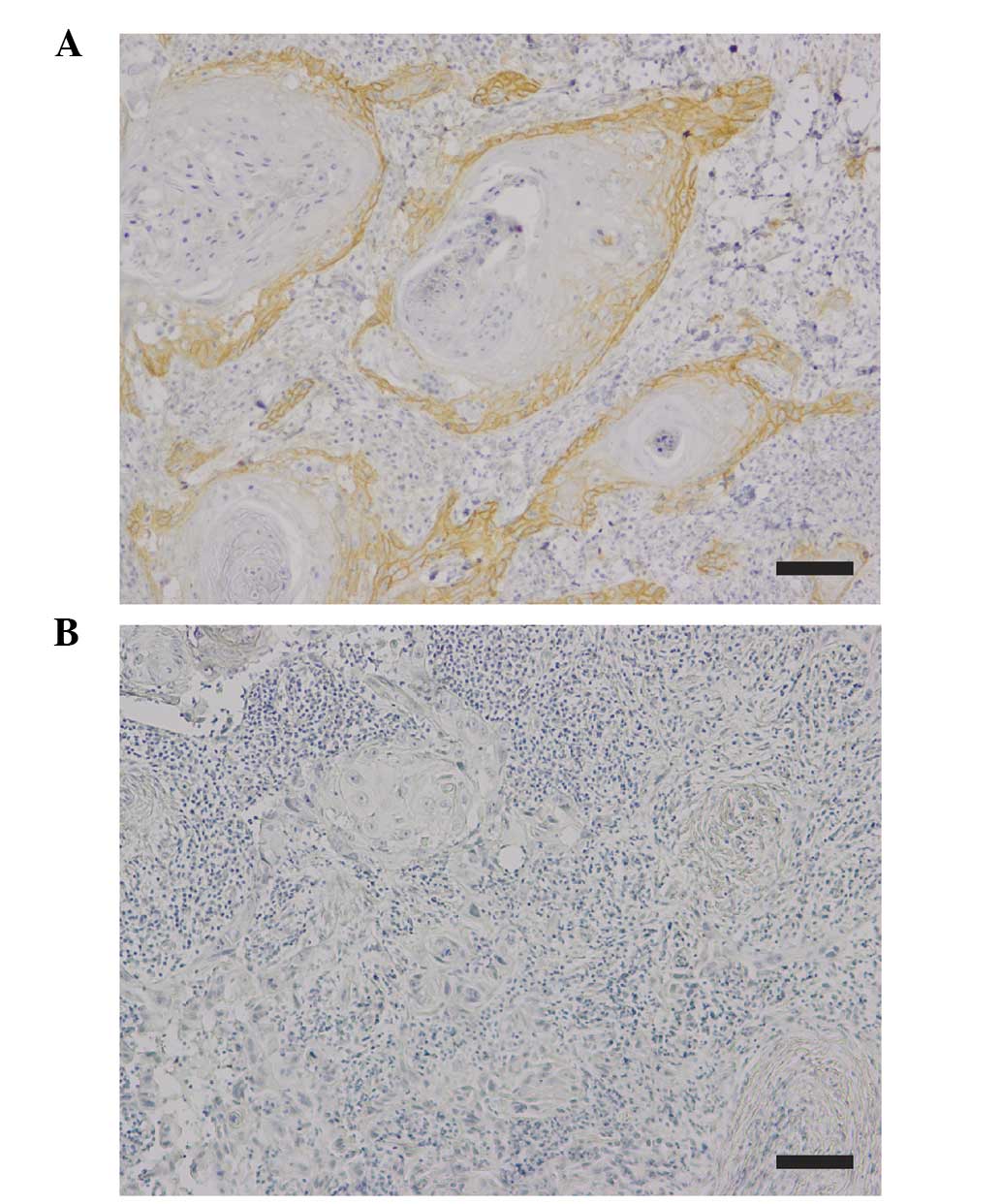
EGFR expression was detected in the cell membrane
and cytoplasm of the tumor cells (Fig.
1). According to the criteria for EGFR immunohistochemical
evaluation (26), 58.3% (14/24) of
the total cases investigated were positive for EGFR expression.
Table I illustrates the correlations
between EGFR expression and certain clinicopathological features.
EGFR expression in high-grade invasive OSCC (mode of invasion,
grade 4D) was significantly downregulated compared with that of
low-grade invasive OSCC (mode of invasion, grades 3 and 4C;
P=0.039). By contrast, EGFR expression did not correlate with tumor
size (P=0.991), local recurrence (P=0.071) histological
differentiation (P=0.137) or lymph node metastasis (P=0.456).
 | Table I.Correlation between EGFR expression
and clinicopathological features of oral squamous cell carcinoma
patients. |
Table I.
Correlation between EGFR expression
and clinicopathological features of oral squamous cell carcinoma
patients.
|
| EGFR expression |
|
|---|
|
|
|
|
|---|
| Clinicopathological
feature | High (n=14) | Low (n=10) | P-value |
|---|
| Tumor category |
|
| 0.991 |
| T1 | 3 | 2 |
|
| T2 | 8 | 6 |
|
| T3 | 1 | 0 |
|
| T4 | 2 | 2 |
|
| Node category |
|
| 0.456 |
|
Negative | 13 | 7 |
|
|
Positive | 1 | 3 |
|
| Local recurrence |
|
| 0.071 |
|
Negative | 10 | 4 |
|
|
Positive | 4 | 6 |
|
| Differentiation |
|
| 0.137 |
|
Well | 10 | 7 |
|
|
Moderate | 3 | 0 |
|
|
Poor | 1 | 3 |
|
| Mode of invasion,
grade |
|
|
0.039a |
| 3 | 7 | 1 |
|
| 4C | 5 | 3 |
|
| 4D | 2 | 6 |
|
EGFR is differentially expressed in
the 3 different grades of invasive OSCC cell lines
As demonstrated by immunohistochemical analysis,
only invasiveness (mode of invasion) was inversely correlated with
the expression of EGFR (Table I).
Therefore, EGFR expression in the 3 different grades of invasive
human OSCC lines (OSC-19, OSC-20 and HOC313) was analyzed at the
mRNA and protein level using qPCR and ELISA, respectively. Data are
presented as a fold-change relative to the mean of 3 replicates of
the NHDF cell line (Fig. 2). Among
the three OSCC cell lines (OSC-19, OSC-20 and HOC313), the EGFR
mRNA expression level ranged from 0.7- to 3.7-fold higher compared
with that of the NHDFs (Fig. 2A). The
expression of EGFR mRNA in the 2 low-grade invasive cell lines
(OSC-20 and OSC-19) was 3.7- and 2.3-fold higher compared with the
NHDFs, respectively. The EGFR mRNA expression in the high-grade
invasive cells (HOC313) was markedly reduced and similar to that of
the NHDFs. By contrast, the 2 low-grade invasive cell lines (OSC-20
and OSC-19) possessed EGFR protein expression levels 3.5- and
2.1-fold higher compared with those of the NHDFs, respectively
(Fig. 2B). The high-grade invasive
cells (HOC313) exhibited the lowest levels of EGFR protein
expression. The quantities of phosphorylated EGFR were assessed in
order to determine the activation status of EGFR. Expression levels
of phosphorylated EGFR ranged from 2.2- to 0.87-fold higher
compared with those of the NHDFs (Fig.
2C). Expression patterns of phosphorylated EGFR were similar to
the expression patterns of EGFR mRNA and protein.
Cetuximab sensitivity is dependent on
OSCC invasiveness
In order to evaluate the significance of EGFR status
in relation to cetuximab sensitivity, the cetuximab sensitivity of
3 human OSCC lines exhibiting varying grades of invasive ability
(OSC-19, OSC-20 and HOC313) was evaluated. Cells were seeded at
clonal density, and the antiproliferative effects of cetuximab were
determined using an MTT assay (Fig.
3). Antiproliferative effects of cetuximab were observed in the
OSC-20 and OSC-19 cell lines. In the OSC-20 and OSC-19 cells
(low-grade invasive OSCC), following incubation with cetuximab
(0.005 µg/ml and 0.01 µg/ml) for 48 and 72 h, proliferation was
significantly inhibited compared with non-treated cells
(P<0.05). However, the varying concentrations of cetuximab
exerted no significant antiproliferative effects on the HOC313 cell
line (P>0.05).
An EMT phenotype is detectable in the
high-grade invasive OSCC cell line
In order to evaluate whether EGFR expression was
associated with the features of EMT, levels of N-cadherin,
E-cadherin, vimentin and Snail were investigated in 3 different
invasive human OSCC cell lines (OSC-19, OSC-20 and HOC313; Fig. 4A). The expression levels of
EMT-associated genes in the OSC-19 and HOC313 cells are presented
as fold-changes relative to the OSC-20 cells. The HOC313 cells
(high-grade invasive cells with loss of EGFR expression) exhibited
a mesenchymal phenotype manifested through loss of E-cadherin and
acquisition of N-cadherin, vimentin and Snail expression compared
with the OSC-20 and OSC-19 cell lines (Fig. 4A). In order to examine whether TGF-β
altered the expression of the EMT-associated genes and EGFR, the 2
low-grade invasive cell lines (OSC-20 and OSC-19) were exposed to
TGF-β (Fig. 4B). Expression levels
are presented as fold differences relative to the control vehicle
at 48 and 72 h, respectively. Serial examination of EMT markers
(loss of E-cadherin and upregulation of N-cadherin, vimentin and
Snail) over a specific time course (48 and 72 h) revealed that
TGF-β treatment induced EMT in the OSC-20 cells (Fig. 4B). By contrast, TGF-β was not
effective in the induction of EMT in the OSC-19 cells. Notably,
total EGFR mRNA in the OSC-20 and OSC-19 cells was reduced
following EMT induction by TGF-β treatment.
 | Figure 4.EGFR and EMT-associated gene
expression in 3 varying grades of invasive human OSCC line. (A)
Relative mRNA expression levels of EMT-associated genes,
E-cadherin, N-cadherin, vimentin and Snail, in OSC-20, OSC-19 and
HOC313 cell lines. The high-grade invasive OSCC cell line (HOC313)
demonstrated upregulation of EMT-associated genes. Expression
levels in OSC-19 and HOC313 are displayed as fold-changes relative
to the expression levels in OSC-20. (B) Relative mRNA expression
levels of EGFR and EMT-associated genes, E-cadherin, N-cadherin,
vimentin and Snail, in OSC-20 and OSC-19 cell lines. TGF-β induced
EMT-associated gene expression in low-grade invasive OSCC cell
lines (OSC-20 and OSC-19). The cells were treated with human TGF-β
or a control vehicle for 48 and 72 h. Expression levels are
displayed as fold-changes relative to the control vehicle at 48 and
72 h, respectively. EGFR, epidermal growth factor receptor; EMT,
epithelial-mesenchymal transition; OSCC, oral squamous cell
carcinoma; mRNA, messenger RNA; TGF-β, transforming growth factor
β. |
Discussion
EGFR (a tyrosine kinase receptor from the ErbB
family) is an oncogene that has been identified in a number of
malignancies, including cancer of the breast, prostate, pulmonary
system, bladder, and head and neck (30). EGFR is overexpressed and appears to be
the dominant controlling factor underlying the malignant phenotype
in head and neck squamous cell carcinomas, via adjustment of the
molecules involved in invasive angiogenic and lymphangiogenic
processes (12). By analyzing the
immunomarker for EGFR in the present study, positive staining for
EGFR expression was identified in 58.3% of the total cases
analyzed. The results of the present study were consistent with
previously reported data (12–14).
Furthermore, to the best of our knowledge, the present study
demonstrated for the first time that invasiveness (mode of
invasion) was inversely correlated with EGFR expression in OSCC
patients. The present study also revealed that there is a loss of
EGFR expression in the high-grade invasive OSCC line (HOC313
cells). The results of the present study suggested that loss of
EGFR expression was associated with acquirement of an invasive
phenotype in OSCC. This potential association will require further
investigation in future studies.
The present study analyzed the correlation between
EGFR expression and the sensitivity to cetuximab using 3 human OSCC
cell lines (OSC-19, OSC-20 and HOC313), possessing varying grades
of invasive abilities. Variability in the responses of certain cell
lines to cetuximab treatment was identified. Cetuximab was observed
to suppress cell growth by ~23% in the most treatment-sensitive
cell line (OSC-20), however, it exerted no effect on the HOC313
cell line. The OSC-20 cell line demonstrated a marked increase in
EGFR mRNA and protein levels compared with the HOC313 cell line.
However, the HOC313 cell line, which was unaffected by cetuximab
treatment, demonstrated the lowest levels of EGFR expression. When
evaluating the affect of EGFR expression on the cetuximab treatment
responses, a significant difference in EGFR expression levels was
observed between cetuximab-sensitive (OSC-20) and -resistant cells
(HOC313). Cetuximab-resistant cell lines exhibited reduced levels
of EGFR expression, indicating that the cetuximab treatment
response may be dependent on EGFR expression status. In addition,
increased levels of phosphorylated EGFR correlated with cetuximab
sensitivity, revealing a potential role of activated EGFR in
patient responses to cetuximab. A previous study has suggested that
cells may be sensitive to the inhibition of EGFR only if they are
dependent on EGFR activation for cell survival and growth (31). In agreement with these previous
results, the levels of phosphorylated EGFR have been utilized to
predict gefitinib (an EGFR tyrosine kinase inhibitor) sensitivity
in OSCC patients (32).
The response to EGFR-targeted agents is inversely
correlated with EMT in multiple tumor types without known EGFR
mutations, including non-small cell lung cancer, and carcinoma of
the bladder, colorectal region, pancreas and breast (33–37). In
the present study, the EMT-associated genes, N-cadherin, vimentin
and Snail, were upregulated in cetuximab-resistant OSCC tumor cells
(HOC313). The present study additionally demonstrated that TGF-β
was able to induce a low-grade invasive OSCC cell line (OSC-20) to
undergo an EMT-associated gene switch, which resulted in a loss of
EGFR expression. The results of the present study indicated that
the net effect of TGF-β signaling was the loss of EGFR activity,
accompanied by a concomitant EMT-associated gene switch of tumor
cells, as well as acquirement of a migratory mesenchymal phenotype.
Further studies are required in order to elucidate the precise
molecular mechanisms underlying the association between EMT and the
regulation of EGFR expression.
In the present study, it was demonstrated that EGFR
status affected the response to cetuximab treatments, therefore,
the level of invasiveness or presence of an EMT phenotype may
potentially assist in the prediction of EGFR status. EMT-associated
alterations in gene expression, including the upregulation of
N-cadherin, vimentin and Snail, have additionally been proposed as
potential biomarkers for the indication of EGFR status and the
prediction of patient response to cetuximab treatment. However,
further in vivo studies using clinical samples are required
in order to confirm these results.
Acknowledgements
The authors would like to thank American Journal
Experts for providing English language editing.
References
|
1
|
Ang KK, Berkey BA, Tu X, Zhang HZ, Katz R,
Hammond EH, Fu KK and Milas L: Impact of epidermal growth factor
receptor expression on survival and pattern of relapse in patients
with advanced head and neck carcinoma. Cancer Res. 62:7350–7356.
2002.PubMed/NCBI
|
|
2
|
Mahipal A, Kothari N and Gupta S:
Epidermal growth factor receptor inhibitors: coming of age. Cancer
Control. 21:74–79. 2014.PubMed/NCBI
|
|
3
|
Loeffler-Ragg J, Schwentner I, Sprinzl GM
and Zwierzina H: EGFR inhibition as a therapy for head and neck
squamous cell carcinoma. Expert Opin Investig Drugs. 17:1517–1531.
2008. View Article : Google Scholar : PubMed/NCBI
|
|
4
|
Bourhis J, Rivera F, Mesia R, Awada A,
Geoffrois L, Borel C, Humblet Y, Lopez-Pousa A, Hitt R, Vega
Villegas ME, et al: Phase I/II study of cetuximab in combination
with cisplatin or carboplatin and fluorouracil in patients with
recurrent or metastatic squamous cell carcinoma of the head and
neck. J Clin Oncol. 24:2866–2872. 2006. View Article : Google Scholar : PubMed/NCBI
|
|
5
|
Burtness B, Goldwasser MA, Flood W, Mattar
B and Forastiere AA: Eastern Cooperative Oncology Group: Phase III
randomized trial of cisplatin plus placebo compared with cisplatin
plus cetuximab in metastatic/recurrent head and neck cancer: An
eastern cooperative oncology group study. J Clin Oncol.
23:8646–8654. 2005. View Article : Google Scholar : PubMed/NCBI
|
|
6
|
Shin DM, Donato NJ, Perez-Soler R, Shin
HJ, Wu JY, Zhang P, Lawhorn K, Khuri FR, Glisson BS, Myers J, et
al: Epidermal growth factor receptor-targeted therapy with C225 and
cisplatin in patients with head and neck cancer. Clin Cancer Res.
7:1204–1213. 2001.PubMed/NCBI
|
|
7
|
Vermorken JB, Mesia R, Rivera F, Remenar
E, Kawecki A, Rottey S, Erfan J, Zabolotnyy D, Kienzer HR, Cupissol
D, et al: Platinum-based chemotherapy plus cetuximab in head and
neck cancer. N Engl J Med. 359:1116–1127. 2008. View Article : Google Scholar : PubMed/NCBI
|
|
8
|
Baselga J, Trigo JM, Bourhis J, et al:
Phase II multicenter study of the antiepidermal growth factor
receptor monoclonal antibody cetuximab in combination with
platinum-based chemotherapy in patients with platinum-refractory
metastatic and/or recurrent squamous cell carcinoma of the head and
neck. J Clin Oncol. 23:5568–5577. 2005. View Article : Google Scholar : PubMed/NCBI
|
|
9
|
Herbst RS, Arquette M, Shin DM, et al:
Phase II multicenter study of the epidermal growth factor receptor
antibody cetuximab and cisplatin for recurrent and refractory
squamous cell carcinoma of the head and neck. J Clin Oncol.
23:5578–5587. 2005. View Article : Google Scholar : PubMed/NCBI
|
|
10
|
Vermorken JB, Trigo J, Hitt R, et al:
Open-label, uncontrolled, multicenter phase II study to evaluate
the efficacy and toxicity of cetuximab as a single agent in
patients with recurrent and/or metastatic squamous cell carcinoma
of the head and neck who failed to respond to platinum-based
therapy. J Clin Oncol. 25:2171–2177. 2007. View Article : Google Scholar : PubMed/NCBI
|
|
11
|
O-charoenrat P, Rhys-Evans PH, Archer DJ
and Eccles SA: C-erbB receptors in squamous cell carcinomas of the
head and neck: Clinical significance and correlation with matrix
metalloproteinases and vascular endothelial growth factors. Oral
Oncol. 38:73–80. 2002. View Article : Google Scholar : PubMed/NCBI
|
|
12
|
O-charoenrat P, Rhys-Evans PH, Modjtahedi
H and Eccles SA: The role of c-erbB receptors and ligands in head
and neck squamous cell carcinoma. Oral Oncol. 38:627–640. 2002.
|
|
13
|
Sarkis SA, Abdullah BH, Abdul Majeed BA
and Talabani NG: Immunohistochemical expression of epidermal growth
factor receptor (EGFR) in oral squamous cell carcinoma in relation
to proliferation, apoptosis, angiogenesis and lymphangiogenesis.
Head Neck Oncol. 2:132010. View Article : Google Scholar : PubMed/NCBI
|
|
14
|
Ryott M, Wangsa D, Heselmeyer-Haddad K,
Lindholm J, Elmberger G, Auer G, Avall Lundqvist E, Ried T and
Munck-Wikland E: EGFR protein overexpression and gene copy number
increases in oral tongue squamous cell carcinoma. Eur J Cancer.
45:1700–1708. 2009. View Article : Google Scholar : PubMed/NCBI
|
|
15
|
Bonner JA, Harari PM, Giralt J, Azarnia N,
Shin DM, Cohen RB, Jones CU, Sur R, Raben D, Jassem J, et al:
Radiotherapy plus cetuximab for squamous-cell carcinoma of the head
and neck. N Engl J Med. 354:567–578. 2006. View Article : Google Scholar : PubMed/NCBI
|
|
16
|
Lynch TJ, Bell DW, Sordella R,
Gurubhagavatula S, Okimoto RA, Brannigan BW, Harris PL, Haserlat
SM, Supko JG, Haluska FG, et al: Activating mutations in the
epidermal growth factor receptor underlying responsiveness of
non-small-cell lung cancer to gefitinib. N Engl J Med.
350:2129–2139. 2004. View Article : Google Scholar : PubMed/NCBI
|
|
17
|
Paez JG, Jänne PA, Lee JC, Tracy S,
Greulich H, Gabriel S, Herman P, Kaye FJ, Lindeman N, Boggon TJ, et
al: EGFR mutations in lung cancer: Correlation with clinical
response to gefitinib therapy. Science. 304:1497–1500. 2004.
View Article : Google Scholar : PubMed/NCBI
|
|
18
|
Lemos-González Y, de la Páez Cadena M,
Rodríguez-Berrocal FJ, Rodríguez-Piñeiro AM, Pallas E and Valverde
D: Absence of activating mutations in the EGFR kinase domain in
Spanish head and neck cancer patients. Tumour Biol. 28:273–279.
2007. View Article : Google Scholar : PubMed/NCBI
|
|
19
|
Nantajit D, Lin D and Li JJ: The network
of epithelial-mesenchymal transition: Potential new targets for
tumor resistance. J Cancer Res Clin Oncol. 141:1697–1713. 2015.
View Article : Google Scholar : PubMed/NCBI
|
|
20
|
Banyard J and Bielenberg DR: The role of
EMT and MET in cancer dissemination. Connect Tissue Res. 1–11.
2015.
|
|
21
|
Hsu DS, Lan HY, Huang CH, Tai SK, Chang
SY, Tsai TL, Chang CC, Tzeng CH, Wu KJ, Kao JY and Yang MH:
Regulation of excision repair cross-complementation group 1 by
Snail contributes to cisplatin resistance in head and neck cancer.
Clin Cancer Res. 16:4561–4571. 2010. View Article : Google Scholar : PubMed/NCBI
|
|
22
|
Skvortsova I, Skvortsov S, Raju U, Stasyk
T, Riesterer O, Schottdorf EM, Popper BA, Schiestl B, Eichberger P,
Debbage P, et al: Epithelial-to-mesenchymal transition and c-myc
expression are the determinants of cetuximab-induced enhancement of
squamous cell carcinoma radioresponse. Radiother Oncol. 96:108–115.
2010. View Article : Google Scholar : PubMed/NCBI
|
|
23
|
Greene FL and Sobin LH: A worldwide
approach to the TNM staging system: collaborative efforts of the
AJCC and UICC. J Surg Oncol. 99:269–272. 2009. View Article : Google Scholar : PubMed/NCBI
|
|
24
|
Thompson L: World Health Organization
classification of tumours: pathology and genetics of head and neck
tumours. Ear Nose Throat J. 85:742006.PubMed/NCBI
|
|
25
|
Yamamoto E, Kohama G, Sunakawa H, Iwai M
and Hiratsuka H: Mode of invasion, bleomycin sensitivity and
clinical course in squamous cell carcinoma of the oral cavity.
Cancer. 51:2175–2180. 1983. View Article : Google Scholar : PubMed/NCBI
|
|
26
|
Bernardes VF, Gleber-Netto FO, Sousa SF,
Silva TA and Aguiar MC: Clinical significance of EGFR, Her-2 and
EGF in oral squamous cell carcinoma: A case control study. J Exp
Clin Cancer Res. 29:402010. View Article : Google Scholar : PubMed/NCBI
|
|
27
|
Yokoi T, Hirata S, Nishimura F, Miyakawa
A, Odajima T and Kohama G: Some properties of a newly established
human cell line derived from an oral squamous carcinoma. Tumor Res.
25:93–103. 1990.
|
|
28
|
Yokoi T, Homma H and Odajima T:
Establishment and characterization of OSC-19 cell line in serum-
and protein-free culture. Tumor Res. 24:1–17. 1988.
|
|
29
|
Ishisaki A, Oida S, Momose F, Amagasa T,
Rikimaru K, Ichijo H and Sasaki S: Identification and
characterization of autocrine-motility-factor-like activity in oral
squamous-cell-carcinoma cells. Int J Cancer. 59:783–788. 1994.
View Article : Google Scholar : PubMed/NCBI
|
|
30
|
Oliveira S, van Bergen en Henegouwen PM,
Storm G and Schiffelers RM: Molecular biology of epidermal growth
factor receptor inhibition for cancer therapy. Expert Opin Biol
Ther. 6:605–617. 2006. View Article : Google Scholar : PubMed/NCBI
|
|
31
|
Pernas FG, Allen CT, Winters ME, Yan B,
Friedman J, Dabir B, Saigal K, Mundinger GS, Xu X, Morris JC, et
al: Proteomic signatures of epidermal growth factor receptor and
survival signal pathways correspond to gefitinib sensitivity in
head and neck cancer. Clin Cancer Res. 15:2361–2372. 2009.
View Article : Google Scholar : PubMed/NCBI
|
|
32
|
Hamakawa H, Nakashiro K, Sumida T,
Shintani S, Myers JN, Takes RP, Rinaldo A and Ferlito A: Basic
evidence of molecular targeted therapy for oral cancer and salivary
gland cancer. Head Neck. 30:800–809. 2008. View Article : Google Scholar : PubMed/NCBI
|
|
33
|
Yauch RL, Januario T, Eberhard DA, Cavet
G, Zhu W, Fu L, Pham TQ, Soriano R, Stinson J, Seshagiri S, et al:
Epithelial versus mesenchymal phenotype determines in vitro
sensitivity and predicts clinical activity of erlotinib in lung
cancer patients. Clin Cancer Res. 11:8686–8698. 2005. View Article : Google Scholar : PubMed/NCBI
|
|
34
|
Thomson S, Petti F, Sujka-Kwok I, Epstein
D and Haley JD: Kinase switching in mesenchymal-like non-small cell
lung cancer lines contributes to EGFR inhibitor resistance through
pathway redundancy. Clin Exp Metastasis. 25:843–854. 2008.
View Article : Google Scholar : PubMed/NCBI
|
|
35
|
Thomson S, Buck E, Petti F, Griffin G,
Brown E, Ramnarine N, Iwata KK, Gibson N and Haley JD: Epithelial
to mesenchymal transition is a determinant of sensitivity of
non-small-cell lung carcinoma cell lines and xenografts to
epidermal growth factor receptor inhibition. Cancer Res.
65:9455–9462. 2005. View Article : Google Scholar : PubMed/NCBI
|
|
36
|
Adam L, Zhong M, Choi W, Qi W, Nicoloso M,
Arora A, Calin G, Wang H, Siefker-Radtke A, McConkey D, et al:
miR-200 expression regulates epithelial-to-mesenchymal transition
in bladder cancer cells and reverses resistance to epidermal growth
factor receptor therapy. Clin Cancer Res. 15:5060–5072. 2009.
View Article : Google Scholar : PubMed/NCBI
|
|
37
|
Barr S, Thomson S, Buck E, Russo S, Petti
F, Sujka-Kwok I, Eyzaguirre A, Rosenfeld-Franklin M, Gibson NW,
Miglarese M, et al: Bypassing cellular EGF receptor dependence
through epithelial-to-mesenchymal-like transitions. Clin Exp
Metastasis. 25:685–693. 2008. View Article : Google Scholar : PubMed/NCBI
|