Introduction
The gastrointestinal (GI) tract is one of the most
common extranodal sites of non-Hodgkin's lymphoma (NHL), accounting
for 20–40% of all NHL cases (1).
Approximately 50% of all primary GI lymphomas are identified in the
stomach, and 14–38% of cases are found in the small intestine,
including duodenal lymphoma (2).
Duodenal lymphomas account for 0.8–2% of all GI lymphomas (3,4) and
symptoms include anorexia, abdominal pain and weight loss (5). The median survival time of primary GI
lymphoma patients following diagnosis is reported to be 54 months
(3). Among GI lymphomas, accurate
diagnosis of submucosal tumor (SMT)-like tumors is complicated as
it is difficult to obtain a sufficient sample of submucosal tissue
for diagnostic examination. Submucosal tumor (SMT)-like duodenal
tumors are sometimes found to be malignant lymphomas. Although
endoscopic ultrasonography (EUS), a procedure that uses ultrasound
to endoscopically examine the wall of the GI tract, is one of the
most useful modalities for the diagnosis of SMT, it is not always
effective in distinguishing between malignant lymphomas and other
types of tumors. As histological analyses are essential for the
diagnosis of lymphoma, endoscopic ultrasound-guided fine-needle
aspiration (EUS-FNA) has recently been used as an endoscopic tissue
sampling method for these duodenal tumors (6). However, this technique remains
inadequate for immunohistological analyses due to limitations
imposed by a large sample size (7,8). Recently,
mucosal incision-assisted biopsy (MIAB), a novel technique used to
obtain tissue samples from SMT-like tumors using the endoscopic
submucosal dissection (ESD) method, has been reported for the
histological diagnosis of gastric GI stromal tumors (GISTs)
(9). The present study demonstrates,
to the best of our knowledge, the first case with the application
of MIAB as a sampling method for the histological diagnosis of
SMT-like duodenal malignant lymphoma.
Case report
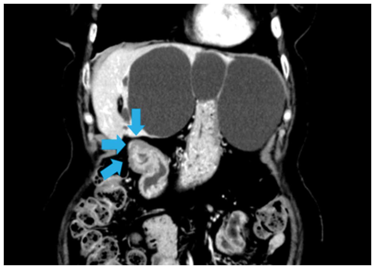
In August 2013, an 81-year-old woman presented to
Kagawa University Hospital (Kagawa, Japan) with abdominal
distension. An enhanced computed tomography scan revealed a
duodenal tumor associated with increased wall thickness (Fig. 1). A positron emission tomography scan
also indicated an abnormal uptake of tracer in the duodenum
(Fig. 2). The endoscopic examination
showed the presence of an SMT, ~25 mm in diameter, located in the
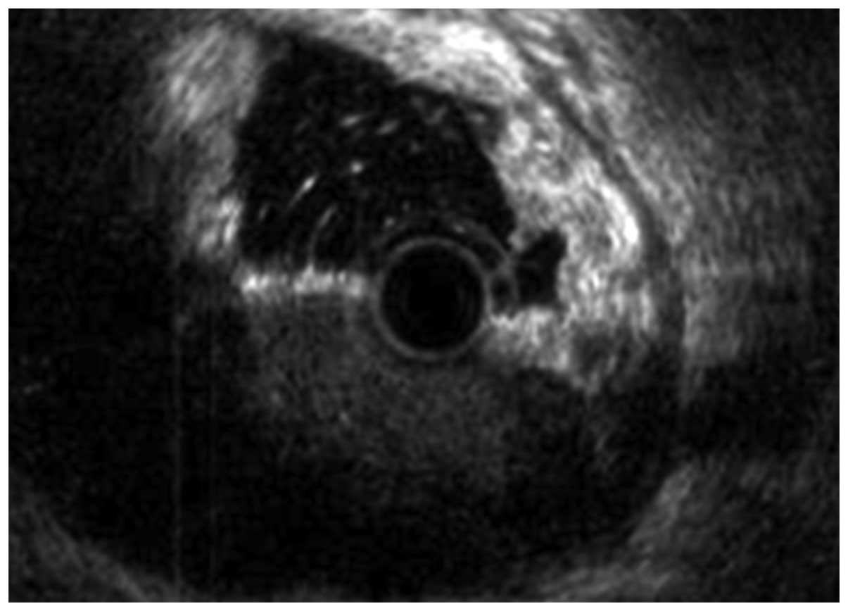
superior duodenal angle (Fig. 3). EUS
revealed that the lesion originated from the fourth layer
(muscularis propria) of the duodenum (Fig. 4). EUS-FNA was attempted for tissue
sampling, but failed due to the difficult location of the tumor.
Therefore, after obtaining the patient's informed consent, a MIAB
was performed. A 10-cm mucosal incision exposed the surface of the
SMT apex (Fig. 5), and tissue samples
were successfully obtained, followed by closure of the mucosal
incision using hemoclips (Fig. 6). No
complications were detected after MIAB. The pathological
examination of the biopsy samples revealed the presence of a
diffuse proliferation of atypical lymphocytes, and the expression
of cluster of differentiation (CD)20 and CD79a, but no expression
of CD3 in the tumor specimens (Fig.
7). The patient was diagnosed with diffuse large B-cell
lymphoma and immediately received eight cycles of treatment with a
3-week chemotherapy regimen (375 mg/m2 rituximab, day 1;
30mg/m2 tetrahydropyranyl adriamycin, day 2; 500
mg/m2 cyclophosphamide, day 2; 1.0 mg/m2
vincristine, day 2; and 30 mg/m2 prednisolone, days
2–7). The patient was followed up 27 months following MIAB, and
exhibited a complete response.
Discussion
Tissue sampling is essential for the histological
diagnosis of SMTs, as they comprise both benign and malignant
lesions (8,10). EUS-FNA has been established as a
preferred method for tissue sampling due to its accuracy and
safety, but it occasionally fails to provide adequate samples for
histological diagnosis (8). In a
previous study, the diagnostic yield from EUS-FNA was reported to
provide adequate samples in 117/141 (83.0%) patients with gastric
SMTs. In 29/117 cases, tissue samples were adequate for a possible
diagnosis by cytological examination, but inadequate for
histological diagnosis (8). Hence,
adequate samples were only obtained in 88/141 (62.4%) cases. In the
present study, adequate samples were required to diagnose a
malignant lymphoma. Additionally, due to the location of the
SMT-like mass in the supraduodenal angle, it was technically
difficult to perform EUS-FNA. Therefore, a MIAB was performed to
diagnose this duodenal tumor.
Previous studies have demonstrated the endoscopic
histological diagnosis of GISTs using ESD techniques, including
MIAB (9,11). Lee et al (11) revealed that endoscopic biopsies using
ESD techniques were sufficient for staining of all biopsy specimens
obtained from SMT-like tumors, including leiomyoma, lipoma and
ectopic pancreas. One of the most advantageous aspects of
performing MIAB is the ability to harvest sufficient quantities of
tissue samples for the definitive diagnosis of lymphoma (9,11).
However, potential disadvantages are procedure-related
complications, including bleeding and perforation. In particular,
procedure-related bleeding by MIAB is a frequent complication that
is easily controlled by hemostatic procedures (12). With regard to potential late
complications, such as post-operative bleeding and perforation,
mucosal closure with hemoclips after tissue sampling has been used
successfully (13).
In conclusion, the present study demonstrated, for
the first time, a case using MIAB as a sampling method for the
histological diagnosis of SMT-like primary duodenal malignant
lymphoma. This case suggests that MIAB may be a novel, valuable,
tissue sampling method for the diagnosis of SMT-like duodenal
tumors.
References
|
1
|
Yoo CC, Levine MS, McLarney JK, Rubesin SE
and Herlinger H: Value of barium studies for predicting primary
versus secondary non-Hodgkin's gastrointestinal lymphoma. Abdom
Imaging. 25:368–372. 2000. View Article : Google Scholar : PubMed/NCBI
|
|
2
|
Yaranal PJ, Harish SG and Purushotham B:
Primary intestinal lymphoma: A clinicopathological study. Indian J
Cancer. 51:306–308. 2014. View Article : Google Scholar : PubMed/NCBI
|
|
3
|
Cirillo M, Federico M, Curci G, Tamborrino
E, Piccinini L and Silingardi V: Primary gastrointestinal lymphoma:
A clinicopathological study of 58 cases. Haematologica. 77:156–161.
1992.PubMed/NCBI
|
|
4
|
Domizio P, Owen RA, Shepherd NA, Talbot IC
and Norton AJ: Primary lymphoma of the small intestine. A
clinicopathological study of 119 cases. Am J Surg Pathol.
17:429–442. 1993. View Article : Google Scholar : PubMed/NCBI
|
|
5
|
Boddie AW Jr, Eisenberg BL, Mullins JD and
Schlichtemeier AL: The diagnosis and treatment of obstructive
jaundice secondary to malignant lymphoma: A problem in
multidisciplinary management. J Surg Oncol. 14:111–123. 1980.
View Article : Google Scholar : PubMed/NCBI
|
|
6
|
Larghi A, Fuccio L, Chiarello G, Attili F,
Vanella G, Paliani GB, Napoleone M, Rindi G, Larocca LM, Costamagna
G and Ricci R: Fine-needle tissue acquisition from subepithelial
lesions using a forward-viewing linear echoendoscope. Endoscopy.
46:39–45. 2014.PubMed/NCBI
|
|
7
|
Hoda KM, Rodriguez SA and Faigel DO:
EUS-guided sampling of suspected GI stromal tumors. Gastrointest
Endosc. 69:1218–1223. 2009. View Article : Google Scholar : PubMed/NCBI
|
|
8
|
Mekky MA, Yamao K, Sawaki A, Mizuno N,
Hara K, Nafeh MA, Osman AM, Koshikawa T, Yatabe Y and Bhatia V:
Diagnostic utility of EUS-guided FNA in patients with gastric
submucosal tumors. Gastrointest Endosc. 71:913–919. 2010.
View Article : Google Scholar : PubMed/NCBI
|
|
9
|
Ihara E, Matsuzaka H, Honda K, Hata Y,
Sumida Y, Akiho H, Misawa T, Toyoshima S, Chijiiwa Y, Nakamura K
and Takayanagi R: Mucosal-incision assisted biopsy for suspected
gastric gastrointestinal stromal tumors. World J Gastrointest
Endosc. 5:191–196. 2013. View Article : Google Scholar : PubMed/NCBI
|
|
10
|
Turhan N, Aydog G, Ozin Y, Cicek B, Kurt M
and Oguz D: Endoscopic ultrasonography-guided fine-needle
aspiration for diagnosing upper gastrointestinal submucosal
lesions: A prospective study of 50 cases. Diagn Cytopathol.
39:808–817. 2011. View
Article : Google Scholar : PubMed/NCBI
|
|
11
|
Lee HL, Kwon OW, Lee KN, Jun DW, Eun CS,
Lee OY, Jeon YC, Han DS, Yoon BC, Choi HS, et al: Endoscopic
histologic diagnosis of gastric GI submucosal tumors via the
endoscopic submucosal dissection technique. Gastrointest Endosc.
74:693–695. 2011. View Article : Google Scholar : PubMed/NCBI
|
|
12
|
Kobara H, Mori H, Fujihara S, Nishiyama N,
Kobayashi M, Kamata H and Masaki T: Bloc biopsy by using submucosal
endoscopy with a mucosal flap method for gastric subepithelial
tumor tissue sampling (with video). Gastrointest Endosc.
77:141–145. 2013. View Article : Google Scholar : PubMed/NCBI
|
|
13
|
Kataoka M, Kawai T, Yagi K, Sugimoto H,
Yamamoto K, Hayama Y, Nonaka M, Aoki T, Fukuzawa M, Fukuzawa M, et
al: Mucosal cutting biopsy technique for histological diagnosis of
suspected gastrointestinal stromal tumors of the stomach. Dig
Endosc. 25:274–280. 2013. View Article : Google Scholar : PubMed/NCBI
|