Introduction
Derived from chromaffin tissue of the sympathetic
nervous system, paragangliomas have the potential to secrete
catecholamines, accounting for several of the predominant symptoms
of functional paragangliomas. The common presentation of urinary
bladder paraganglioma is painless haematura, paroxysmal
hypertension, headache, palpitation and anxiety (1). Metastasis to the lymph nodes and
invasion of adjacent organs indicate the malignant potential
(2). Urinary bladder paragangliomas
are rare neoplasms constituting only 0.06% of all bladder tumors
(1). The preoperative diagnosis and
assessment of malignant potential of paragangliomas, particularly
non-functional paragangliomas, present significant challenges for
urologists (3). Tumor-specific
patterns of chromosomal imbalances characterize almost all forms of
neoplasm, and have been used in the diagnosis and malignancy
determination of many tumors (4). For
example, multiprobe fluorescence in situ hybridization
(FISH) analysis of exfoliated cells in the urine has been approved
for use in the detection of recurrent urothelial carcinoma and the
evaluation of hematuria, as FISH exhibits a higher sensitivity
compared with conventional cytology in detecting urothelial
carcinoma (81 vs. 58%) (5).
To the best of our knowledge, the current study is
the first report of a case of a urinary bladder paraganglioma in
which chromosome duplications were detected by fluorescence in
situ hybridization (FISH) in exfoliated cells in the urine.
This may be helpful to its preoperative diagnosis and malignant
potential determination.
Case report
A 34-year-old male complaining of paroxysmal gross
hematuria lasting for 7 months was admitted to Tongji Hospital
(Wuhan, China). His medical and family history were unremarkable.
General examination, including blood pressure and heart rate,
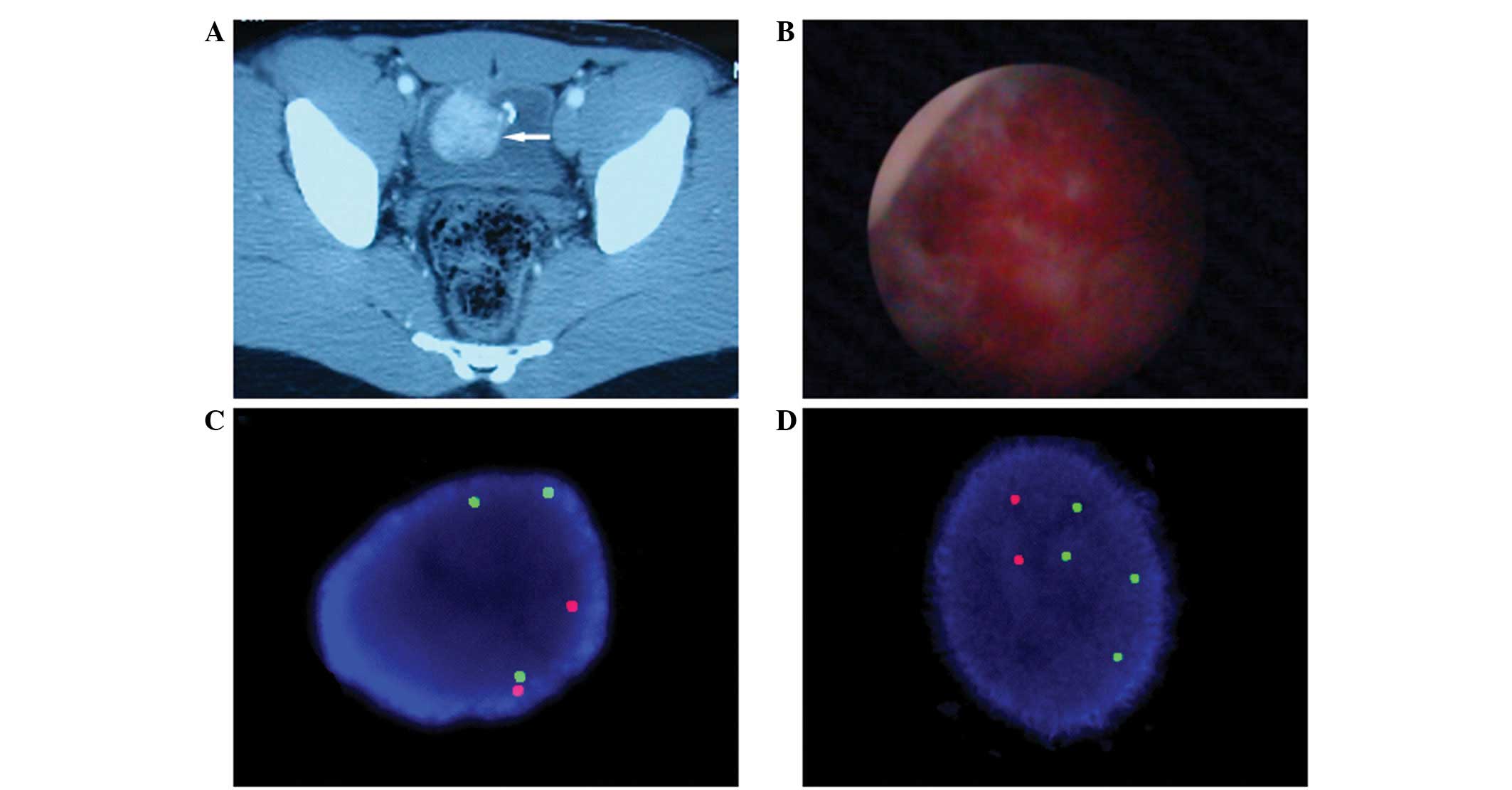
yielded results within the normal range. Abdominal computed
tomography (CT) imaging revealed a 3.6×3.9-cm mass on the dome of
the bladder, which exhibited intense uniform enhancement on a
contrast study (Fig. 1A). There was
no indication of lymph node involvement or invasion to the
surrounding structures. Cystoscopy revealed a 3×4-cm submucosal
neoplasm on the dome of the bladder, partly covered by normal
urothelium (Fig. 1B). A biopsy of the
neoplasm was conducted, revealing a highly vascular tumor. FISH of
exfoliated cells in the urine indicated the presence of
duplications of chromosomes 3 and 17 (Fig. 1C and D).
Upon diagnosis of the bladder tumor, a transurethral
resection was performed. During the surgery, no hypertension
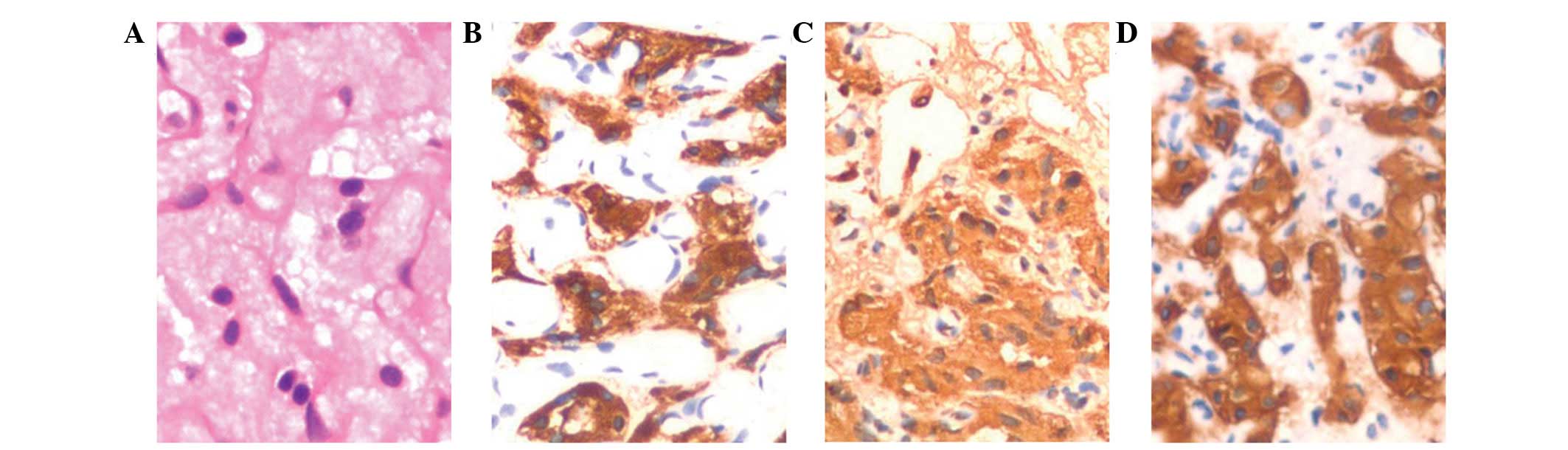
occurred. On microscopic examination of the resected specimen,
nested spindle cells were observed (Fig.
2A). Immunohistochemical staining revealed positive reactivity
for chromogranin A, S-100 protein, CD56 and synaptophysin, as well
as negative reactivity for cytokeratins 7, 8 and 18, confirming the
diagnosis of paraganglioma (Fig. 2B, C
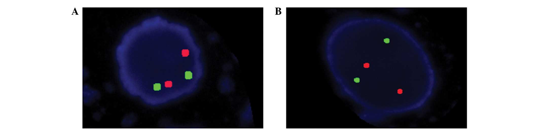
and D). Following surgery, FISH of exfoliated cells in the
urine showed no chromosomal duplications (Fig. 3). Further treatment was avoided as no
tumor cells were observed in the deep muscle layer. At 2 years
after the surgery, the patient remained disease-free. Written
informed consent was obtained from the patient for inclusion in the
present study.
Discussion
Histological and immunohistochemical staining is
often used for the definitive diagnosis of paragangliomas; however,
their determination of malignant potential remains a challenge for
urologists and pathologists (6).
Chromosomal imbalances have become a new parameter to predict the
assessment of malignant potential of paraganglioma (7). As summarized by Schaefer et al
(7), gains or losses of chromosomes
1, 3, 6, 7, 8, 9, 11, 16, 17, 19, 20, 21 and 22 in paragangliomas
have been reported. Furthermore, a gain of 17p has been implicated
in increased probability of progression to malignancy (8). A previous comparative genomic
hybridization study was the first to identify that paraganglioma of
the urinary bladder shares common chromosomal imbalances with other
paragangliomas and related pheochromocytomas (7). Similarly, multiprobe FISH analysis of
exfoliated cells in the urine may identify specific abnormalities
in chromosomes 3 and 17, in addition to others (9). As a non-invasive method, it has been
approved for use in the detection of recurrent urothelial carcinoma
and the evaluation of hematuria (9).
In the current patient, chromosome duplications in paraganglioma
tissue were detected by FISH in exfoliated cells in the urine,
which is helpful to determine the malignant potential of
paraganglioma. However, the precise association between chromosome
duplications and the malignant potential of paraganglioma requires
further research.
In the present case, the submucosal neoplasm of the
urinary bladder with intense uniform enhancement, and the results
of the preoperative histological analysis, were indicative of
hemangioma. However, duplications of chromosomes 3 or 17 have not
been reported in cases of hemangioma of the urinary bladder,
providing a clue for their differential diagnosis. Furthermore,
bladder neoplasms with positive FISH results are commonly
considered to be urothelial carcinomas (9). It is indicated that submucosal tumors,
such as paraganglioma, with chromosome duplications and ulceration,
may shed into the urine and be detected. Thus, if FISH of urine
exfoliated cells from a patient with a neoplasm of the urinary
system, such as a urothelial carcinoma and submucosal tumor,
reveals positive results, then chromosome duplication and
ulceration should be considered, as shown in the current case
report.
In conclusion, the preoperative diagnosis and
malignant potential predication of urinary bladder paraganglioma
present significant challenges for urologists, and is currently
limited to evaluation of specific symptoms, imaging investigations
and histological analysis of tissue biopsies. As in the current
case, chromosome duplications of urinary bladder paraganglioma
detected by FISH in urine exfoliated cells have the potential to
provide additional information. This may facilitate the
differential diagnosis and malignant determination of patients with
urinary bladder paraganglioma.
Acknowledgements
The authors would like to thank Professor Hua Xu
(Department of Urology, Tongji Hospital, Tongji Medical College,
Huazhong University of Science and Technology) for technical
assistance in the analysis of the FISH studies.
References
|
1
|
Leestma JE and Price EB Jr: Paraganglioma
of the urinary bladder. Cancer. 28:1063–1073. 1971. View Article : Google Scholar : PubMed/NCBI
|
|
2
|
Ansari MS, Goel A, Goel S, Durairajan LN
and Seth A: Malignant paraganglioma of the urinary bladder. A case
report. Int Urol Nephrol 2001. 33:343–345. 2001. View Article : Google Scholar
|
|
3
|
Lai Y, Chen D, Yu Z, Ni L and Yang S:
Non-functioning paraganglioma of the urinary bladder: A case report
and review of the literature. Oncol Lett. 7:891–893.
2014.PubMed/NCBI
|
|
4
|
Stallings RL: Are chromosomal imbalances
important in cancer? Trends Genet. 23:278–283. 2007. View Article : Google Scholar : PubMed/NCBI
|
|
5
|
Halling KC, King W, Sokolova IA, Meyer RG,
Burkhardt HM, Halling AC, Cheville JC, Sebo TJ, Ramakumar S,
Stewart CS, et al: A comparison of cytology and fluorescence in
situ hybridization for the detection of urothelial carcinoma. J
Urol. 164:1768–1775. 2000. View Article : Google Scholar : PubMed/NCBI
|
|
6
|
Kang WY, Shen JT and Chai CY:
Paraganglioma of the urinary bladder: A case report. Kaohsiung J
Med Sci. 19:136–140. 2003. View Article : Google Scholar : PubMed/NCBI
|
|
7
|
Schaefer IM, Gunawan B, Füzesi L, Blech M,
Frasunek J and Loertzer H: Chromosomal imbalances in urinary
bladder paraganglioma. Cancer Genet Cytogenet. 203:341–344. 2010.
View Article : Google Scholar : PubMed/NCBI
|
|
8
|
August C, August K, Schroeder S, Bahn H,
Hinze R, Baba HA, Kersting C and Buerger H: CGH and CD 44/MIB-1
immunohistochemistry are helpful to distinguish metastasized from
nonmetastasized sporadic pheochromocytomas. Mod Pathol.
17:1119–1128. 2004. View Article : Google Scholar : PubMed/NCBI
|
|
9
|
Caraway NP and Katz RL: A review on the
current state of urine cytology emphasizing the role of
fluorescence in situ hybridization as an adjunct to
diagnosis. Cancer Cytopathol. 118:175–183. 2010. View Article : Google Scholar : PubMed/NCBI
|