Introduction
Acute myeloid leukemia (AML) is an aggressive and
heterogeneous malignant disease, which accounts for 15–20% of
observed pediatric leukemia cases (1). Utilizing current treatment strategies,
which are similar to those proposed in adult AML treatment
protocols, complete remission rates approach 95% and the overall
survival rate is ~70% in developed countries; however, the survival
rate is markedly lower in developing countries (2). In addition, the prognosis for children
exhibiting therapy resistance or suffering from relapse is worse
than that of patients without relapsed or refractory AML (3). Increasing evidence has indicated that
small populations of leukemia cells consisting of early
stem/progenitor cells, which are termed leukemia stem cells (LSCs)
or leukemia initiating cells, are proposed to be resistant to
currently used chemotherapeutic drugs, and to mediate disease
relapse (4). Effective therapeutic
strategies that specifically eliminate these LSCs may, therefore,
be promising for the cure of this disease.
Previous studies have indicated that normal
hematopoietic stem cells (HSCs) and LSCs are negative for the
expression of lineage markers (Lin), positive for the expression of
cluster of differentiation (CD)34 and negative for the expression
of CD38 (5). Subsequent studies
revealed that a number of markers [CD33 (6), CD44 (7),
CD123 (8), CD133 (9), CD32 (10),
CD47 (11) and C-type lectin-like
molecule-1 (12)] are expressed
aberrantly on LSCs, and may be considered to be specific markers
indicating the presence of AML LSCs. However, CD33 (13), CD123 (13) and CD133 (14,15) are
also reported to be expressed on normal HSCs. Gemtuzumab ozogamicin
(GO), an antibody against CD33 that is conjugated with the
cytotoxic agent calicheamicin, was withdrawn from the market in
2010 in the United States due to low improvement in survival
outcomes and high levels of hepatic toxicity (16). Furthermore, the distribution of these
markers on various cell fractions in AML, including
CD34−, CD34+CD38+ and
CD34+CD38−Lin− cells, remains to
be elucidated. In addition, antigen expression on AML LSCs in
pediatric patients has not been extensively investigated.
Therefore, the high level of inconsistency regarding
antigen expression and the observed failure of clinical treatment
has suggested the need to re-evaluate the expression levels of
various antigens on stem cells, and to investigate the potential
cell differentiation stage selectivity of antigen expression. In
the present study, multicolor flow cytometry was utilized in order
to analyze the expression of the progenitor cell marker human
leukocyte antigen (HLA)-DR, as well as additional well-known stem
cell markers (CD33, CD123, CD133 and CD44) on a number of cell
populations, including CD34−,
CD34+CD38+, CD34+CD38−
and CD34+CD38−Lin− cells, in order
to explore the potential therapeutic benefits for childhood
AML.
Materials and methods
Patient and control samples
A total of 54 patients newly diagnosed with AML
[French-American-British (FAB) subtypes M0-M6] (17) were examined at the Department of
Hematology-Oncology, Children's Hospital of Zhejiang University
School of Medicine (Hangzhou, China), following receipt of informed
consent from their parents or guardians. All of the patients were
diagnosed based on morphologic, immunophenotypic, cytogenetic and
molecular biology, in accordance with the World Health Organization
diagnostic criteria (18). Patients
exhibiting <1% CD34 expression were excluded from the present
study to avoid the misgating of non-leukemia cells. Control bone
marrow samples were obtained from 11 patients with immune
thrombocytopenia following receipt of informed consent, in
accordance with the Declaration of Helsinki (19). Patient characteristics are summarized
in Table I.
 | Table I.Clinicopathological features of AML
patients at diagnosis. |
Table I.
Clinicopathological features of AML
patients at diagnosis.
| Clinicopathological
feature | Value |
|---|
| No. of patients
(male/female) | 54 (34/20) |
| Median age, years
(range) | 5.5 (1.1–14.1) |
| Median WBC count,
×109/l (range) | 15.4 (0.9–460) |
| Median hemoglobin,
g/l (range) | 77.5 (42–124) |
| Median platelet,
×109/l (range) | 43.5 (2–295) |
| Median bone marrow
blast, % (range) | 72 (30–95) |
|
French-American-British subtypes |
|
|
M0-M1 | 4 |
| M2 | 14 |
| M3 | 5 |
|
M4-M5 | 29 |
| M6 | 2 |
| Cytogenetics, %
(n) |
|
|
Favorablea | 27.7 (13/47) |
|
Intermediateb | 46.8 (22/47) |
|
Unfavorablec | 25.5 (12/47) |
| Molecular markers,
% (n) |
|
|
Fms-related tyrosine kinase
3 | 14.3 (4/28) |
| CEBPA
mutations | 0.0 (0/28) |
| c-Kit
mutation | 3.6 (1/28) |
|
Nucleophosmin 1 mutations | 3.6 (1/28) |
Flow cytometry (FCM) analysis
Direct fluorescent immunostaining was performed on
heparinized (Sangor Biotech Co., Ltd., Shangai, China) whole bone
marrow cells (1×10⁶ leukocytes/tube in 100 µl volume)
for 20 min at room temperature in the dark. Subsequently,
erythrocytes were lysed using FACS Lysing Solution (BD Biosciences,
San Jose, CA, USA) for 10 min and washed twice using
phosphate-buffered saline (pH 7.4; Sangon Biotech Co., Ltd.),
supplemented with 5% fetal calf serum (Sijiqing Biotech Ltd.,
Hangzhou, China) and 0.02% sodium azide (Sangon Biotech Co., Ltd.).
Four colors of antibody conjugates [fluorescein isothiocyanate
(FITC)/phycoerythrin (PE)/peridinin chlorophyll
(PerCP)/allophycocyanin (APC)] were systematically applied in
various combinations, as follows: Lin1/CD33/CD34/CD38,
Lin1/CD123/CD34/CD38, Lin1/CD133/CD34/CD38, Lin1/CD44/CD34/CD38 and
Lin1/HLA-DR/CD34/CD38. The Lin 1 antibody cocktail included
antibodies against CD3, CD56, CD19, CD20, CD14 and CD16, labeled
with FITC (cat no. 340546; BD Biosciences). Additional individual
fluorophore-conjugated mouse monoclonal antibodies, purchased from
BD Biosciences include: CD34-PerCP (clone 8G12; cat no. 340430),
CD38-APC (clone HB7; cat no. 345807), CD33-PE (clone P67.6; cat no.
347787), CD123-PE (clone 9F5; cat no. 340545), CD44-PE (clone
G44–26; cat no. 555479) and HLA-DR-PE (clone L243; cat no. 347367).
CD133-PE (clone AC133; cat no. 130-080-801) was purchased from
Miltenyi Biotech (Bergisch Gladbach, Germany). Isotype-matched
control antibodies were utilized for each antibody reaction.
Antigen expression level expressed as the percentage of positive
cells among the total gated cells. For each case, positivity was
defined as ≥20% cell antigen expression in gated cells. Data
acquisition was performed and analyzed with a FACSCalibur™ flow
cytometer using CellQuest 2000 software (BD Biosciences).
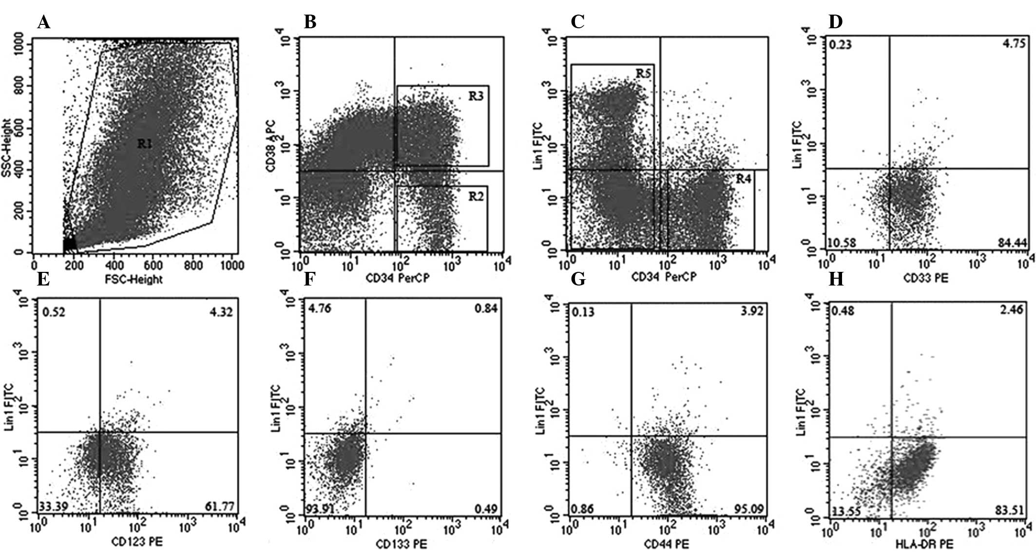
Cell fraction definitions
CD34− cells were considered to represent
a more mature cell population compared with CD34+ cells.
CD34+ cells were additionally gated as
CD34+CD38+ progenitor cells, and more
immature CD34+CD38− cells.
CD34+CD38−Lin− cells demonstrated
increased enrichment for stem cells compared with
CD34+CD38− cells. The gating strategy is
shown in Fig. 1.
Statistical analysis
Data were analyzed using SPSS version 19.0 (IBM
SPSS, Armonk, NY, USA). Variables were presented as the median and
range, or the mean ± standard error. For quantitative variables,
the antigen expression differences between two groups were analyzed
using Mann-Whitney U tests. Comparisons of antigen expression
between different cell fractions were performed using the Wilcoxon
signed-rank test. P<0.05 (two-sided) was considered to indicate
a statistically significant difference.
Results
Molecular targets are present on
CD34+CD38− and
CD34+CD38−Lin− stem cells in AML
samples
Patient clinicopathological features are summarized
in Table I. The median age of AML
patients at diagnosis was 5.5 years, with a range of 1.1–14.1
years. A total of 12/47 (25.5%) patients demonstrated unfavorable
cytogenetics at diagnosis (Table
I).
The median proportions of
CD34+CD38− and
CD34+CD38−Lin− stem cells in AML
at diagnosis were 1.28% (range, 0.01–20.32%) and 0.88% (range,
0.01–20.2%), compared with 0.45% (range, 0.01–3.03%) and 0.26%
(range, 0.01–2.72%) in control samples (P<0.05), respectively.
Antigen expression on CD34+CD38− cells and
CD34+CD38−Lin− stem cells is
summarized in Table II. The
expression levels of CD33 on
CD34+CD38−Lin− cells (median, 81.7
vs. 18.3%; P=0.002) and CD34+CD38− cells
(median, 86.5 vs. 32.9%; P=0.0016) in AML samples were
significantly increased compared with those of control samples.
CD123 was found to be positively expressed on
CD34+CD38− cells in 49/54 (90.7%) AML cases,
and on CD34+CD38−Lin− cells in
51/54 (94.4%) AML cases. Compared with control samples, CD123
exhibited increased expression levels on
CD34+CD38− cells (median, 75.9 vs. 21.6%;
P=0.003) and CD34+CD38−Lin− cells
(median, 75.8 vs. 19.1%; P=0.003) in AML samples. CD133 was
additionally present on
CD34+CD38−Lin− AML LSCs, however,
it demonstrated reduced frequency (63% positive cases) and
expression levels (median, 38.2%). In addition, no significant
differences in the rates of expression of CD133 or HLA-DR on
CD34+CD38−Lin− (P=0.3241 for CD133
and P=0.4781 for HLA-DR, respectively) and
CD34+CD38− cells (P=0.1131 for CD133 and
P=0.9423 for HLA-DR, respectively) between AML and control samples
were observed. CD44 was observed to be positively expressed on
CD34+CD38− and
CD34+CD38−Lin− cells in all AML
and control samples; however, significant differences in the level
of expression were identified between AML and control groups in
CD34+CD38− cells (median, 98.5 vs. 85.7%;
P=0.0003) and CD34+CD38−Lin− stem
cells (median, 97.7 vs. 83.1%; P=0.0028).
| Table II.Antigen expression analysis of
leukemia stem cells exhibiting AML at diagnosis. |
Table II.
Antigen expression analysis of
leukemia stem cells exhibiting AML at diagnosis.
| A,
Positive/CD34+CD38− gated |
|---|
|
|---|
| Antigen |
Controla,
% (range) | AMLb, % (range) |
|---|
| CD33 | 32.9
(5.3–92.9) | 86.5
(3.4–100)c |
| CD123 | 21.6
(1.2–84.1) | 76.9
(13.9–100)c |
| CD133 | 15.3
(0.6–86.1) | 47.3 (0–97.7) |
| CD44 | 85.7
(33.7–98.4) | 98.5
(50.4–100)c |
| HLA-DR | 90.5 (67–100) | 93.5 (0.6–100) |
|
| B,
Positive/CD34+CD38−Lin− gated |
|
| Antigen |
Controla,
% (range) | AMLb, % (range) |
|
| CD33 | 18.3 (8.1–88) | 81.7
(3.3–100)c |
| CD123 | 19.1 (0–78.3) | 75.8
(12.9–100)c |
| CD133 | 20.9
(0.2–91.6) | 38.2 (0–97.9) |
| CD44 | 84.4
(60.8–99.4) | 97.7
(48.7–100)c |
| HLA-DR | 90.8
(56.8–94.7) | 80.3 (0.7–100) |
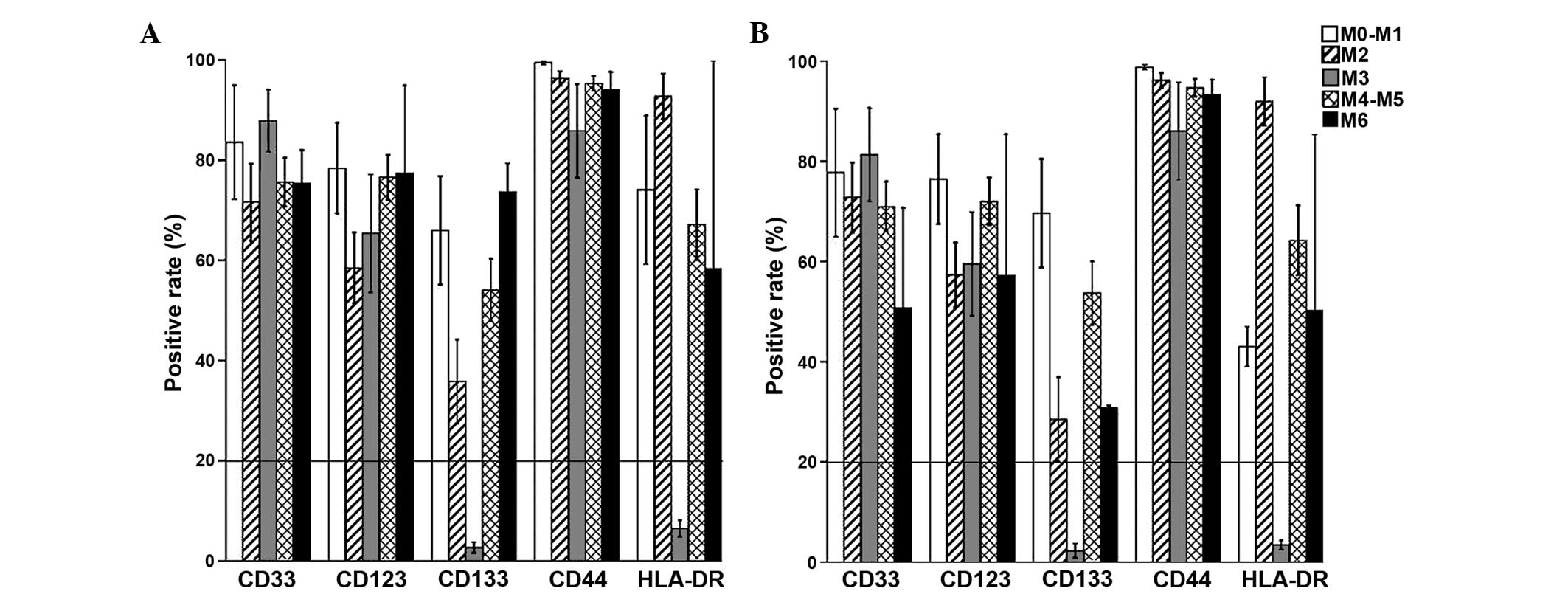
The expression of cell surface markers
on stem cells differs in various subtypes of AML
When AML samples were grouped according to FAB
classification, CD33 demonstrated higher expression levels on AML
LSCs in M3 when compared with other AML subtypes. CD123 was
preferentially expressed in M0-M1, M4-M5 and M6, but not in M2;
similar observations were made for CD133. CD44 demonstrated similar
patterns of expression in all AML subtypes. HLA-DR and CD133
demonstrated low expression levels on more mature M3 leukemia cells
(Fig. 2). Due to the low number of
samples in some subtype of AML, especially for M6 (n=2), we did not
perform statistical analysis of the differences between different
AML subtypes.
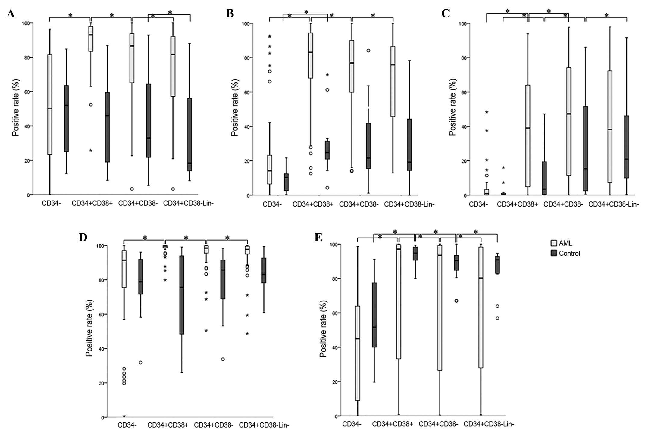
Antigen expression varies depending on
the cell subset in AML
In order to demonstrate the selective expression of
antigens on cells at certain stages of differentiation, cell
subpopulations, characterized by the presence of specific
immunophenotypes, including CD34−,
CD34+CD38+, CD34+CD38−
and CD34+CD38−Lin−, were analyzed
for the expression of various antigens in AML and control samples
(Fig. 3).
In general, the median expression levels of the
antigens on CD34− mature cells were decreased compared
with that of more immature CD34+CD38+,
CD34+CD38− and
CD34+CD38−Lin− cell fractions, in
AML and control samples. CD33 and HLA-DR were preferentially
expressed on CD34+CD38+ leukemia cells in AML
samples. CD133 was preferentially expressed on the most primitive
CD34+CD38−Lin− HSCs, whilst it was
more likely to be highly expressed on
CD34+CD38− leukemia cells than
CD34+CD38−Lin− LSCs in AML. CD123
was preferentially expressed on CD34+CD38+
cells in AML and control samples. As an adhesion molecule, CD44
demonstrated similar expression level on the four cell populations
(CD34-, CD34+CD38+, CD34+CD38- and CD34+CD38-Lin) in control
samples, while it demonstrated increased expression levels on
CD34+CD38− leukemia cells in AML samples.
Discussion
In the present study, the expression of a variety of
membrane markers was investigated in the
CD34+CD38−Lin− cell populations
and additional cell subsets in AML samples, in order to quantify
the antigen expression levels on LSCs and identify the cell subsets
with preferential expression of certain antigens.
CD33 was observed to be highly expressed on AML
LSCs, particularly in cases of the M3 subtype, which is similar to
the results of a previous study that reported high expression of
CD33 on AML blasts in acute promyelocytic leukemia cases (APL)
(20). The relatively more selective
expression of CD33 on CD34+CD38+ and not
CD34+CD38− leukemia cells may lead to the
ineffective elimination of LSCs by CD33-targeted therapy, thus
resulting in disease relapse, and may partly explain why treatment
with GO has proved to be ineffective in the majority of AML
patients (21). In addition, the low
antibody internalization rate and low cell surface density of CD33
presented challenges for the use of CD33 antibodies (22). Furthermore, consistent with Taussig
et al (13), the present study
identified the presence of CD33 on CD34+CD38−
and CD34+CD38−Lin− cells in
control samples. CD33-directed therapy has demonstrated efficacy
in vivo against normal hematopoietic progenitor cells
(23,24). The results of the present study have
provided further evidence to support the hypothesis of Taussig
et al (13), that utilizing
CD33 antigen-targeted therapies may lead to potential HSC killing.
Recently, a number of novel anti-CD33 antibody-based agents,
including Seattle Genetics (SGN)-CD33A and bispecific monoclonal
antibodies, have demonstrated high effectiveness and reduced side
effects in preclinical studies; therefore, they may be useful new
therapeutic tools for the treatment of AML (25,26).
Subsequent clinical trials have demonstrated high efficacy of GO
against APL, and that the use of low-fractionated doses of GO in
combination with chemotherapy may improve survival with less
toxicity and reduce the risk of relapse in AML patients (27,28).
However, the value of CD33-targeted therapy in AML remains to be
elucidated.
CD123, alternatively known as the interleukin-3
receptor-α subunit, has been reported to demonstrate high levels of
expression on leukemia progenitors and LSCs in AML (29). However, it was also observed to be
partially expressed on normal CD34+CD38−
cells in control samples, with a median expression level of 21.6%
in the present study. The current findings are consistent with
those of Taussig et al (13),
who reported that CD123 was expressed on the majority of cord blood
CD34+CD38− cells and regenerating bone marrow
(BM) CD34+CD38− cells, but only on a minority
of normal BM CD34+CD38− cells (13). It was additionally demonstrated that
CD123 expression was expressed on the majority of CD34+
hematopoietic progenitors during hematopoietic differentiation
(30). In the present study, CD123
was observed to be expressed predominantly on
CD34+CD38+ cells in AML samples, particularly
in immature AML FAB subtypes (M0-M1 and M4-M5). Pizzitola et
al (23) reported that anti-CD123
chimeric antigen receptor (CAR) cells demonstrate a reduced
toxicity profile against normal hematopoiesis compared with
anti-CD33 CAR cells, while sharing a similar effect against
leukemia cells. CSL362, an anti-CD123 antibody engineered with
increased affinity for human CD16, is able to potently induce
antibody-dependent cell-mediated cytotoxicity of
CD34+CD38−CD123+ LSCs by natural
killer cells, and inhibits leukemic cell growth in mouse models
(31). However, in clinical trials,
CD123-targeted therapies (https://clinicaltrials.gov/; NCT00401739 and
NCT00397579) in AML have failed to generate favorable clinical
responses within the safety profile (32). Therefore, as a potentially valuable
therapeutic target for AML, it may be more beneficial to focus on
the application of CD123 in immature AML subtypes, including M0-M1
and M4-M5, in future studies.
CD133 has been reported to be expressed on normal
HSCs (14,15), endothelial (33) and neural epithelial cells (34). According to previous studies, CD133 is
preferentially expressed on
CD34+CD38−Lin− HSCs when compared
with alternative more mature cell populations, and is frequently
identified to be more highly expressed in M0-M1 and M4-M5 AML
subtypes (35). However, in the
present study, CD133 was partially expressed on
CD34+CD38−Lin− LSCs in 34/54 AML
cases, with a median expression level of 38.2%. In addition, no
significant difference was identified for CD133 expression between
AML and control samples on
CD34+CD38−Lin− stem cells, which
would make it difficult to use as a target for hematological
malignancies. Therefore, regarding CD133, it is suggested that
further studies should focus on prognostic association analyses in
patients, and that a clinical targeted therapy for leukemia
utilizing this antibody may not provide any benefit.
CD44 has been reported to be broadly expressed on
normal CD34+CD38− cells, more differentiated
hematopoietic cells and cells from a number of other tissues
(7). In line with the results of
previous studies, CD44 was identified to be expressed on
CD34+CD38−Lin− stem cells,
CD34+CD38− cells, more mature
CD34+CD38+ and CD34− cells, in a
similar expression pattern to control sample cells. Although CD44
demonstrated significant differential expression on stem cells
between AML and control samples, it exhibited high expression
levels on CD34+CD38− cells (median 85.7%) and
CD34+CD38−Lin− HSCs (median
83.1%). An anti-human CD44 monoclonal antibody has been reported to
be capable of eliminating human LSCs in mouse models, via
disruption of cell homing capacity to the microenvironment or by
induction of blast differentiation (7). However, to the best of our knowledge, a
follow-up study investigating this antibody has not been performed.
We hypothesize that potential toxicity induced by increased CD44
expression on HSCs and broad expression on other tissues may limit
the use of this antigen as a target for AML or alternative
anticancer therapies in clinical practice.
It has previously been demonstrated that
CD34+CD38− AML LSCs and normal HSCs do not
express HLA-DR (36). In the present
study, although it was observed that HLA-DR is expressed on
CD34+CD38− and
CD34+CD38−Lin− cell populations,
HLA-DR was preferentially expressed on
CD34+CD38+ cells of increased maturity.
In conclusion, CD33 and CD123 may be suitable for
further investigation for use as targeted therapies, particularly
CD123 in the immature AML subtype. It may also be necessary to
identify and compare cell surface antigen expression on AML LSCs
and normal HSCs, which may be of significant importance for the
design of directed therapies and for predicting the clinical
response. Furthermore, it may be important to identify novel
potential cell surface markers in order to complement the current
selection of antigen targets. As a result, personalized strategies
may improve outcomes due to the heterogeneous expression of
antigens in AML.
Acknowledgements
The present study was supported in part by grants
from the National Natural Science Foundation of China (grant no.
30971283), the Zhejiang Provincial Natural Science Foundation of
China (grant no. LZ12H08001) and Leukemia Research Innovative Team
of Zhejiang (grant no. 2011R50015). The authors would additionally
like to thank Mr. Hongqiang Shen, Mrs. Baiqin Qian, Mrs. Sisi Li,
Mrs. Ping Chen and Mr. Ning Zhao (Department of
Hematology-Oncology, Children's Hospital, Zhejiang University
School of Medicine, Hangzhou, Zhejiang, China) for their excellent
technical support.
References
|
1
|
Linabery AM and Ross JA: Trends in
childhood cancer incidence in the U.S. (1992–2004). Cancer.
112:416–432. 2008. View Article : Google Scholar : PubMed/NCBI
|
|
2
|
Barth M, Raetz E and Cairo MS: The future
role of monoclonal antibody therapy in childhood acute leukaemias.
Br J Haematol. 159:3–17. 2012. View Article : Google Scholar : PubMed/NCBI
|
|
3
|
Gorman MF, Ji L, Ko RH, et al: Outcome for
children treated for relapsed or refractory acute myelogenous
leukemia (rAML): A Therapeutic Advances in Childhood Leukemia
(TACL) Consortium study. Pediatr Blood Cancer. 55:421–429. 2010.
View Article : Google Scholar : PubMed/NCBI
|
|
4
|
Huntly BJ and Gilliland DG: Leukaemia stem
cells and the evolution of cancer-stem-cell research. Nat Rev
Cancer. 5:311–321. 2005. View
Article : Google Scholar : PubMed/NCBI
|
|
5
|
Bonnet D and Dick JE: Human acute myeloid
leukemia is organized as a hierarchy that originates from a
primitive hematopoietic cell. Nat Med. 3:730–737. 1997. View Article : Google Scholar : PubMed/NCBI
|
|
6
|
Hauswirth AW, Florian S, Printz D, Sotlar
K, Krauth MT, Fritsch G, Schernthaner GH, Wacheck V, Selzer E,
Sperr WR and Valent P: Expression of the target receptor CD33 in
CD34+/CD38−/CD123+ AML stem cells.
Eur J Clin Invest. 37:73–82. 2007. View Article : Google Scholar : PubMed/NCBI
|
|
7
|
Jin L, Hope KJ, Zhai Q, Smadja-Joffe F and
Dick JE: Targeting of CD44 eradicates human acute myeloid leukemic
stem cells. Nat Med. 12:1167–1174. 2006. View Article : Google Scholar : PubMed/NCBI
|
|
8
|
Jordan CT, Upchurch D, Szilvassy SJ,
Guzman ML, Howard DS, Pettigrew AL, Meyerrose T, Rossi R, Grimes B,
Rizzieri DA, et al: The interleukin-3 receptor alpha chain is a
unique marker for human acute myelogenous leukemia stem cells.
Leukemia. 14:1777–1784. 2000. View Article : Google Scholar : PubMed/NCBI
|
|
9
|
Toren A, Bielorai B, Jacob-Hirsch J,
Fisher T, Kreiser D, Moran O, Zeligson S, Givol D, Yitzhaky A,
Itskovitz-Eldor J, et al: CD133-positive hematopoietic stem cell
‘stemness’ genes contain many genes mutated or abnormally expressed
in leukemia. Stem Cells. 23:1142–1153. 2005. View Article : Google Scholar : PubMed/NCBI
|
|
10
|
Saito Y, Kitamura H, Hijikata A, et al:
Identification of therapeutic targets for quiescent,
chemotherapy-resistant human leukemia stem cells. Sci Transl Med.
2:17ra92010. View Article : Google Scholar : PubMed/NCBI
|
|
11
|
Majeti R, Chao MP, Alizadeh AA, Pang WW,
Jaiswal S, Gibbs KD Jr, van Rooijen N and Weissman IL: CD47 is an
adverse prognostic factor and therapeutic antibody target on human
acute myeloid leukemia stem cells. Cell. 138:286–299. 2009.
View Article : Google Scholar : PubMed/NCBI
|
|
12
|
van Rhenen A, van Dongen GA, Kelder A,
Rombouts EJ, Feller N, Moshaver B, Stigter-van Walsum M, Zweegman
S, Ossenkoppele GJ and Jan Schuurhuis G: The novel AML stem cell
associated antigen CLL-1 aids in discrimination between normal and
leukemic stem cells. Blood. 110:2659–2666. 2007. View Article : Google Scholar : PubMed/NCBI
|
|
13
|
Taussig DC, Pearce DJ, Simpson C,
Rohatiner AZ, Lister TA, Kelly G, Luongo JL, Danet-Desnoyers GA and
Bonnet D: Hematopoietic stem cells express multiple myeloid
markers: Implications for the origin and targeted therapy of acute
myeloid leukemia. Blood. 106:4086–4092. 2005. View Article : Google Scholar : PubMed/NCBI
|
|
14
|
Yin AH, Miraglia S, Zanjani ED,
Almeida-Porada G, Ogawa M, Leary AG, Olweus J, Kearney J and Buck
DW: AC133, a novel marker for human hematopoietic stem and
progenitor cells. Blood. 90:5002–5012. 1997.PubMed/NCBI
|
|
15
|
Hess DA, Wirthlin L, Craft TP, Herrbrich
PE, Hohm SA, Lahey R, Eades WC, Creer MH and Nolta JA: Selection
based on CD133 and high aldehyde dehydrogenase activity isolates
long-term reconstituting human hematopoietic stem cells. Blood.
107:2162–2169. 2006. View Article : Google Scholar : PubMed/NCBI
|
|
16
|
Scott AM, Wolchok JD and Old LJ: Antibody
therapy of cancer. Nat Rev Cancer. 12:278–287. 2012. View Article : Google Scholar : PubMed/NCBI
|
|
17
|
Bennett JM, Catovsky D, Daniel MT,
Flandrin G, Galton DA, Gralnick HR and Sultan C: Proposals for the
classification of the acute leukaemias. French-American-British
(FAB) co-operative group. Br J Haematol. 33:451–458. 1976.
View Article : Google Scholar : PubMed/NCBI
|
|
18
|
Heerema-McKenney A and Arber DA: Acute
myeloid leukemia. Hematol Oncol Clin North Am. 23:633–654. 2009.
View Article : Google Scholar : PubMed/NCBI
|
|
19
|
Human Experimentation. Code of Ethics of
the World Medical Association (Declaration of Helsinki). Can Med
Assoc J. 91:6191964.PubMed/NCBI
|
|
20
|
Paietta E: Expression of cell-surface
antigens in acute promyelocytic leukaemia. Best Pract Res Clin
Haematol. 16:369–385. 2003. View Article : Google Scholar : PubMed/NCBI
|
|
21
|
Beckman RA, Weiner LM and Davis HM:
Antibody constructs in cancer therapy: Protein engineering
strategies to improve exposure in solid tumors. Cancer.
109:170–179. 2007. View Article : Google Scholar : PubMed/NCBI
|
|
22
|
Laszlo GS, Estey EH and Walter RB: The
past and future of CD33 as therapeutic target in acute myeloid
leukemia. Blood Rev. 28:143–153. 2014. View Article : Google Scholar : PubMed/NCBI
|
|
23
|
Pizzitola I, Anjos-Afonso F,
Rouault-Pierre K, Lassailly F, Tettamanti S, Spinelli O, Biondi A,
Biagi E and Bonnet D: Chimeric antigen receptors against CD33/CD123
antigens efficiently target primary acute myeloid leukemia cells
in vivo. Leukemia. 28:1596–1605. 2014. View Article : Google Scholar : PubMed/NCBI
|
|
24
|
Dutour A, Marin V, Pizzitola I,
Valsesia-Wittmann S, Lee D, Yvon E, Finney H, Lawson A, Brenner M,
Biondi A, et al: In vitro and in vivo antitumor
effect of anti-CD33 chimeric receptor-expressing EBV-CTL against
CD33 acute myeloid leukemia. Adv Hematol. 2012:6830652012.
View Article : Google Scholar : PubMed/NCBI
|
|
25
|
Laszlo GS, Gudgeon CJ, Harrington KH,
Dell'Aringa J, Newhall KJ, Means GD, Sinclair AM, Kischel R,
Frankel SR and Walter RB: Cellular determinants for preclinical
activity of a novel CD33/CD3 bispecific T-cell engager (BiTE)
antibody, AMG 330, against human AML. Blood. 123:554–561. 2014.
View Article : Google Scholar : PubMed/NCBI
|
|
26
|
Kung Sutherland MS, Walter RB, Jeffrey SC,
Burke PJ, Yu C, Kostner H, Stone I, Ryan MC, Sussman D, Lyon RP, et
al: SGN-CD33A: A novel CD33-targeting antibody-drug conjugate using
a pyrrolobenzodiazepine dimer is active in models of drug-resistant
AML. Blood. 122:1455–1463. 2013. View Article : Google Scholar : PubMed/NCBI
|
|
27
|
Castaigne S, Pautas C, Terré C, Raffoux E,
Bordessoule D, Bastie JN, Legrand O, Thomas X, Turlure P, Reman O,
et al: Acute Leukemia French Association: Effect of gemtuzumab
ozogamicin on survival of adult patients with de-novo acute
myeloid leukaemia (ALFA-0701): A randomised, open-label, phase 3
study. Lancet. 379:1508–1516. 2012. View Article : Google Scholar : PubMed/NCBI
|
|
28
|
Burnett AK, Russell NH, Hills RK, Kell J,
Freeman S, Kjeldsen L, Hunter AE, Yin J, Craddock CF, Dufva IH, et
al: Addition of gemtuzumab ozogamicin to induction chemotherapy
improves survival in older patients with acute myeloid leukemia. J
Clin Oncol. 30:3924–3931. 2012. View Article : Google Scholar : PubMed/NCBI
|
|
29
|
Du X, Ho M and Pastan I: New immunotoxins
targeting CD123, a stem cell antigen on acute myeloid leukemia
cells. J Immunother. 30:607–613. 2007. View Article : Google Scholar : PubMed/NCBI
|
|
30
|
Testa U, Fossati C, Samoggia P, Masciulli
R, et al: Expression of growth factor receptors in unilineage
differentiation culture of purified hematopoietic progenitors.
Blood. 88:3391–3406. 1996.PubMed/NCBI
|
|
31
|
Busfield SJ, Biondo M, Wong M, Ramshaw HS,
Lee EM, Ghosh S, Braley H, Panousis C, Roberts AW, He SZ, et al:
Targeting of acute myeloid leukemia in vitro and in
vivo with an anti-CD123 mAb engineered for optimal ADCC.
Leukemia. 28:2213–2221. 2014. View Article : Google Scholar : PubMed/NCBI
|
|
32
|
Mahnke YD, Brodie TM, Sallusto F, Roederer
M and Lugli E: The who's who of T-cell differentiation: Human
memory T-cell subsets. Eur J Immunol. 43:2797–2809. 2013.
View Article : Google Scholar : PubMed/NCBI
|
|
33
|
Peichev M, Naiyer AJ, Pereira D, Zhu Z,
Lane WJ, Williams M, Oz MC, Hicklin DJ, Witte L, Moore MA and Rafii
S: Expression of VEGFR-2 and AC133 by circulating human CD34(+)
cells identifies a population of functional endothelial precursors.
Blood. 95:952–958. 2000.PubMed/NCBI
|
|
34
|
Pfenninger CV, Roschupkina T, Hertwig F,
Kottwitz D, Englund E, Bengzon J, Jacobsen SE and Nuber UA: CD133
is not present on neurogenic astrocytes in the adult subventricular
zone, but on embryonic neural stem cells, ependymal cells, and
glioblastoma cells. Cancer Res. 67:5727–5736. 2007. View Article : Google Scholar : PubMed/NCBI
|
|
35
|
Miraglia S, Godfrey W, Yin AH, Atkins K,
Warnke R, Holden JT, Bray RA, Waller EK and Buck DW: A novel
five-transmembrane hematopoietic stem cell antigen: Isolation,
characterization, and molecular cloning. Blood. 90:5013–5021.
1997.PubMed/NCBI
|
|
36
|
van Gosliga D, Schepers H, Rizo A, van der
Kolk D, Vellenga E and Schuringa JJ: Establishing long-term
cultures with self-renewing acute myeloid leukemia stem/progenitor
cells. Exp Hematol. 35:1538–1549. 2007. View Article : Google Scholar : PubMed/NCBI
|