Introduction
Pulmonary tumor thrombotic microangiopathy (PTTM),
which was established as a distinct clinicopathological entity by
von Herbay et al in 1990, is an uncommon cancer-related
complication (1). Its primary
histopathological characteristic is intimal proliferation in
pulmonary small arteries and arterioles with or without tumor
emboli, resulting in vascular stenosis. In the majority of cases,
the causative lesion is gastric poorly differentiated
adenocarcinoma, including signet ring cell carcinoma (1–3). PTTM
caused by lung cancer is extremely rare (1,4).
In the present report, an autopsy case of PTTM
caused by lung adenocarcinoma is examined and the pathogenesis of
this complication is discussed.
Patients and methods
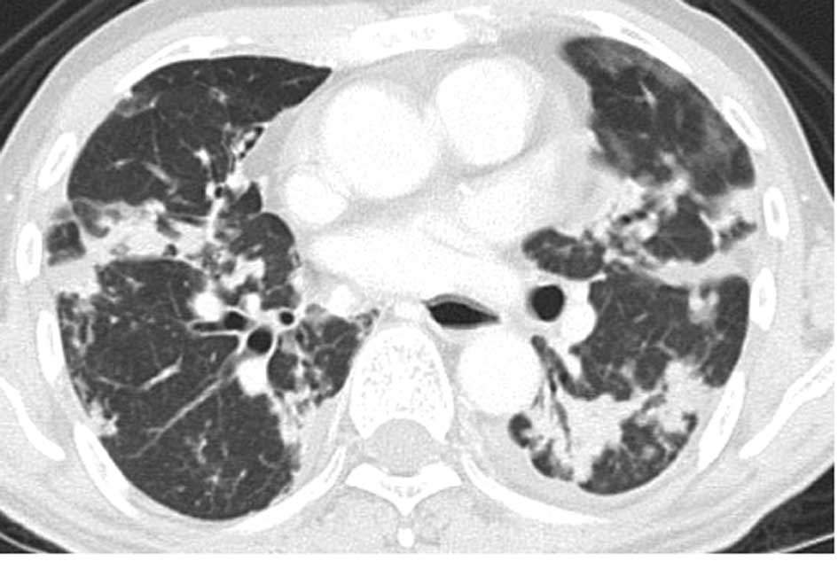
A 62-year-old Japanese man with a history of
cigarette smoking (30 years; one pack/day) presented with a
persistent cough and shortness of breath lasting 1 year. A chest
computed tomography (CT) showed multiple small nodules in the
bilateral lungs (Fig. 1).
Laboratory tests revealed elevated serum CA19-9 (5,455 U/ml; range
<37) and carcinoembryonic antigen (4,306 ng/ml; range <5).
Transbronchial biopsy from the right lung nodule revealed non-small
cell carcinoma and an abdominal CT showed metastases in the
bilateral adrenal glands and lumbar vertebrae. The patient
subsequently received four cycles of chemotherapy (cisplatin and
vinorelbine), following which the above-mentioned tumor markers
decreased (CA19-9, 692 U/ml and CEA, 823 ng/ml). However, 5 months
after the initial diagnosis two nodular lesions were found in S7
and S8 of the liver by abdominal CT. The lesions were diagnosed as
metastases from lung carcinoma by clinical imaging findings. As a
result, four cycles of chemotherapy (docetaxel) were added. Nine
months after the initial diagnosis, the patient complained of a
cough and shortness of breath again, and a chest CT showed an
increase in the size and number of lung nodules. Radiation therapy
was scheduled; however, the patient succumbed to sudden
dyspnea.
Formalin-fixed, paraffin-embedded tissue blocks were
cut into 3-μm sections, deparaffinized and rehydrated. Each section
was stained with H&E and Alcian blue, and used for
immunohistochemical analyses. Immunohistochemical analyses were
performed using an autostainer (XT system Benchmark; Ventana
Medical System, Tucson, AZ, USA) according to the manufacturer’s
instructions. Primary antibodies were used against the following
antigens: vascular endothelial growth factor (VEGF; A-20; Santa
Cruz Biotechnology, Santa Cruz, CA, USA) and osteopontin (OPN;
10A16; Immuno-Biological Laboratories, Japan). The present study
was approved by the Ethics Committee of our university.
Results
At autopsy, gross examination identified multiple
yellowish-white nodules with central necrosis, measuring 5 mm to 3
cm, in the bilateral lungs (Fig.
2). Histopathological examination of the lung nodules showed
infiltrative growth of variably sized and irregular-shaped
epithelial cell nests with ductal formation (Fig. 3). These atypical epithelial cells
had round to oval large nuclei with nucleolus and eosinophilic
cytoplasm (Fig. 3). Alcian blue
staining revealed intracytoplasmic mucin in the tumor cells. A
poorly differentiated adenocarcinoma with multiple intrapulmonary
metastases was diagnosed. The largest nodule was situated in the
middle lobe of the right lung, which was thought to be the origin
of the metastatic lesions.
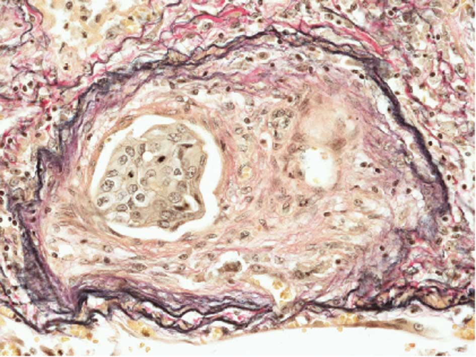
Additionally, prominent fibrocellular and/or
fibromuscular intimal proliferation of small arteries and
arterioles with or without tumor emboli, which caused marked
luminal stenosis, was identified (Fig.
4). These characteristic histopathological findings were
consistent with PTTM caused by lung adenocarcinoma (1). Lymphatic tumor invasion was also
observed.
Metastatic adenocarcinoma was identified in the
liver, bilateral adrenal glands and kidneys, and lumbar
vertebrae.
The immunohistochemical analyses revealed that the
tumor cells were positive for VEGF and OPN (Fig. 5)
Discussion
The suggested mechanism of PTTM involves tumor cells
invading the pulmonary vascular system where they occlude the small
arteries and arterioles, activate coagulation systems and release
inflammatory mediators and growth factors. This process results in
thrombosis, fibrocellular and/or fibromuscular intimal
proliferation and luminal stenosis, which are the characteristic
histopathological findings of PTTM and which differ from
conventional vascular invasion by cancer cells (1,2).
Clinically, patients with PTTM often present with progressive
dyspnea and severe pulmonary hypertension of unknown etiology, and
develop acute cor pulmonale (1–3). Only
four cases of PTTM caused by lung carcinoma have been reported
(1,4), and the histopathological typing of
these cases is adenocarcinoma, as in the present case (Table I).
 | Table IClinicopathological characteristics of
pulmonary tumor thrombotic microangiopathy caused by lung
cancer. |
Table I
Clinicopathological characteristics of
pulmonary tumor thrombotic microangiopathy caused by lung
cancer.
| Case no. | Age/Gender | Histology | Refs. |
|---|
| 1 | 46/M | Adenocarcinoma
(mucinous) | 1 |
| 2 | 54/M | Adenocarcinoma
(mucinous) | 1 |
| 3 | 58/M | Adenocarcinoma
(mucinous) | 1 |
| 4 | 46/F | Adenocarcinoma | 4 |
| Present case | 62/M | Adenocarcinoma | |
Studies have suggested that certain molecules
produced by tumor cells are involved in the onset and/or
development of PTTM (2,3). One candidate is VEGF, a glycoprotein
involved in the proliferation of endothelial cells and tumor
angiogenesis. VEGF expression in tumor cells in cases of PTTM
caused by gastric cancer have been reported (2,3), and
one case of PTTM caused by lung adenocarcinoma expressing VEGF has
also been documented (4).
Another candidate is OPN, a phosphoglycoprotein
produced by various carcinoma cells, which is known to promote
adhesion, migration and proliferation of endothelial cells and
fibroblasts (5). Takahashi et
al speculated that OPN may promote intimal cell proliferation
in cooperation with VEGF in the pathogenesis of PTTM caused by
gastric cancer (3). This is the
first reported case of PTTM caused by lung adenocarcinoma showing
the possible involvement of both VEGF and OPN in promoting intimal
proliferation and pathogenesis in PTTM.
PTTM rapidly progresses and is in most cases fatal
since almost all patients with PTTM die within 1 week from the
onset. Therefore, ante mortem diagnosis of PTTM is extremely
difficult (4,6). Miyano et al reported a notable
case of PTTM caused by gastric cancer, which was diagnosed by lung
biopsy. In their study, VEGF expression in tumor cells was
confirmed by immunohistochemistry; the increased serum VEGF level
became normalized after chemotherapy, resulting in a favorable
clinical course (6). Their findings
indicate that early clinical detection of PTTM may lead to timely
and appropriate management as well as a more favorable
prognosis.
PTTM is found in 3.3% of autopsies of patients with
malignant tumors (1). The present
case suggests that lung adenocarcinoma causes PTTM and shares a
common mechanism with gastric adenocarcinoma. Therefore, it is
necessary for both oncologists and pathologists to take particular
note of PTTM.
References
|
1
|
Von Herbay A, Illes A, Waldherr R and Otto
HF: Pulmonary tumor thrombotic microangiopathy with pulmonary
hypertension. Cancer. 66:587–592. 1990.PubMed/NCBI
|
|
2
|
Sakashita N, Yokose C, Fujii K, et al:
Pulmonary tumor thrombotic microangiopathy resulting from
metastatic signet ring cell carcinoma of the stomach. Pathol Int.
57:383–387. 2007. View Article : Google Scholar : PubMed/NCBI
|
|
3
|
Takahashi F, Kumasaka T, Nagaoka T, et al:
Osteopontin expression in pulmonary tumor thrombotic
microangiopathy caused by gastric carcinoma. Pathol Int.
59:752–756. 2009. View Article : Google Scholar : PubMed/NCBI
|
|
4
|
Uruga H, Morokawa N, Enomoto T, et al: A
case of pulmonary tumor thrombotic microangiopathy associated with
lung adenocarcinoma diagnosed by CT-guided lung biopsy. Nihon
Kokyuki Gakkai Zasshi (In Japanese). 46:928–933. 2008.PubMed/NCBI
|
|
5
|
Denhardt DT, Noda M, O’Regan AW, Pavlin D
and Berman JS: Osteopontin as a means to cope with environmental
insults: regulation of inflammation, tissue remodeling, and cell
survival. J Clin Invest. 107:1055–1061. 2001. View Article : Google Scholar : PubMed/NCBI
|
|
6
|
Miyano S, Izumi S, Takeda Y, et al:
Pulmonary tumor thrombotic microangiopathy. J Clin Oncol.
25:597–599. 2007. View Article : Google Scholar : PubMed/NCBI
|