Spandidos Publications Logo
  • About
    • About Spandidos
    • Aims and Scopes
    • Abstracting and Indexing
    • Editorial Policies
    • Reprints and Permissions
    • Job Opportunities
    • Terms and Conditions
    • Contact
  • Journals
    • All Journals
    • Oncology Letters
      • Oncology Letters
      • Information for Authors
      • Editorial Policies
      • Editorial Board
      • Aims and Scope
      • Abstracting and Indexing
      • Bibliographic Information
      • Archive
    • International Journal of Oncology
      • International Journal of Oncology
      • Information for Authors
      • Editorial Policies
      • Editorial Board
      • Aims and Scope
      • Abstracting and Indexing
      • Bibliographic Information
      • Archive
    • Molecular and Clinical Oncology
      • Molecular and Clinical Oncology
      • Information for Authors
      • Editorial Policies
      • Editorial Board
      • Aims and Scope
      • Abstracting and Indexing
      • Bibliographic Information
      • Archive
    • Experimental and Therapeutic Medicine
      • Experimental and Therapeutic Medicine
      • Information for Authors
      • Editorial Policies
      • Editorial Board
      • Aims and Scope
      • Abstracting and Indexing
      • Bibliographic Information
      • Archive
    • International Journal of Molecular Medicine
      • International Journal of Molecular Medicine
      • Information for Authors
      • Editorial Policies
      • Editorial Board
      • Aims and Scope
      • Abstracting and Indexing
      • Bibliographic Information
      • Archive
    • Biomedical Reports
      • Biomedical Reports
      • Information for Authors
      • Editorial Policies
      • Editorial Board
      • Aims and Scope
      • Abstracting and Indexing
      • Bibliographic Information
      • Archive
    • Oncology Reports
      • Oncology Reports
      • Information for Authors
      • Editorial Policies
      • Editorial Board
      • Aims and Scope
      • Abstracting and Indexing
      • Bibliographic Information
      • Archive
    • Molecular Medicine Reports
      • Molecular Medicine Reports
      • Information for Authors
      • Editorial Policies
      • Editorial Board
      • Aims and Scope
      • Abstracting and Indexing
      • Bibliographic Information
      • Archive
    • World Academy of Sciences Journal
      • World Academy of Sciences Journal
      • Information for Authors
      • Editorial Policies
      • Editorial Board
      • Aims and Scope
      • Abstracting and Indexing
      • Bibliographic Information
      • Archive
    • International Journal of Functional Nutrition
      • International Journal of Functional Nutrition
      • Information for Authors
      • Editorial Policies
      • Editorial Board
      • Aims and Scope
      • Abstracting and Indexing
      • Bibliographic Information
      • Archive
    • International Journal of Epigenetics
      • International Journal of Epigenetics
      • Information for Authors
      • Editorial Policies
      • Editorial Board
      • Aims and Scope
      • Abstracting and Indexing
      • Bibliographic Information
      • Archive
    • Medicine International
      • Medicine International
      • Information for Authors
      • Editorial Policies
      • Editorial Board
      • Aims and Scope
      • Abstracting and Indexing
      • Bibliographic Information
      • Archive
  • Articles
  • Information
    • Information for Authors
    • Information for Reviewers
    • Information for Librarians
    • Information for Advertisers
    • Conferences
  • Language Editing
Spandidos Publications Logo
  • About
    • About Spandidos
    • Aims and Scopes
    • Abstracting and Indexing
    • Editorial Policies
    • Reprints and Permissions
    • Job Opportunities
    • Terms and Conditions
    • Contact
  • Journals
    • All Journals
    • Biomedical Reports
      • Information for Authors
      • Editorial Policies
      • Editorial Board
      • Aims and Scope
      • Abstracting and Indexing
      • Bibliographic Information
      • Archive
    • Experimental and Therapeutic Medicine
      • Information for Authors
      • Editorial Policies
      • Editorial Board
      • Aims and Scope
      • Abstracting and Indexing
      • Bibliographic Information
      • Archive
    • International Journal of Epigenetics
      • Information for Authors
      • Editorial Policies
      • Editorial Board
      • Aims and Scope
      • Abstracting and Indexing
      • Bibliographic Information
      • Archive
    • International Journal of Functional Nutrition
      • Information for Authors
      • Editorial Policies
      • Editorial Board
      • Aims and Scope
      • Abstracting and Indexing
      • Bibliographic Information
      • Archive
    • International Journal of Molecular Medicine
      • Information for Authors
      • Editorial Policies
      • Editorial Board
      • Aims and Scope
      • Abstracting and Indexing
      • Bibliographic Information
      • Archive
    • International Journal of Oncology
      • Information for Authors
      • Editorial Policies
      • Editorial Board
      • Aims and Scope
      • Abstracting and Indexing
      • Bibliographic Information
      • Archive
    • Medicine International
      • Information for Authors
      • Editorial Policies
      • Editorial Board
      • Aims and Scope
      • Abstracting and Indexing
      • Bibliographic Information
      • Archive
    • Molecular and Clinical Oncology
      • Information for Authors
      • Editorial Policies
      • Editorial Board
      • Aims and Scope
      • Abstracting and Indexing
      • Bibliographic Information
      • Archive
    • Molecular Medicine Reports
      • Information for Authors
      • Editorial Policies
      • Editorial Board
      • Aims and Scope
      • Abstracting and Indexing
      • Bibliographic Information
      • Archive
    • Oncology Letters
      • Information for Authors
      • Editorial Policies
      • Editorial Board
      • Aims and Scope
      • Abstracting and Indexing
      • Bibliographic Information
      • Archive
    • Oncology Reports
      • Information for Authors
      • Editorial Policies
      • Editorial Board
      • Aims and Scope
      • Abstracting and Indexing
      • Bibliographic Information
      • Archive
    • World Academy of Sciences Journal
      • Information for Authors
      • Editorial Policies
      • Editorial Board
      • Aims and Scope
      • Abstracting and Indexing
      • Bibliographic Information
      • Archive
  • Articles
  • Information
    • For Authors
    • For Reviewers
    • For Librarians
    • For Advertisers
    • Conferences
  • Language Editing
Login Register Submit
  • This site uses cookies
  • You can change your cookie settings at any time by following the instructions in our Cookie Policy. To find out more, you may read our Privacy Policy.

    I agree
Search articles by DOI, keyword, author or affiliation
Search
Advanced Search
presentation
Oncology Letters
Join Editorial Board Propose a Special Issue
Print ISSN: 1792-1074 Online ISSN: 1792-1082
Journal Cover
March 2013 Volume 5 Issue 3

Full Size Image

Sign up for eToc alerts
Recommend to Library

Journals

International Journal of Molecular Medicine

International Journal of Molecular Medicine

International Journal of Molecular Medicine is an international journal devoted to molecular mechanisms of human disease.

International Journal of Oncology

International Journal of Oncology

International Journal of Oncology is an international journal devoted to oncology research and cancer treatment.

Molecular Medicine Reports

Molecular Medicine Reports

Covers molecular medicine topics such as pharmacology, pathology, genetics, neuroscience, infectious diseases, molecular cardiology, and molecular surgery.

Oncology Reports

Oncology Reports

Oncology Reports is an international journal devoted to fundamental and applied research in Oncology.

Experimental and Therapeutic Medicine

Experimental and Therapeutic Medicine

Experimental and Therapeutic Medicine is an international journal devoted to laboratory and clinical medicine.

Oncology Letters

Oncology Letters

Oncology Letters is an international journal devoted to Experimental and Clinical Oncology.

Biomedical Reports

Biomedical Reports

Explores a wide range of biological and medical fields, including pharmacology, genetics, microbiology, neuroscience, and molecular cardiology.

Molecular and Clinical Oncology

Molecular and Clinical Oncology

International journal addressing all aspects of oncology research, from tumorigenesis and oncogenes to chemotherapy and metastasis.

World Academy of Sciences Journal

World Academy of Sciences Journal

Multidisciplinary open-access journal spanning biochemistry, genetics, neuroscience, environmental health, and synthetic biology.

International Journal of Functional Nutrition

International Journal of Functional Nutrition

Open-access journal combining biochemistry, pharmacology, immunology, and genetics to advance health through functional nutrition.

International Journal of Epigenetics

International Journal of Epigenetics

Publishes open-access research on using epigenetics to advance understanding and treatment of human disease.

Medicine International

Medicine International

An International Open Access Journal Devoted to General Medicine.

Journal Cover
March 2013 Volume 5 Issue 3

Full Size Image

Sign up for eToc alerts
Recommend to Library

  • Article
  • Citations
    • Cite This Article
    • Download Citation
    • Create Citation Alert
    • Remove Citation Alert
    • Cited By
  • Similar Articles
    • Related Articles (in Spandidos Publications)
    • Similar Articles (Google Scholar)
    • Similar Articles (PubMed)
  • Download PDF
  • Download XML
  • View XML
Case Report

Primary spinal glioblastoma: A case report and review of the literature

  • Authors:
    • Nuno Morais
    • Lino Mascarenhas
    • J. P. Soares-Fernandes
    • Ana  Silva
    • Zita Magalhães
    • J. A. Moreira Da Costa
  • View Affiliations / Copyright

    Affiliations: Department of Neurosurgery, Hospital de Braga, Braga, Portugal, Department of Neurosurgery, Centro Hospitalar Vila Nova de Gaia/Espinho, Vila Nova de Gaia, Portugal, Department of Neuroradiology, Hospital de Braga, Braga, Portugal, Department of Anatomic Pathology, Hospital de Braga, Braga, Portugal
  • Pages: 992-996
    |
    Published online on: December 14, 2012
       https://doi.org/10.3892/ol.2012.1076
  • Expand metrics +
Metrics: Total Views: 0 (Spandidos Publications: | PMC Statistics: )
Metrics: Total PDF Downloads: 0 (Spandidos Publications: | PMC Statistics: )
Cited By (CrossRef): 0 citations Loading Articles...

This article is mentioned in:


Abstract

Primary spinal glioblastoma (GBM) is a rare disease, with an aggressive course and a poor prognosis. We report a case of a 19-year-old male with a 4-week history of progressive weakness in both lower limbs, which progressed to paraparesis with a left predominance and difficulty in initiating urination over a week. Spine magnetic resonance imaging (MRI) showed an intramedullary expansile mass localised between T6 and T11. We performed a laminotomy and laminoplasty between T6 and T11 and the tumour was partially removed. Histopathological study was compatible with GBM. The patient was administered focal spine radiotherapy with chemotherapy with temozolamide. Serial MRI performed after the initial surgery demonstrated enlargement of the enhancing mass from T3 to T12 and subarachnoid metastatic deposits in C2 and C4, the pituitary stalk, interpeduncular cistern, left superior cerebellar peduncle and hydrocephalus. We review the literature with regard to the disease and treatment options, and report the unique features of this case. Primary spinal GBM is an extremely rare entity with a poor prognosis and a short survival time. An aggressive management of the different complications as they arise and improvement of current modes of treatment and new treatment options are required to improve survival and ensure better quality of life.

Introduction

Astrocytoma is the most common primary tumour of the central nervous system (1). Spinal neoplasms in the adult population are mostly extradural (55%) and intradural extra-medullary tumours (40%), whereas intramedullary tumours account for 5% of all spinal cord tumours, excluding metastatic lesions (2). Of these, ∼30% are tumours of low malignancy, including slow-growing astrocytomas and ependymomas. Spinal cord astrocytomas are rare, representing ∼1% of all primary central nervous system tumours and 6 to 8% of all spinal cord tumours (3). Few spinal cord astrocytomas are anaplastic in nature; most are slow-growing lesions. The ratio of low-grade to high-grade astrocytomas in the spinal cord is ∼3 to 1 (4,5). Glioblastomas (GBMs) represent ∼7.5% of all intramedullary gliomas and 1 to 3% of all spinal cord tumours (4,6). Moreover, GBM has a predilection of development at the cervical or cervicothoracic region in >60% of cases (1,7–9). Clinical presentation is associated with the region of spinal cord involved, irrespective of tumour type. Unlike their intracranial counterpart, intramedullary GBMs have received scant attention in the literature, with <200 cases reported. Even with aggressive management, these tumours are generally associated with a dismal outcome.

Case report

A 19-year-old male was transferred to our institution in May 2010 with a 4-week history of progressive weakness in both lower limbs, which progressed to paraparesis with a left predominance and difficulty in initiating urination over a week. Examination revealed spastic paraparesis (right/left: grade 4+ and 3+, respectively) and hypoesthesia below the T10 sensory dermatome.

The study was approved by the Ethics Committee of Hospital de Braga, Braga, Portugal. Informed consent was obtained from the patient’s family.

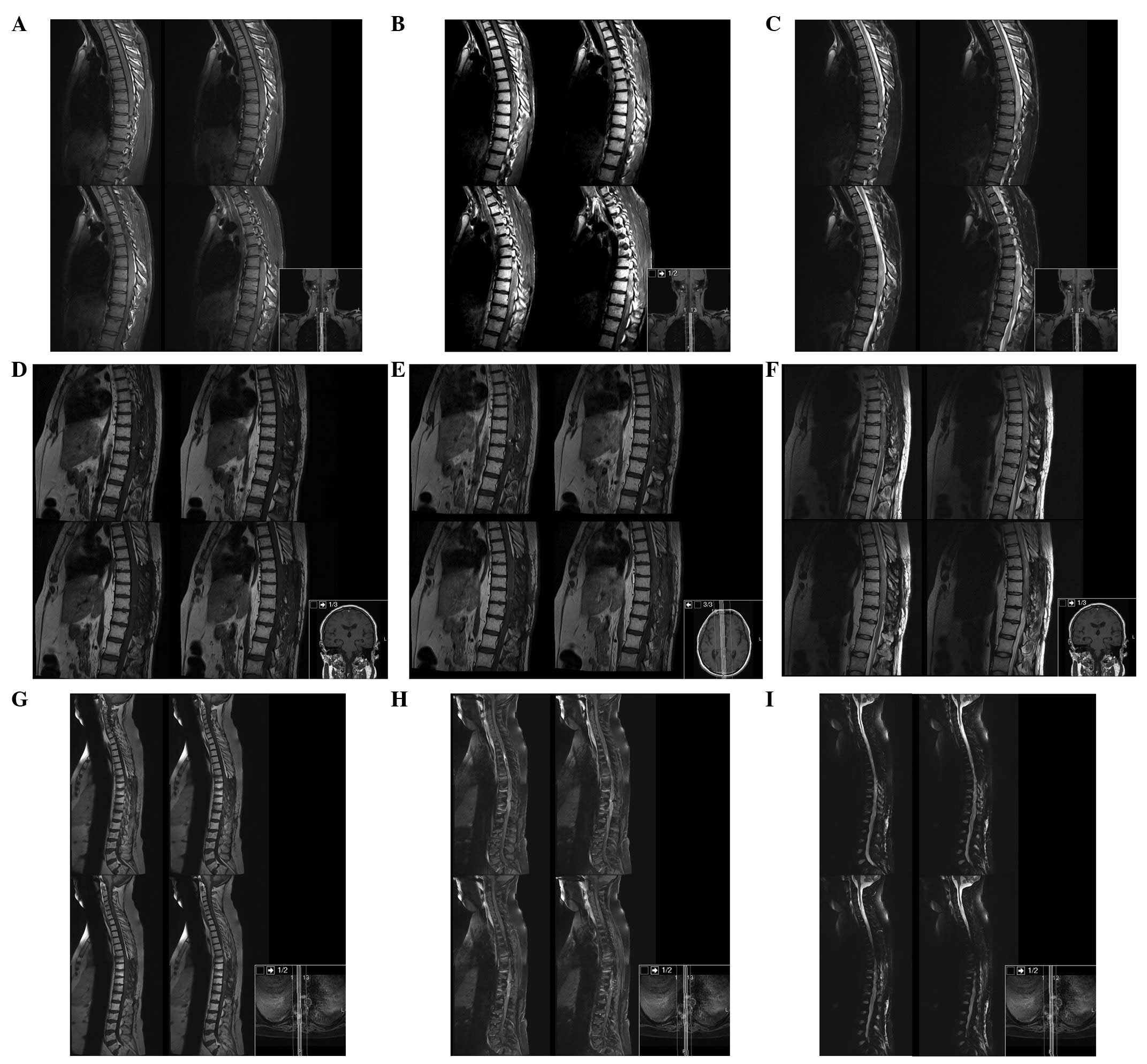
The patient underwent brain and spine magnetic resonance imaging (MRI). The brain and cervical spine were negative for masses and signal intensity alterations, whereas from T1 to L1 there was a marked spinal cord signal intensity and morphology alteration, with notable spinal cord expansion between T6 and T11 and contrast enhancement between T6 and T9 (Fig. 1).

Figure 1

Spine MRI. (A) T1, (B) T1 with contrast and (C) T2-weighted dorsal spine sagittal view MRI at presentation, demonstrating the presence of the tumour between T1 and L1. (D) T1, (E) T1 with contrast and (F) T2-weighted dorsal spine sagittal view MRI 3 months after surgery. (G) T1, (H) T1 with contrast and (I) T2-weighted dorsal spine sagittal view MRI 17 months after surgery, demonstrating enlargement of the enhancing mass from T3 to T12 with subarachnoid metastatic deposit in C2 and C4. MRI, magnetic resonance imaging.

We performed a laminotomy and laminoplasty between T6 and T11, and partial tumour removal under motor-evoked potential monitoring. We were unable to distinguish the tumour margin from the spinal cord and decided to partially remove the mass.

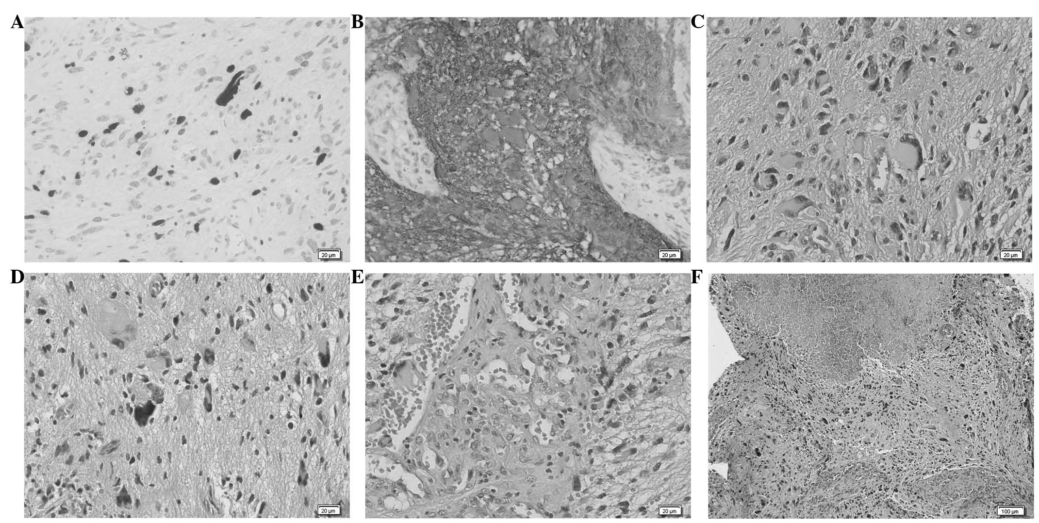
Histopathological study confirmed the diagnosis of GBM (Fig. 2), with histological findings of pleomorphism, atypical cells with high cellularity, vascular proliferation and necrosis. As demonstrated by immunohistochemistry, glial fibrillary acidic protein (GFAP) and S100 Protein were consistently expressed by tumour cells. The neoplasm also showed a high MIB1/Ki-67 labelling index.

Figure 2

Histological findings of glioblastoma. (A) High MIB-1 /Ki-67 labelling index. (B) Marked immunoreactivity for GFAP. (C and D) Pleomorphic glial tumour cells with marked nuclear atypia. (E) Vascular proliferation was observed throughout the lesion. (F) Presence of extensive areas of necrosis. (A–E) Magnification, ×400. (F) Magnification, ×100. (C–F) Hematoxylin/eosin staining. GFAP, glial fibrillary acidic protein.

Post-operatively the patient had transient neurological deterioration with worsening of paraparesis, but with intensive rehabilitation his condition returned to baseline. Thoracoabdominal CT scan for extra-neuronal metastases was negative. The patient was administered spinal radiotherapy between T1 and L1 (45 Gy in 28 fractions) with chemotherapy with temozolamide (completed 6 cycles).

Serial MRI at 3, 6 and 17 months was performed (Fig. 3). Six months after surgery the patient deteriorated, becoming completely paraplegic and losing bladder function. Cranial and spine MRI revealed enlargement of the residual tumour from T3 to T12 with cranial extension of oedema to the obex, and subarachnoid metastatic deposits in C2, C4 and in the pituitary stalk, with hydrocephalus. We proposed cranio-spinal irradiation, but as the patient was stable and without signs or symptoms of increased intracranial pressure (ICP) the patient and his family decided to withhold this treatment and wait.
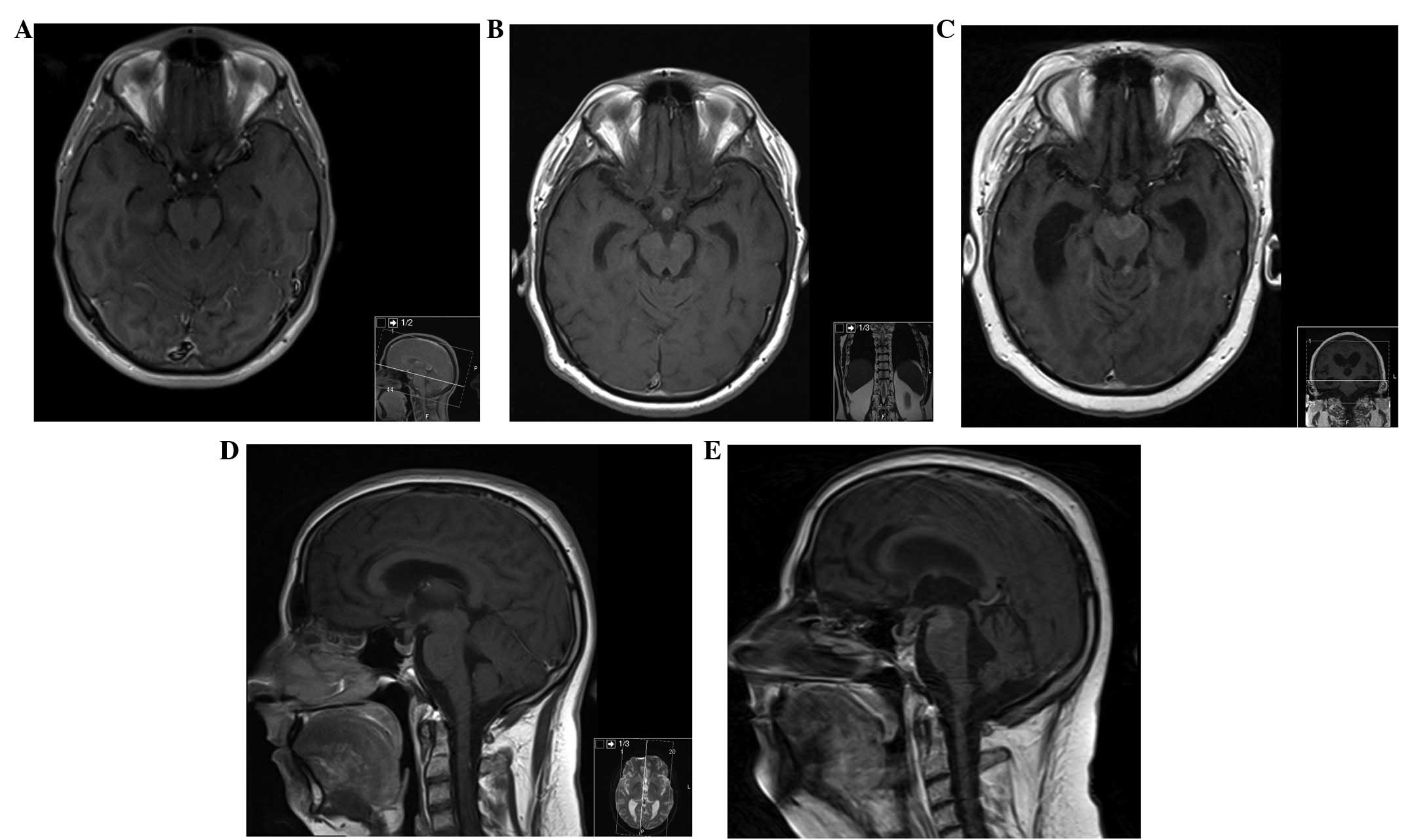
Figure 3

MRI of the brain. (A) Axial section, contrast-enhanced T1-weighted MRI of the brain, at presentation, demonstrating no lesions. (B and D) Axial and sagittal section, respectively, contrast-enhanced T1-weighted MRI of the brain, at 6 months after initial presentation, showing a metastatic deposit in the pituitary stalk. (C) Axial and (E) sagittal section contrast-enhanced T1-weighted MRI of the brain, at 17 months after initial presentation, demonstrating new metastatic deposits in the interpeduncular cistern and in the left superior cerebellar peduncle. MRI, magnetic resonance imaging.

Neuroaxis MRI performed 17 months after surgery revealed an enlargement of the enhancement mass from T3 to T12 with less perilesional oedema and a new metastatic deposit in the interpeduncular cistern and left superior cerebellar peduncle.

On the follow-up 20 months after surgery, the patient presented with a left third cranial nerve paralysis and bilateral mydriasis without signs of increased ICP or mental status change. CT scan showed larger ventricles and enlargement of the pituitary stalk and left superior cerebellar peduncle metastatic deposits. Given the disease progression and the Karnofsky score (40%), we did not consider further treatment and the patient succumbed to the disease 1 month later.

Discussion

Intramedullary GBM is a rare disease entity. It develops primarily from the spinal cord or as a secondary metastasis from the brain, which covers up to 25% of the total occurrences. Intramedullary GBM has a predilection to develop from the cervical region in primary cases, and has a tendency to develop at a young age (<30 years old). Despite the best treatment (surgery and adjuvant therapy), the estimated survival barely exceeds 6 to 16 months (1,8–11).

Certain patients with malignant spinal cord astrocytomas develop hydrocephalus (1,7), which is thought to be due to increased protein concentration in the cerebrospinal fluid (CSF), occlusion of the CSF channel in the subarachnoid space at the skull base and brain surface, arachnoiditis and bleeding of spinal cord tumours (6,7).

Seeding of an intracranial GBM along the spine occurs in 25% of cases, but the reverse process is extremely uncommon (1,3). Patients with malignant spinal cord astrocytomas may develop disseminated disease, mostly via the leptomeningeal route (1,6); however, no definite evidence has been elucidated. Sites of intracranial metastases include the subarachnoid space, ventricles, cerebellum, hypothalamus, brain stem, thalamus and septum pellucidum. Surgical manipulation of GBM has not been shown to increase the tumour seeding into the CSF (3). Continuous spread to contiguous regions is rare, with the most common sites metastases being extra-neuronal, including the lungs, lymph nodes, bone, liver and pleura (12). In our case, metastases were observed in the subarachnoid space in C2 and C4, pituitary stalk, interpeduncular cistern and left superior cerebellar peduncle.

All current therapeutic measures have produced disappointing results and few data concerning their real value are available, with survival times between 6 and 16 months with a mean survival period of 12 months after diagnosis (1,4,7–10) Radical surgery is suggested for confirmation of the diagnosis and for cytoreduction of the tumour as an adjunct to radiotherapy and chemotherapy.

Most authors suggest focal spine radiotherapy and chemo-therapy with temozolamide, while others recommend a more aggressive approach with whole-brain irradiation in addition to focal spine irradiation, even if there is no evidence of intracranial dissemination. Others suggest intrathecal administration of interferon-β via an Ommaya reservoir in conjunction with cranio-spinal irradiation (3,7).

MRI is considered the gold standard imaging modality to diagnose intramedullary tumours (13,14), and gadolinium-enhanced MRI of the entire neuroaxis is advocated to rule out metastasis, evaluate treatment efficacy and detect relapse (15).

To the best of our knowledge, only 16 cases of spinal GBM involving the conus medullaris have been previously reported (1,4,13–19,20–24), making this case the first with spinal and intracranial metastasis with hydrocephalus and the third most longest survival (21 months).

In conclusion, primary spinal GBM is an extremely rare entity. Despite aggressive treatment with radical surgery, radiotherapy and chemotherapy, this disease progresses rapidly with a poor prognosis and a short survival time. We advocate an aggressive management of the different complications as they arise (progression, metastasis, hydrocephalus) to extend the patient’s survival as long as possible with the best quality of life. Improvement of current modes of treatment and new treatment options (chemotherapy protocols, gene therapy) are required to improve survival and ensure better quality of life.

Abbreviations:

GBM

glioblastoma

CSF

cerebrospinal fluid

MRI

magnetic resonance imaging

ICP

intracranial pressure

References

1 

Cohen AR, Wisoff JH, Allen JC and Epstein F: Malignant astrocytomas of the spinal cord. J Neurosurg. 70:50–54. 1989. View Article : Google Scholar : PubMed/NCBI

2 

Balériaux DL: Spinal cord tumors. Eur Radiol. 9:1252–1258. 1999.

3 

Johnson D and Schwarz S: Intracranial metastases from malignant spinal-cord astrocytoma. Case report. J Neurosurg. 66:621–625. 1987. View Article : Google Scholar : PubMed/NCBI

4 

Medhkour A and Chan M: Extremely rare glioblastoma multiforme of the conus medullaris with holocord and brain stem metastases, leading to cranial nerve deficit and respiratory failure: A case report and review of the literature. Surg Neurol. 63:576–582. 2005. View Article : Google Scholar

5 

Stein BM: Surgery of intramedullary spinal cord tumors. Clin Neurosurg. 26:529–542. 1979.PubMed/NCBI

6 

Ciappetta P, Salvati M, Capoccia G, Artico M, Raco A and Fortuna A: Spinal glioblastoma: report of seven cases and review of the literature. Neurosurgery. 28:302–306. 1991. View Article : Google Scholar : PubMed/NCBI

7 

Asano N, Kitamura K, Seo Y, et al: Spinal cord glioblastoma multiforme with intracranial dissemination - case report. Neurol Med Chir (Tokyo). 30:489–494. 1990. View Article : Google Scholar : PubMed/NCBI

8 

Grisold W, Pernetzky G and Jellinger K: Giant-cell glioblastoma of the thoracic cord. Acta Neurochir (Wien). 58:121–126. 1981. View Article : Google Scholar

9 

Guidetti B, Mercuri S and Vagnozzi R: Long-term results of the surgical treatment of 129 intramedullary spinal gliomas. J Neurosurg. 54:323–330. 1981. View Article : Google Scholar : PubMed/NCBI

10 

Alvisi C, Cerisoli M and Giulioni M: Intramedullary spinal gliomas: long-term results of surgical treatments. Acta Neurochir (Wien). 70:169–179. 1984. View Article : Google Scholar : PubMed/NCBI

11 

Kopelson G and Linggood RM: Intramedullary spinal cord astrocytoma versus glioblastoma: the prognostic importance of histological grade. Cancer. 50:732–735. 1982. View Article : Google Scholar : PubMed/NCBI

12 

Russell DS and Rubinstein LJ: Glioblastoma. Pathology of Tumours of the Nervous System. 5th edition. Edward Arnold; London: pp. 426–452. 1998

13 

Bonde V, Balasubramaniam S and Goel A: Glioblastoma multiforme of the conus medullaris with holocordal spread. J Clin Neurosci. 15:601–603. 2008. View Article : Google Scholar : PubMed/NCBI

14 

Stecco A, Quirico C, Giampietro A, Sessa G, Boldorini R and Carriero A: Glioblastoma multiforme of the conus medullaris in a child: description of a case and literature review. AJNR Am J Neuroradiol. 26:2157–2160. 2005.PubMed/NCBI

15 

Mori K, Imai S, Shimizu J, Taga T, Ishida M and Matsusue Y: Spinal glioblastoma multiforme of the conus medullaris with holocordal and intracranial spread in a child: a case report and review of the literature. Spine J. 12:e1–e6. 2012. View Article : Google Scholar : PubMed/NCBI

16 

Andrews AA, Enriques L, Renaudin J and Tomiyasu U: Spinal intramedullary glioblastoma with intracranial seeding. Report of a case. Arch Neurol. 35:244–245. 1978. View Article : Google Scholar : PubMed/NCBI

17 

Eden KC: Dissemination of a glioma of the spinal cord in the leptomeninges. Brain. 61:298–310. 1938. View Article : Google Scholar

18 

Kawanishi M, Kuroiwa T, Nagasawa S, Ohta T, Oketa M and Onomura T: A case of spinal glioblastoma with intracranial dissemination. No Shinkei Geka. 21:1109–1112. 1993.(In Japanese).

19 

O’Connell JE: The subarachnoid dissemination of spinal tumours. J Neurol Neurosurg Psychiatry. 9:55–62. 1946.

20 

Santi M, Mena H, Wong K, Koeller K, Olsen C and Rushing EJ: Spinal cord malignant astrocytomas. Clinicopathologic features in 36 cases. Cancer. 98:554–561. 2003. View Article : Google Scholar : PubMed/NCBI

21 

Scarrow AM, Rajendran P and Welch WC: Glioblastoma multiforme of the conus medullaris. Clin Neurol Neurosurg. 102:166–167. 2000. View Article : Google Scholar : PubMed/NCBI

22 

Shirato H, Kamada T, Hida K, et al: The role of radiotherapy in the management of spinal cord glioma. J Radiat Oncol Biol Phys. 33:323–328. 1995. View Article : Google Scholar : PubMed/NCBI

23 

Strik HM, Effenberger O, Schäfer O, Risch U, Wickboldt J and Meyermann R: A case of spinal glioblastoma multiforme: immunohistochemical study and review of the literature. J Neurooncol. 50:239–243. 2000. View Article : Google Scholar : PubMed/NCBI

24 

Tashiro K, Tachibana S and Tsura M: Clinicopathological studies of spinal cord neoplasm with disseminating intracranial metastasis possibly producing akinetics mutism. No To Shinkei. 28:1311–1318. 1976.(In Japanese).

Related Articles

  • Abstract
  • View
  • Download
  • Twitter
Copy and paste a formatted citation
Spandidos Publications style
Morais N, Mascarenhas L, Soares-Fernandes JP, Silva A, Magalhães Z and Moreira Da Costa JA: Primary spinal glioblastoma: A case report and review of the literature. Oncol Lett 5: 992-996, 2013.
APA
Morais, N., Mascarenhas, L., Soares-Fernandes, J.P., Silva, A., Magalhães, Z., & Moreira Da Costa, J.A. (2013). Primary spinal glioblastoma: A case report and review of the literature. Oncology Letters, 5, 992-996. https://doi.org/10.3892/ol.2012.1076
MLA
Morais, N., Mascarenhas, L., Soares-Fernandes, J. P., Silva, A., Magalhães, Z., Moreira Da Costa, J. A."Primary spinal glioblastoma: A case report and review of the literature". Oncology Letters 5.3 (2013): 992-996.
Chicago
Morais, N., Mascarenhas, L., Soares-Fernandes, J. P., Silva, A., Magalhães, Z., Moreira Da Costa, J. A."Primary spinal glioblastoma: A case report and review of the literature". Oncology Letters 5, no. 3 (2013): 992-996. https://doi.org/10.3892/ol.2012.1076
Copy and paste a formatted citation
x
Spandidos Publications style
Morais N, Mascarenhas L, Soares-Fernandes JP, Silva A, Magalhães Z and Moreira Da Costa JA: Primary spinal glioblastoma: A case report and review of the literature. Oncol Lett 5: 992-996, 2013.
APA
Morais, N., Mascarenhas, L., Soares-Fernandes, J.P., Silva, A., Magalhães, Z., & Moreira Da Costa, J.A. (2013). Primary spinal glioblastoma: A case report and review of the literature. Oncology Letters, 5, 992-996. https://doi.org/10.3892/ol.2012.1076
MLA
Morais, N., Mascarenhas, L., Soares-Fernandes, J. P., Silva, A., Magalhães, Z., Moreira Da Costa, J. A."Primary spinal glioblastoma: A case report and review of the literature". Oncology Letters 5.3 (2013): 992-996.
Chicago
Morais, N., Mascarenhas, L., Soares-Fernandes, J. P., Silva, A., Magalhães, Z., Moreira Da Costa, J. A."Primary spinal glioblastoma: A case report and review of the literature". Oncology Letters 5, no. 3 (2013): 992-996. https://doi.org/10.3892/ol.2012.1076
Follow us
  • Twitter
  • LinkedIn
  • Facebook
About
  • Spandidos Publications
  • Careers
  • Cookie Policy
  • Privacy Policy
How can we help?
  • Help
  • Live Chat
  • Contact
  • Email to our Support Team