Introduction
Liver cancer is one of the most common types of
malignant tumor in humans; its incidence is the fifth highest
amongst the malignant tumors (1,2). China
has a particularly high prevalence of liver cancer and the number
of resulting annual mortalities account for ~45% of the global
mortality rate due to liver cancer. The traditional treatment
strategy for liver cancer includes surgery, radiotherapy and
chemotherapy. Surgery has the highest potential for reducing tumor
burden; however, numerous patients are already in the middle or
advanced stages of the disease when they enroll in treatment, and
are therefore past the optimum stage for undergoing surgery.
Although systemic chemotherapy may be used in the treatment of
advanced liver cancer, patients often find the side effects
difficult to tolerate. The total effective dose of liver cancer
radiotherapy is 60 Gy, however, healthy liver tissue can only
tolerate a limit of 40 Gy and, therefore, radiotherapy cannot be
used as the conventional treatment strategy for liver cancer. Thus,
there is a requirement for the development of radical treatment
strategies for liver cancer.
Transcatheter arterial chemotherapy (TAC) has become
an important method in the treatment of liver cancer patients in
the middle or advanced stages of the disease, as it is minimally
invasive, uses high local concentrations of chemotherapeutic agents
and exhibits small systemic side effects. TAC therapy is based on
the evidence that blood is primarily supplied to healthy liver
tissues via the hepatic portal vein, while the hepatic artery
accounts for >80% of the blood supplied to liver cancer
(3–5).
As research into oncobiology has increased, the
development of tumor gene therapy has broadened the treatment
prospects for cancer patients (6).
Liver cancer is characterized by the type of the gene mutation
present; the majority of cases of liver cancer are caused by the
deletion of the p53 tumor suppressor gene (also known as the
wild-type p53 gene, wt-p53). It is reported that ~50% of cases of
human cancer are associated with the p53 gene mutation (i.e.,
mutated-type p53 gene), and cannot express normally functioning p53
(7,8), resulting in malignancy. Previous
studies have identified that the introduction of wt-p53 into tumor
cells may result in cells which are able to effectively perform p53
functions, such as proliferation inhibition, induction of
apoptosis, inhibition of angiogenesis, delayed metastasis and
increased sensitivity to chemotherapeutic agents; thus, wt-p53
itself may act as a therapeutic agent against the cancer (9). The transfection efficiency of wt-p53
into tumor cells is associated with the local intratumoral
concentration of the p53 gene; the higher the concentration of the
p53 gene, the higher the transfection efficiency and the greater
efficacy towards the tumor cells (10). This p53-based gene therapy has
evolved from basic research to clinical practice; for example, the
Ad-p53 Gendicine has been approved as part of biological cancer
therapy in China (11). However,
how to reduce the risks of gene therapy in clinical practice,
improve the transfection efficiency of the exogenous p53 gene in
liver tumor cells, increase Ad-p53 target specificity, reduce the
immune response and increase the efficiency of gene therapy require
further investigation.
Progress has occurred in the field of
ultrasound-mediated gene transfection, and sufficient gene
transfection efficiency has been achieved (12,13).
The effective transfection efficiency and expression levels of the
exogenous genes in tumor cells is the key to the success of gene
therapy, and changes in the cell membrane and capillary wall
permeability are prerequisites of gene transfection. The
microbubble ultrasound contrast agents may reduce the ultrasonic
cavitation threshold, enhance the ultrasonic cavitation effects,
and further improve cell membrane and capillary wall permeability,
therefore, significantly enhancing the transfection efficiency and
expression levels of the exogenous gene in the tumor cells
(14). The ultrasound contrast
agent SonoVue® is a type of sonicated albumin
microbubble; the albumin coat of the microbubble may maintain the
original biological characteristics, combining with the proteins
and antisense oligonucleotides (15,16),
therefore, it may be used as a vector in gene therapy. If this
microbubble could be combined with target therapeutic genes, and if
ultrasonic irradiation could be used to destruct the bubble when
the therapeutic gene was delivered to the lesions via blood flow,
then the therapeutic gene and microbubble coat would be locally
released into the tumor tissue spaces through the
permeability-increased blood vessel walls. In addition, the shock
wave generated by the microbubble rupture may promote the entry of
the gene into the surrounding tumor tissues. Rat experiments
conducted by Skyba et al (17) and Shohet et al (18) demonstrated that, following the
adhesion of the gene to the ultrasound contrast agent, the
ultrasonic irradiation enhanced the gene transfection
efficiency.
The present study aimed to explore the method of
combined interventional Ad-p53 introduction and ultrasound
irradiation (CIAIUI), using the ultrasonic microbubble agent
SonoVue®, for infusion into liver lesions via the
hepatic artery. The present study hypothesized that the addition of
ultrasound irradiation would improve the intratumoral transfection
efficiency of the Ad-p53 gene. Additionally, the study aimed to
observe the inhibition efficiency of vascular endothelial growth
factor (VEGF) and matrix metalloprotein 2 (MMP-2) and explore the
underlying molecular mechanism of this process.
Materials and methods
Animals
Fifteen male and fifteen female chinchilla rabbits
(weight, 2.5–3.0 kg) were provided by the Experimental Animal
Center of the Lanzhou Institute of Biological Products (Lanzhou,
China) The VX2 tumor line was provided by Professor Hongxin Zhang
(Department of Interventional Radiology, the Fourth Military
Medical University, Xi’an, China). This study was approved by the
ethics committee of The First Affiliated Hospital of Xinxiang
Medical University (Xinxiang, China).
Animal model of liver cancer
The frozen VX2 tumor cell line was recovered and the
tumor cell concentration was adjusted to 109 cells/l.
The prepared tumor cell suspension (dose, 0.5 ml) was inoculated
into the rabbit hind lateral muscle and two weeks later the rabbits
had developed sufficient tumors. The tumor-bearing rabbits were
anesthetized with 50 mg/kg pentobarbital sodium and the tumor was
resected under sterile conditions, cut into three 1-mm sections,
and suspended in saline to create a tumor tissue block suspension
at a concentration of 5×10¹⁰ sections/l. Following
general anesthesia with 50 mg/kg pentobarital sodium, the rabbits
were fixed to anatomical plates, the skin of the upper abdomen was
sterilely prepared and ~3-cm longitudinal incisions were made layer
by layer to expose and fix the liver. An inoculation needle was
used to inject 1-ml tumor tissue block suspension into the rabbit
liver under direct vision with a needle depth of ~2 cm. Gauze was
used for local hemostasis compression for 2 min and the abdominal
wall was sutured layer by layer. Three days later, each rabbit
model was intramuscularly injected with 400,000 U/day penicillin to
prevent infection, and the tumor growth was ultrasonically
monitored on days 7, 10 and 14 following inoculation.
Ad-p53 infusion
The rabbit models that survived for 15 days were
randomly divided into three groups (n=10 per group). The rabbits
were anesthetized with 50 mg/kg pentobarbital sodium, fixed and,
under the sterile conditions, the skin and the subcutaneous tissues
of the groin area were incised layer by layer until the femoral
artery was exposed. A radial artery puncture needle was used to
puncture the femoral artery and, under the guidance of Coroskop TOP
digital subtraction machine (Siemens AG, Munich, Germany) and with
the aid of a microwire, a microcatheter was placed in the
tumor-bearing artery. In group one (control group), 10 ml saline
was infused through the catheter; in group two (p53 group), 10 ml
Ad-p53 was infused through the microcatheter [(1×10¹¹
viral particles (vp); SiBiono GeneTech Co., Ltd., Shenzhen, China];
in group three [p53 plus ultrasonic irradiation (p53+US) group], 10
ml Ad-p53 and 0.05 ml/kg ultrasound contrast agent
(SonoVue®; Bracco, Milan, Italy) were infused through
the microcatheter (1×10¹¹ vp) under ultrasonic
irradiation (using a Vivid 7 Ultrasound with an M3S probe operating
at a frequency of 3.4 MHz and a mechanical index of 1.0; GE
Healthcare, Freiburg, Germany). Postoperatively, the femoral artery
was ligated and the incision was sutured.
Determination of serum VEGF levels by
enzyme-linked immunosorbent assay (ELISA)
Blood samples were collected from the ear vein one
day prior to and four days after the Ad-p53 perfusion; the serum
was separated by centrifugation and stored at −80°C. Serum VEGF
levels were determined using a double-antibody sandwich assay, with
the specific experimental procedures performed according to the
manufacturer’s instructions (Senxiong Biotech Industry Co., Ltd.,
Shanghai, China). Following the termination of staining, the
optical density (OD) values were detected using a microplate reader
(Bioworld Technology, Nanjing, China) at a wavelength of 495 nm.
The absolute value of serum VEGF was calculated according to the
standard curve.
Immunohistochemistry
On the fourth day after Ad-p53 infusion, the rabbits
were anesthetized with 50 mg/kg pentobarbital sodium to obtain
liver cancer specimens. Five liver specimens from each group were
fixed in 4% paraformaldehyde and a routine paraffin-embedded method
(19) was performed to obtain 5-μm
serial sections. Immunohistochemistry was performed on the serial
sections, which were stained for with monoclonal mouse anti-rabbit
p53 (1:500; cat. no. ab2462), polyclonal rabbit anti-human MMP2
(1:500; cat. no. ab16033), monoclonal mouse anti-mouse VEGF (1:500;
cat. no. ab1316) and monoclonal rabbit anti-human glyceraldehyde
3-phosphate dehydrogenase (1:500; cat. no. ab128915) antibodies,
which were all purchased from Abcam (Cambridge, UK). Light
microscopy was performed at ×400 magnification for each
pathological chip in five randomly selected, unrepeated fields.
Subsequently, the pathological image analysis system was used to
measure the integral OD value of each field under the same
conditions.
Western blot
On the fourth day after Ad-p53 infusion, the
remaining five liver specimens from each group were frozen in
liquid nitrogen, ground and the total proteins were extracted using
RIPA buffer (Thermo Fisher Scientific, Waltham, MA, USA)
(containing protease inhibitors). Following SDS-PAGE gel
electrophoresis, the total proteins were transferred to a
polyvinylidene difluoride membrane, blocked with 5% skimmed milk
and incubated with p53 antibody (dilution, 1:1000) at 4°C
overnight. The total proteins were washed with Tris-buffered saline
plus Tween-20 solution (Sigma-Aldrich, Munich, Germany), conjugated
with horseradish peroxidase secondary antibody for one hour at room
temperature, and stained using an enhanced chemiluminescence (ECL),
fixation and development technique with an ECL substrate (cat. no.
32106; ECL, Thermo Fisher Scientific).
Statistical analysis
SPSS statistical software (version 11.0) was used to
perform the statistical analyses and all data are expressed as the
mean ± standard error of the mean. Multi-group comparisons were
performed using one-way analysis of variance, and the pre- and
post-treatment data from each group was compared using the unpaired
t-test. P<0.05 indicated a statistically significant
difference.
Results
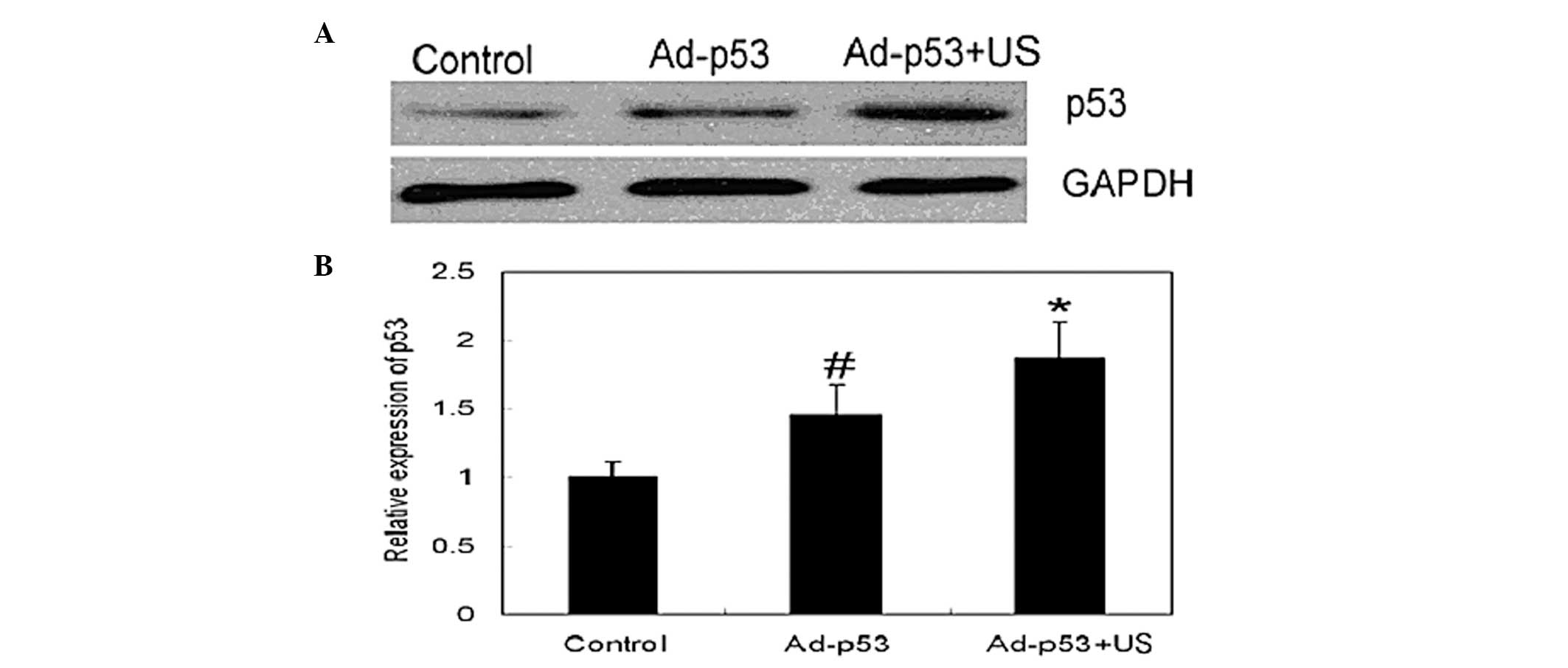
Effect of different Ad-p53 administration
methods on the p53 expression
To observe whether different administration methods
would affect the transfection efficiency of Ad-p53, western
blotting was used to detect the wt-p53 expression levels in the
different groups (Fig. 1). The
results demonstrated that p53 expression was higher in the p53
treatment group compared with the control group and the p53
expression was significantly higher in the p53+US group compared
with in the p53 group, indicating that ultrasonic irradiation may
improve the transfection and expression efficiency of Ad-p53.
Additionally, to observe the morphological characteristics of p53
expression, immunohistochemistry was performed (Fig. 2); the results demonstrated that p53
was distributed throughout the cytoplasm. Furthermore, pathological
image analysis techniques were used to determine that the
p53-positive products (VEGF and MMP2) were most highly expressed in
the p53+US group followed by the p53 group, while the control group
exhibited the lowest expression.
Effect of Ad-p53 therapy on serum VEGF
levels
The growth and invasion of tumors is closely
associated with the angiogenesis of tumor tissues. As the key
nutritional factor in angiogenesis, VEGF is closely associated with
tumor growth, invasion and metastasis; therefore, an ELISA was used
to detect the serum levels of VEGF in the three groups (Table I). No significant difference was
identified in the serum VEGF levels among the three groups prior to
the treatment. Following Ad-p53 treatment, no significant
difference was identified in the serum VEGF levels between the
control and p53 group; however, the serum VEGF level in the p53+US
group was significantly lower than in the control group.
 | Table IImpact of different Ad-p53
administration methods on the serum VEGF expression levels before
and after treatment. |
Table I
Impact of different Ad-p53
administration methods on the serum VEGF expression levels before
and after treatment.
| Serum VEGF expression
level, (pg/ml) mean ± SEM |
|---|
|
|
|---|
| Group (n=10) | Pre-treatment | Post-treatment |
|---|
| Control | 396.96±85.95 | 559.44±10.90 |
| Ad-p53 | 510.75±51.17 | 410.86±34.60* |
| Ad-p53 + US | 404.71±37.09 | 224.14±15.41** |
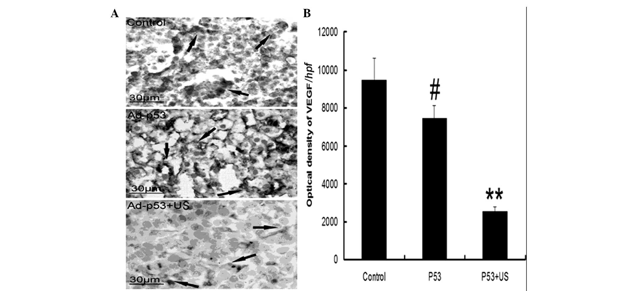
Effect of Ad-p53 therapy on the protein
expression levels of MMP2 and VEGF
The invasion and metastasis of tumor tissues
involves the destruction of the basement membranes, which surround
the tumor tissues. The MMP family is the predominant family of
proteins involved in basement membrane destruction. Additionally,
angiogenesis is considered to be closely associated with the
invasion and metastasis of tumors. In the present study, combined
with pathological image analysis, immunohistochemistry was
performed to semi-quantitatively evaluate the protein expression
levels of MMP2 and VEGF. The results demonstrated that MMP2
expression was significantly enhanced in tumor tissues (Fig. 3) and that Ad-p53 infusion therapy
could partially alleviate the MMP2 expression level; although no
statistically significant difference in MMP2 expression was
demonstrated in the Ad-p53 group compared with the control group
(P>0.05), the expression level of MMP2 in the p53+US group was
significantly reduced compared with the control group (P<0.05).
Similarly, VEGF expression levels were significantly enhanced in
tumor tissues (Fig. 4) and p53
infusion therapy significantly reduced VEGF expression compared
with the control group (P<0.05). In the p53+US group, the tumor
tissue expression level of VEGF was significantly reduced compared
with the control group (P<0.01).
Discussion
To improve the expression level of the therapeutic
p53 gene in tumor cells, the present study combined TAC and
ultrasonic-irradiating therapeutic genes bearing ultrasound
contrast agent. The results of the three groups of abovementioned
animal experiments demonstrated that CIAIUI may significantly
increase the expression of p53 protein in tumor tissues, as p53
protein expression levels were significantly higher in the p53+US
group compared with the p53 and control groups. The results also
demonstrated that tumor invasion and metastasis were associated
with angiogenesis, and VEGF was the factor most closely associated
with angiogenesis. As the majority of VEGF is secreted by the tumor
cells, it may increase the permeability of the vascular walls
(20,21). The present study identified that
serum VEGF levels increased in a rabbit model and were highly
expressed in tumor tissues; however, VEGF expression was reduced in
the peripheral blood and tumor tissues of the p53 group, and
significantly reduced in the p53+US group. This reduction in VEGF
expression may be due to the high expression levels of p53
promoting tumor cell apoptosis, which reduces the excessive
proliferation of tumor cells, therefore, decreasing VEGF expression
levels. The present study also demonstrated that the invasion
process was associated with the erosion and destruction of the
basement membrane by tumor tissues. Previous studies have
identified that the metastasis-associated protein family MMP is
involved in this process, among which MMP2 was of the most
importance (22,23). The present study identified that
MMP2 protein expression levels were significantly increased in the
control group, but downregulated in the Ad-p53 treatment groups.
The expression of MMP2 was significantly downregulated in the
p53+US group compared with the control group, which is consistent
with the VEGF and wt-p53 expression patterns determined in the
present study. The abovementioned results indicated that
CIAIUI-administered Ad-p53 may significantly increase p53
expression levels in tumors.
Although VX2 hepatic carcinoma is an artificial
metastatic squamous cell carcinoma that exhibits different
biological characteristics from primary liver cancer, its
advantages of stable biological characteristics and easy
duplication mean that it is currently the most commonly used large
animal cancer model (24,25). Therefore, VX2 cells were suitable
for achieving the aim of the present study. In conclusion, CIAIUI
via IIHTBA may significantly improve therapeutic gene expression
levels within tumor cells. Furthermore, it is an efficient and safe
method for targeted gene introduction, possesses advantages that
are absent from traditional gene transfection methods and may
provide basic understanding towards gene therapy in the future.
References
|
1
|
Jemal A, Center MM, DeSantis C and Ward
EM: Global patterns of cancer incidence and mortality rates and
trends. Cancer Epidemiol Biomarkers Prev. 19:1893–1907. 2010.
View Article : Google Scholar : PubMed/NCBI
|
|
2
|
El-Serag HB and Rudolph KL: Hepatocellular
carcinoma: epidemiology and molecular carcinogenesis.
Gastroenterology. 132:2557–2576. 2007. View Article : Google Scholar : PubMed/NCBI
|
|
3
|
Lencioni R: Chemoembolization for
hepatocellular carcinoma. Semin Oncol. 39:503–509. 2012. View Article : Google Scholar : PubMed/NCBI
|
|
4
|
Lencioni R and Crocetti L: Local-regional
treatment of hepatocellular carcinoma. Radiology. 262:43–58. 2012.
View Article : Google Scholar
|
|
5
|
Liapi E and Geschwind JF:
Chemoembolization for primary and metastatic liver cancer. Cancer
J. 16:156–162. 2010. View Article : Google Scholar : PubMed/NCBI
|
|
6
|
Sangro B and Prieto J: Gene therapy for
liver cancer: clinical experience and future prospects. Curr Opin
Mol Ther. 12:561–569. 2010.PubMed/NCBI
|
|
7
|
Ji X, Ma L, Huang Q, et al: Network effect
of Wt-mutant p53 interactions and implications on p53 gene therapy.
Curr Pharm Des. 20:1259–1267. 2014. View Article : Google Scholar
|
|
8
|
Nault JC and Zucman-Rossi J: Genetics of
hepatobiliary carcinogenesis. Semin Liver Dis. 31:173–187. 2011.
View Article : Google Scholar : PubMed/NCBI
|
|
9
|
Brito AF, Abrantes AM, Pinto-Costa C, et
al: Hepatocellular carcinoma and chemotherapy: the role of p53.
Chemotherapy. 58:381–386. 2012. View Article : Google Scholar : PubMed/NCBI
|
|
10
|
Gu T, Li CX, Feng Y, Wang Q, Li CH and Li
CF: Trans-arterial gene therapy for hepatocellular carcinoma in a
rabbit model. World J Gastroenterol. 13:2113–2117. 2007.PubMed/NCBI
|
|
11
|
Zhang XZ, Lin H, Yang XY, et al: Quality
control of clinical-grade recombinant adenovirus used in gene
therapy. Zhonghua Yi Xue Za Zhi. 84:849–852. 2004.(In Chinese).
PubMed/NCBI
|
|
12
|
Chen YC, Liang HD, Zhang QP, Blomley MJ
and Lu QL: Pluronic block copolymers: novel functions in
ultrasound-mediated gene transfer and against cell damage.
Ultrasound Med Biol. 32:131–137. 2006. View Article : Google Scholar
|
|
13
|
Yu H, Xu L and Chen S: A transfer
efficiency model for ultrasound mediated drug/gene transferring
into cells. Ultrason Sonochem. 21:113–120. 2014. View Article : Google Scholar
|
|
14
|
Lawrie A, Brisken AF, Francis SE,
Cumberland DC, Crossman DC and Newman CM: Microbubble-enhanced
ultrasound for vascular gene delivery. Gene Ther. 7:2023–2027.
2000. View Article : Google Scholar
|
|
15
|
He Y, Geng Q, Liu H and Han X: First
experience using 4-dimensional hysterosalpingo-contrast sonography
with SonoVue for assessing fallopian tube patency. J Ultrasound
Med. 32:1233–1243. 2013. View Article : Google Scholar : PubMed/NCBI
|
|
16
|
Porter TR, Iversen PL, Li S and Xie F:
Interaction of diagnostic ultrasound with synthetic
oligonucleotide-labeled perfluorocarbon-exposed sonicated dextrose
albumin microbubbles. J Ultrasound Med. 15:577–584. 1996.PubMed/NCBI
|
|
17
|
Skyba DM, Price RJ, Linka AZ, Skalak TC
and Kaul S: Direct in vivo visualization of intravascular
destruction of microbubbles by ultrasound and its local effects on
tissue. Circulation. 98:290–293. 1998. View Article : Google Scholar : PubMed/NCBI
|
|
18
|
Shohet RV, Chen SY, Zhou Y, et al:
Echocardiographic destruction of albumin microbubbles directs gene
delivery to the myocardium. Circulation. 101:2554–2556. 2000.
View Article : Google Scholar : PubMed/NCBI
|
|
19
|
Yoke-Kqueen C, Ab Mutalib NS, Sidik SM,
Learn-Han L and Geok-Chin T: p53 codon 72 polmorphisms and random
amplified polymorphic DNA analysis of non-melanoma skin cancer
through archival formalin-fixed paraffin-embedded tissue. Oncol
Rep. 27:753–763. 2012.
|
|
20
|
Clarke JM and Hurwitz HI: Understanding
and targeting resistance to anti-angiogenic therapies. J
Gastrointest Oncol. 4:253–263. 2013.PubMed/NCBI
|
|
21
|
Sia D, Alsinet C, Newell P and Villanueva
A: VEGF signaling in cancer treatment. Curr Pharm Des.
20:2834–2842. 2014. View Article : Google Scholar
|
|
22
|
Hadler-Olsen E, Winberg JO and
Uhlin-Hansen L: Matrix metalloproteinases in cancer: their value as
diagnostic and prognostic markers and therapeutic targets. Tumour
Biol. 34:2041–2051. 2013. View Article : Google Scholar : PubMed/NCBI
|
|
23
|
Pytliak M, Vargová V and Mechírová V:
Matrix metalloproteinases and their role in oncogenesis: a review.
Onkologie. 35:49–53. 2012. View Article : Google Scholar : PubMed/NCBI
|
|
24
|
Sun H, Xu L, Fan T, et al: Targeted
hyperthermia after selective embolization with ferromagnetic
nanoparticles in a VX2 rabbit liver tumor model. Int J
Nanomedicine. 8:3795–3804. 2013. View Article : Google Scholar : PubMed/NCBI
|
|
25
|
Lee EW, Wong D, Tafti BA, et al:
Irreversible electroporation in eradication of rabbit VX2 liver
tumor. J Vasc Interv Radiol. 23:833–840. 2012. View Article : Google Scholar : PubMed/NCBI
|