Introduction
Lung cancer is the leading cause of
cancer-associated mortalities in males and females worldwide
(1,2).
The two primary types of lung cancer, small cell lung cancer and
non-small cell lung cancer (NSCLC), account for 90% of all the
diagnosed lung cases. NSCLC, which includes squamous cell
carcinoma, adenocarcinoma and large cell carcinoma, constitutes
~80% of all lung cancer types (3–5). The
incidence, progression and underlying mechanisms of tumor
metastasis have been a focus of investigation. Although advances
have been made in the treatment and diagnosis of NSCLC over the
last several years, the overall prognoses for patients with NSCLC
remain poor (6). A number of studies
have attempted to identify novel prognostic and predictive
biomarkers for the diagnosis and treatment of NSCLC (7,8).
Previous studies have established that tumor growth,
development, invasion and metastasis depend upon sufficient
nutrient supply and metabolism, and that water molecules are
important in the modulation of the tumor microenvironment and
metabolism (9,10). Increasing evidence has demonstrated
that aquaporins (AQPs), a family of small transmembrane proteins
responsible for water transport, are involved in the incidence and
progression of cancer (11–13). In particular, AQP1 has been found to
be implicated in the incidence and progression of tumors (11,12),
whilst AQP5 has been proposed to enhance tumor growth and be
associated with tumor metastasis (13).
AQPs are widely distributed in a variety of organs,
and primarily regulate the transport of water molecules by osmotic
pressure. In addition, AQPs regulate the transmembrane transport of
other small molecules, including glycerol and urea. In total, 12
types of AQPs are expressed in humans, and AQP1, 3, 4 and 5 are
expressed in the respiratory system. AQP5 is primarily distributed
in the epithelial cells of type I alveoli and glandular epithelial
cells of the submucosal glands. Its main functions are water
transport and involvement in glandular secretion and airway
hyperactivity (14). Previous studies
have established that AQP1, 3 and 5 are associated with malignant
tumors. Furthermore, it has been demonstrated that AQP1 and 3
promote the metastasis of tumors (15,16). Woo
et al (13) revealed that AQP5
enhanced the proliferation of tumor cells. In addition, high AQP5
expression has been identified to be associated with the metastasis
of rectal, uterine and ovarian cancer (17–19).
However, only a limited number of studies have
investigated the role of AQP5 in cancer. The aims of the present
study were to investigate the expression of AQP5 and determine its
role in NSCLC.
Patients and methods
Study participants
In total, 145 patients were selected by Professor
Ying Wang from the Sichuan Cancer Hospital (Chengdu, China) between
August 2006 and February 2008. The patients were recruited through
oncologist referrals, having been diagnosed with NSCLC following
presentation with lung cancer symptoms, such as chest pain,
shortness of breath, hemoptysis and wheezing. The inclusion
criteria were as follows: Patients with NSCLC; patients treated
with surgery; and patients agreed to participate the present study.
All the patients provided written informed consent for
participation. The study experiments conformed to the ethical
guidelines of the 1975 Declaration of Helsinki (20), and were approved by the Institutional
Review Board of the Sichuan Cancer Hospital.
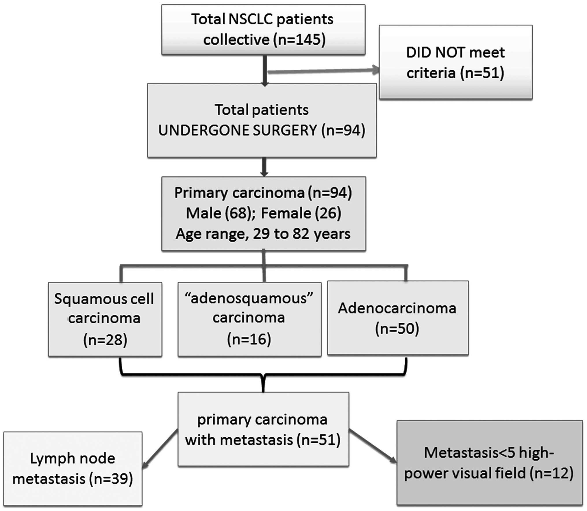
Of the 145 NSCLC patients, 51 patients were excluded
due to having received prior radiation therapy or chemotherapy,
having incomplete clinical information, or unavailability of
paraffin-embedded tissue sections. The remaining 94 patients with
primary NSCLC (male, 68; female, 26; age range, 29–82 years) were
included in the present study (Fig.
1). These patients had not received prior radiation therapy or
chemotherapy. Complete clinical information and paraffin-embedded
sections were obtained. The pathological staging of the
histological types was based on the standard histological
classification of lung cancer, established by the World Health
Organization in 2004 (21). Following
the collection of clinical samples for the present study, the
presence or absence of metastasis was assessed.
Patient follow-up
The patients were routinely followed-up on an
outpatient basis, according to a standard protocol. Briefly, in the
first year following treatment/surgery, visits were conducted every
3 months; in the second and third years, every 6 months; and until
the end of the fifth year, once every 12 months. Patients who did
not attend the outpatient program were contacted by telephone
and/or letter in order to obtain follow-up data, until the visit
cut-off date on August 30, 2012. The rate of missed visits was
4.6%. The period of survival was calculated from the date of
diagnosis to the date of the final follow-up or the date of
mortality due to recurrence or metastasis.
Immunohistochemical staining
AQP5 expression in the biopsy tissue specimens was
detected by immunohistochemical analysis. The biopsy tissue
specimens were collected from the NSCLC patients who had undergone
surgical removal of the tumor. Tumor sections measuring 5 µm in
thickness were initially deparaffinized and rehydrated in a graded
series of alcohols: Slides were submerged in xylene two times for 5
min, before soaking in 100% ethanol two times for 5 min, 95%
ethanol for 5 min and in 80% ethanol for 5 min. Following
rehydration, an antigen retrieval process was performed under high
pressure (22). The sections were
then was hed twice in phosphate-buffered saline (PBS)-Tween 20 for
2 min each. The sections were allowed to cool prior to blocking
with 20% goat serum in PBS for 15 min. Next, the sections were
incubated at 37°C for 1 h, and then overnight at 4°C with a
polyclonal rabbit antibody against AQP5 (dilution, 1:100; Santa
Cruz Biotechnology, Inc., Dallas, TX, USA; catalog no. SC-28628).
Subsequent to was hing three times with PBS-Tween 20 for 2 min
each, the sections were incubated for 10 min at room temperature in
a peroxidase blocking solution. Next, the sections were rinsed
three times with PBS-Tween 20 for 2 min each and incubated with a
biotinylated goat anti-rabbit IgG secondary antibody (dilution,
1:100; Santa Cruz Biotechnology, Inc.; catalog no. SC-2040) in PBS.
Subsequent to was hing, the sections were incubated in a
horseradish peroxidase-streptavidin solution (dilution, 1:100;
Sigma-Aldrich, St. Louis, MO, USA) for 30 min at 37°C. The sections
were again rinsed three times in PBS-Tween 20 for 2 min each and
incubated in a peroxidase substrate solution (Sigma-Aldrich).
Subsequently, the sections were rinsed in running tap water for 2–5
min, followed by dehydration with 95% ethanol for 1 min and twice
with 100% ethanol for 3 min each. The sections were then cleared
twice in 100% xylene for 5 min each and then covered with neutral
gum (Bioworld Technology, Inc., St. Louis Park, MN, USA). The
primary and secondary antibodies and 3,3′diaminobenzidine
tetrahydrochloride (DAB) were obtained from Santa Cruz
Biotechnology, Inc. All the steps were treated with
deoxyribonuclease (Life Technologies, Grand lsland, NY, USA). This
method was used to detect the expression of AQP5 in the lung cancer
tissues. Microscopic immunohistochemical analysis of the tissue
sections was performed using an Olympus BX41 microscope (Olympus
Corporation, Tokyo, Japan).
Scoring of immunohistochemical
staining results
Scoring of the AQP5 expression levels and analysis
of all the specimens were performed by technicians who were blinded
to the clinical information. The appearance of yellowish-brown
granules in the cytoplasm was regarded to be a positive result for
the expression of AQP5. The staining intensity of the AQP5-positive
cells was scored based on the percentage of stained tumor cells as
follows: i) 0, unstained; ii) 1, pale yellow staining; iii) 2,
yellowish-brown staining; and iv) 3, brown staining. Scores were
also assigned according to the percentage of AQP5-positive cells as
follows: i) 4, >75%; ii) 3, 51–75%; iii) 2, 11–50%; iv) 1, ≤10%;
and v) 0, negative. The two scores obtained for each case were then
multiplied and assigned to one of the following groups: i) 0–2,
(−); ii) 3–5, (+); iii) 5–8, (++); and iv) 9–12, (+++) (23).
Statistical analysis
To avoid bias, sample collection and data analysis
were conducted without knowledge of the subject status. SPSS
version 15.0 statistical software (SPSS, Inc., Chicago, IL, USA)
was used for data analysis. The χ2 test was used to
compare differences in AQP5 expression, the Kaplan-Meier method was
used for the analysis of the survival data and the log-rank test
was used to compare the survival curves. P<0.05 was considered
to indicate a statistically significant difference.
Results
Study population and baseline
data
Fig. 1 shows the
screening, randomization and participant flow of the different
groups. Of the 145 randomly selected patients, 94 were found to
conform to the selection criteria. Of the 94 cases of NSCLC (male,
68; female, 26; mean age, 58.68 years; age range, 29–82 years), 28
presented with squamous cell carcinoma, 16 with adenosquamous
carcinoma and 50 with adenocarcinoma (Table I). Overall, there were 15 cases of
highly-differentiated carcinoma, 33 cases of
moderately-differentiated carcinoma, 30 cases of
poorly-differentiated carcinoma and 16 cases of adenosquamous
carcinoma that could not be staged. In addition, 51 cases of
primary carcinoma were accompanied by metastasis, including 39
cases with lymph node-specific metastasis (requirement for
metastasis, ≥5x high-power visual field). In accordance with the
revised standard for the 2009 lung cancer international
tumor-node-metastasis (TNM) staging (24), 15 cases were at stage I, 33 cases were
at stage II, 36 cases were at stage III and 10 cases were at stage
IV (Table I). Patients with stages
III and IV NSCLC did not undergo surgical treatment.
 | Table I.Clinicopathological characteristics of
NSCLC patients (n=94). |
Table I.
Clinicopathological characteristics of
NSCLC patients (n=94).
| Clinicopathological
characteristics | n (%) |
|---|
| Gender |
|
| Male | 68 (72.3) |
|
Female | 26 (27.7) |
| Histological
type |
|
| Squamous
cell carcinoma | 28 (29.8) |
|
Adenocarcinoma | 50 (53.2) |
|
Adenosquamous carcinoma | 16 (17.0) |
|
Differentiationa |
|
| Well | 15 (16.0) |
|
Moderate | 33 (35.1) |
| Poor | 30 (31.9) |
| TNM staging |
|
| I | 15 (16.0) |
| II | 33 (35.1) |
| III | 36 (38.3) |
| IV | 10 (10.6) |
Association between AQP5 expression
percentage and the clinical pathological features of NSCLC
AQP5 expression was associated with the histological
type and TNM staging of the NSCLCs. The percentage of AQP5
expression in the adenocarcinomas was significantly higher compared
with that in the squamous carcinomas (P=0.002). Of the 16 cases of
adenosquamous carcinoma, 11 were positive for the expression of
AQP5, which yielded an expression percentage of 68.8%. This was
significantly higher than the 31.2% expression percentage (5 cases)
of the squamous carcinoma group (P<0.034). The positive
expression percentages of AQP5 in stages III and IV NSCLCs were
significantly higher compared with those in stages I and II NSCLCs
(P=0.027). The expression percentages of AQP5 in the highly-,
moderately- and poorly-differentiated tumors were 46.7, 51.5 and
60.0%, respectively, and no statistically significant differences
were observed (P=0.904; Table
II).
| Table II.AQP5 expression in different
pathological tissues. |
Table II.
AQP5 expression in different
pathological tissues.
|
|
| AQP5 expression |
|
|
|---|
|
|
|
|
|
|
|---|
| Pathological
features | n | – | + | ++ | +++ | χ2 | P-value |
|---|
| Histological
type |
|
|
|
|
|
|
|
| Squamous
cell carcinoma | 28 | 20 | 5 | 2 | 1 | 14.38 | 0.002 |
|
Adenocarcinoma | 50 | 16 | 8 | 14 | 12 |
|
|
|
Adenosquamous carcinoma | 16 | 4 | 3 | 6 | 3 |
|
|
Differentiationa |
|
|
|
|
|
|
|
| Well | 15 | 8 | 3 | 2 | 2 | 2.16 | 0.904 |
|
Moderate | 33 | 16 | 6 | 6 | 5 |
|
|
| Poor | 30 | 12 | 4 | 8 | 6 |
|
|
| TNM staging |
|
|
|
|
|
|
|
| I | 15 | 11 | 1 | 2 | 1 | 18.73 | 0.027 |
| II | 33 | 14 | 5 | 9 | 5 |
|
|
| III | 36 | 16 | 5 | 4 | 11 |
|
|
| IV | 10 | 1 | 3 | 5 | 1 |
|
|
Association between AQP5 expression
and NSCLC metastasis
The AQP5 expression percentages in primary
carcinomas accompanied by lymph node metastasis were significantly
higher compared with those in primary carcinomas not accompanied by
lymph node metastasis (P=0.024). The AQP5 expression percentages in
metastatic carcinoma cases did not exhibit a statistically
significant difference compared with the primary carcinoma cases
(P=0.377; Table III). In primary
carcinoma cases with high AQP5 expression, the metastatic
carcinomas exhibited augmented AQP5 expression. Therefore, AQP5 may
be involved in the metastasis of NSCLC. In addition, the results
indicated that NSCLCs with elevated AQP5 expression are more likely
to be associated with lymph node metastasis.
| Table III.AQP5 expression in primary and
metastatic carcinomas. |
Table III.
AQP5 expression in primary and
metastatic carcinomas.
|
|
| AQP5
expression |
|
|
|---|
|
|
|
|
|
|
|---|
| Groups | n | – | + | ++ | +++ | χ2 | P-value |
|---|
| Lymph node
metastasis |
|
|
|
|
|
|
|
| No | 43 | 25 | 6 | 9 | 3 | 9.40 | 0.024 |
|
Yes | 51 | 17 | 8 | 11 | 15 |
|
|
| Tumor type |
|
|
|
|
|
|
|
| Primary
carcinoma | 39 | 16 | 7 | 5 | 11 | 3.09 | 0.377 |
|
Metastatic carcinoma | 39 | 11 | 5 | 10 | 13 |
Association between AQP5 expression
and survival rate of NSCLC patients
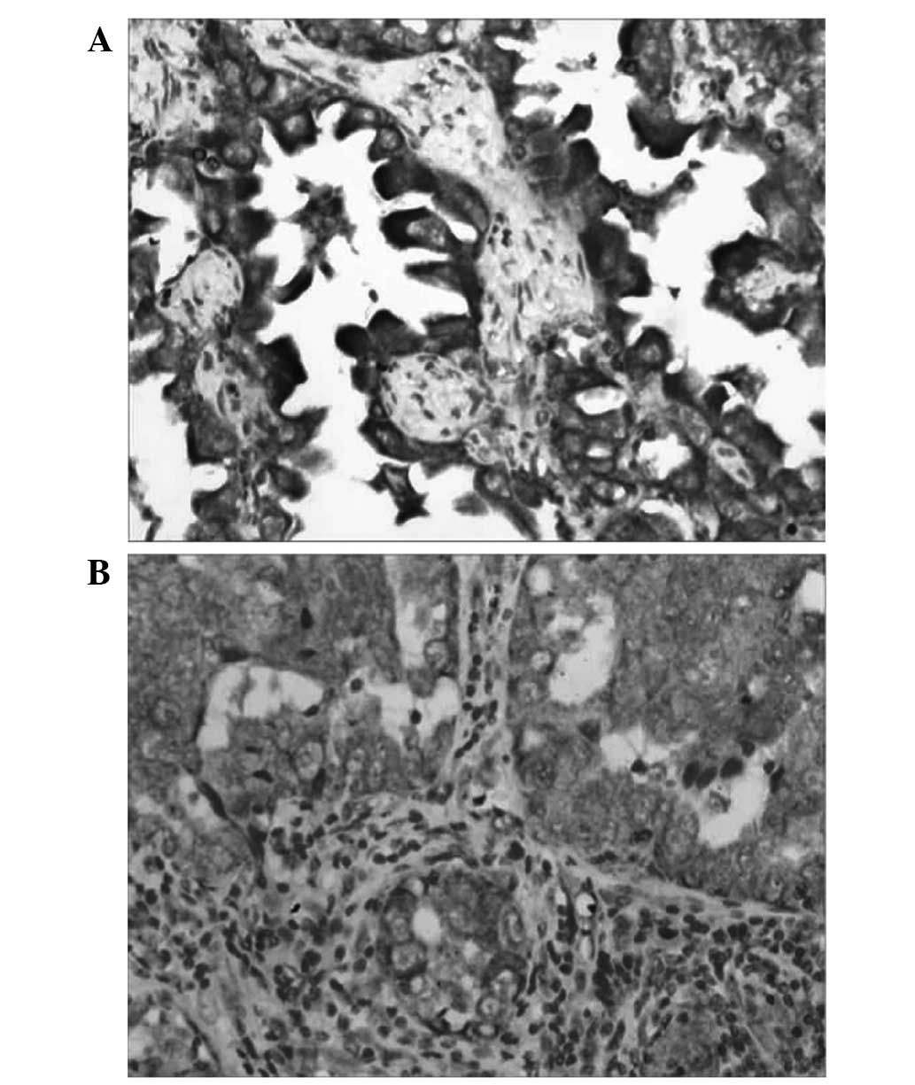
AQP5 expression was evaluated in tissue samples by
immunohistochemical analysis. The appearance of yellowish-brown
granules in the cytoplasm was regarded as a positive result for the
expression of AQP5 (Fig. 2). The
staining intensity of the AQP5-positive cells was scored, as well
as the percentage of AQP5-positive cells. The two scores obtained
for each case were multiplied and assigned to one of the following
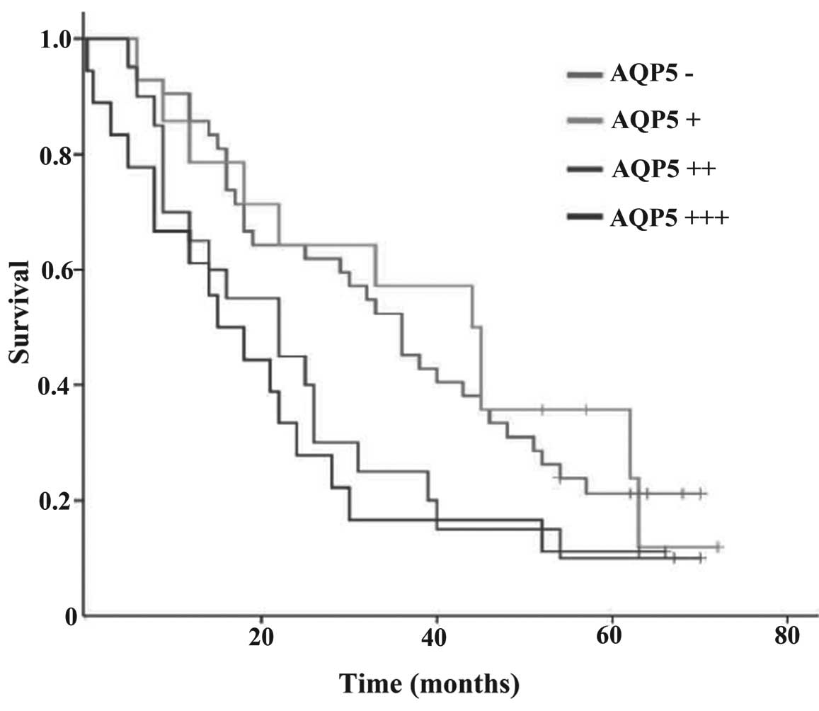
groups: (−), (+), (++), and (+++). The results of the present study
revealed that the five-year survival rate of NSCLC patients with
scores of (−), (+), (++), and (+++) were 21.43, 28.57, 10.00 and
11.11%, respectively. The log-rank test demonstrated a clear
association between the expression of AQP5 and the survival rate of
NSCLC patients (P=0.051; Fig. 3). An
upregulation in the expression of AQP5 was correlated with a
decrease in the survival rate of NSCLC patients.
Discussion
The present study examined the expression of AQP5 in
94 cases of NSCLC primary carcinoma and 51 cases of metastatic
carcinoma. The results revealed that AQP5 expression in NSCLCs was
associated with lymph mode metastasis. AQP5 expression in NSCLC
primary carcinomas accompanied by lymph node metastasis was
markedly higher compared with those without lymph node metastasis.
When the AQP5 expression levels in primary carcinomas was compared
with that in metastatic carcinomas, no statistically significant
difference was identified. In cases of primary carcinoma with high
AQP5 expression, the metastatic carcinomas also exhibited augmented
AQP5 expression. This indicated that NSCLCs with elevated AQP5
expression levels are more likely to be associated with lymph node
metastasis, and that AQP5 plays an important role in the metastasis
of NSCLCs. In addition, the present study revealed that AQP5
expression was associated with the NSCLC clinical stage. The
expression levels of AQP5 in stage III and IV tumors were
significantly higher compared with stage I and II tumors. This
observation indicated that a high level of AQP5 was associated with
the promotion of lung cancer metastasis. Furthermore, AQP5 was
revealed to be closely associated with the incidence and
progression of adenocarcinomas. The positive expression percentage
of AQP5 in adenocarcinomas was 68.0%, which was markedly higher
than the value of 28.6% in squamous carcinomas. In the 16 cases of
squamous carcinoma, the positive expression percentage of AQP5 in
adenocarcinomas was 68.8%, which was notably higher than the value
of 31.2% in squamous carcinomas. These results indicate an
association between AQP5 and adenocarcinomas and a role for AQP5 in
the incidence and progression of adenocarcinomas. Whether the
elevated expression levels of AQP5 in adenocarcinomas can be used
as an indirect marker during clinical and pathological diagnosis
requires further investigation. In addition, the mechanisms
involved in the promotion of tumor metastasis are under
investigation. AQP5 phosphorylation has been demonstrated to be an
important event involved in the promotion, incidence and
progression of tumors (13). Previous
studies have established that AQP5 can activate the RAS/ERK/RB
pathway in rectal cancer cells and enhance the incidence and
progression of cancer. Furthermore, an association with the
metastasis of rectal cancer was also identified (17,25).
In conclusion, the present study revealed an
association between high AQP5 expression and the high invasiveness
and metastatic percentage of NSCLC, as well as with the incidence
and progression of adenocarcinomas. Histopathological analysis
revealed an association between AQP5 and the metastasis, incidence
and progression of NSCLC.
References
|
1
|
Jemal A, Siegel R, Ward E, Murray T, Xu J
and Thun MJ: Cancer statistics, 2007. CA Cancer J Clin. 57:43–66.
2007. View Article : Google Scholar : PubMed/NCBI
|
|
2
|
Stewart BW and Wild CP: Word Cancer Report
2014. IARC Nonserial Publication; 2014
|
|
3
|
Esposito L, Conti D, Ailavajhala R, Khalil
N and Giordano A: Lung cancer: Are we up to the challenge? Curr
Genomics. 11:513–518. 2010. View Article : Google Scholar : PubMed/NCBI
|
|
4
|
Maziak DE, Marman BR, MacKay JA and Evans
WK: Cancer Care Ontario Practice Guidelines Initiative Lung Cancer
Disease Site Group: Photodynamic therapy in non-small cell lung
cancer: a systematic review. Ann Thorac Surg. 77:1484–1491. 2004.
View Article : Google Scholar : PubMed/NCBI
|
|
5
|
Hoffman PC, Mauer AM and Vokes EE: Lung
cancer. Lancet. 355:479–485. 2000. View Article : Google Scholar : PubMed/NCBI
|
|
6
|
Schreiber G and McCrory DC: Performance
characteristics of different modalities for diagnosis of suspected
lung cancer: summary of published evidence. Chest. 123 (Suppl
1):115S–128S. 2003. View Article : Google Scholar : PubMed/NCBI
|
|
7
|
Aggarwal C, Somaiah N and Simon GR:
Biomarkers with predictive and prognostic function in non-small
cell lung cancer: ready for prime time? J Natl Compr Canc Netw.
8:822–832. 2010.PubMed/NCBI
|
|
8
|
Akhtar S, Meeran SM, Katiyar N and Katiyar
SK: Grape seed proanthocyanidins inhibit the growth of human
non-small cell lung cancer xenografts by targeting insulin-like
growth factor binding protein-3, tumor cell proliferation and
angiogenic factors. Clin Cancer Res. 15:821–831. 2009. View Article : Google Scholar : PubMed/NCBI
|
|
9
|
Wang W, Li Q, Yang T, Bai G, Li D, Li Q
and Sun H: Expression of AQP5 and AQP8 in human colorectal
carcinoma and their clinical significance. World J Surg Oncol.
10:2422012. View Article : Google Scholar : PubMed/NCBI
|
|
10
|
Jung HJ, Park JY, Jeon HS and Kwon TH:
Aquaporin-5: a marker protein for proliferation and migration of
human breast cancer cells. PLoS ONE. 6:e284922011. View Article : Google Scholar : PubMed/NCBI
|
|
11
|
Saadoun S, Papadopoulos MC, Hara-Chikuma M
and Verkman AS: Impairment of angiogenesis and cell migration by
targeted aquaporin-1 gene disruption. Nature. 434:786–792. 2005.
View Article : Google Scholar : PubMed/NCBI
|
|
12
|
Burghardt B, Elkaer ML, Kwon TH, Racz GZ,
Varga G, Steward MC and Nielsen S: Distribution of aquaporin water
channels AQP1 and AQP5 in the ductal system of the human pancreas.
Gut. 52:1008–1016. 2003. View Article : Google Scholar : PubMed/NCBI
|
|
13
|
Woo J, Lee J, Chae YK, Kim MS, Baek JH,
Park JC, et al: Over expression of AQP5, a putative oncogene,
promotes cell growth and transformation. Cancer Lett. 264:54–62.
2008. View Article : Google Scholar : PubMed/NCBI
|
|
14
|
Verkman AS: More than just water channels:
unexpected cellular roles of aquaporins. J Cell Sci. 118:3225–3232.
2005. View Article : Google Scholar : PubMed/NCBI
|
|
15
|
Liu YL, Matsuzaki T, Nakazawa T, Murata S,
Nakamura N, Kondo T, Iwashina M, Mochizuki K, Yamane T, Takata K
and Katoh R: Expression of aquaporin 3 (AQP3) in normal and
neoplastic lung tissues. Hum Pathol. 38:171–178. 2007. View Article : Google Scholar : PubMed/NCBI
|
|
16
|
Hoque MO, Soria JC, Woo J, Lee T, Lee J,
Jang SJ, Upadhyay S, Trink B, Monitto C, Desmaze C, Mao L,
Sidransky D and Moon C: Aquaporin 1 is overexpressed in lung cancer
and stimulates NIH-3T3 cell proliferation and anchorage-independent
growth. Am J Pathol. 168:1345–1353. 2006. View Article : Google Scholar : PubMed/NCBI
|
|
17
|
Kang SK, Chae YK, Woo J, Kim MS, Park JC,
Lee J, Soria JC, Jang SJ, Sidransky D and Moon C: Role of human
aquaporin 5 in colorectal carcinogenesis. Am J Pathol. 173:518–525.
2008. View Article : Google Scholar : PubMed/NCBI
|
|
18
|
Zhang T, Zhao C, Chen D and Zhou Z:
Overexpression of AQP5 in cervical cancer: correlation with
clinicopathological features and prognosis. Med Oncol.
29:1998–2004. 2012. View Article : Google Scholar : PubMed/NCBI
|
|
19
|
Yang JH, Shi YF, Cheng Q and Deng L:
Expression and localization of aquaporin-5 in the epithelial
ovarian tumors. Gynecol Oncol. 100:294–299. 2006. View Article : Google Scholar : PubMed/NCBI
|
|
20
|
Declaration of Helsinki, . 29th World
Medical Association General Assembly. Tokyo, Japan: 1975
|
|
21
|
Travis WD, Brambilla E, Muller-Hermelink
HK and Harris CC: World Health Organization Classification of
Tumours: Pathology and Genetics of Tumours of the Lung,
PleuraThymus and Heart. IARC Press; Lyon, France: 2004
|
|
22
|
Fowler CB, Cunningham RE, Waybright TJ,
Blonder J, Veenstra TD, O'Leary TJ and Mason JT: Elevated
hydrostatic pressure promotes protein recovery from formalin-fixed,
paraffin-embedded tissue surrogates. Lab Invest. 88:185–195. 2008.
View Article : Google Scholar : PubMed/NCBI
|
|
23
|
Xu LZ and Yang WT: Standard for appraisal
of results of immunohistochemical reactions. Zhong Guo Ai Zheng Za
Zhi. 6:2292311996.(In Chinese).
|
|
24
|
Goldstraw P and Crowley JJ: IASLC
International Staging Project: The International Association for
the Study of Lung Cancer International Staging Project on Lung
Cancer. J Thorac Oncol. 1:281–286. 2006. View Article : Google Scholar
|
|
25
|
Woo J, Lee J, Kim MS, Jang SJ, Sidransy D
and Moon C: The effect of aquaporin 5 overexpression on the Ras
signaling pathway. Biochem Biophys Res Commun. 367:291–298. 2008.
View Article : Google Scholar : PubMed/NCBI
|