Introduction
Fallopian tube malignancy (FTM) is a rare disease
that comprises only 0.14 to 1.8% of female genital malignancies
(1). The incidence rate in the
United States is estimated to be on average 3.6/million/year
(2). Although rare, FTM is a
disease that has increased 4.5-fold over the past 40 years
(3). Even though the staging and
therapeutic strategy for fallopian tube carcinoma are based on
those of ovarian carcinoma, it is difficult to conduct a
large-scale clinical study exclusively on fallopian tube carcinoma
because of its rarity, and the pattern of metastasis as well as
time to recurrence are not clear. Wethington et al(4) compared the outcome of fallopian tube
carcinoma and ovarian carcinoma based on the Surveillance,
Epidemiology, and End Results (SEER) database of the National
Cancer Institute, and showed that fallopian tube carcinoma was
associated with a more favorable long-term outcome than ovarian
carcinoma. However, the biological behaviors of fallopian tube
carcinoma such as location of recurrence and time to recurrence
remain unknown (4).
This study was a retrospective, multicenter study
that aimed to investigate the clinicopathological features of FTM
and to elucidate the biological behaviors of this disease.
Materials and methods
All patients with FTM treated between January 2001
and December 2011 were eligible for this study. Institutional
review board approval was obtained at each of the 7 participating
academic centers prior to data acquisition, and informed consent
was obtained from the patients or the guardians. Cases were defined
as FTM using diagnostic criteria as documented by Sedlis (5). Briefly, these criteria were as
follows: i) the main tumor arises from the endosalpinx; ii) the
histological pattern reproduces the endothelium of the tube mucosa;
iii) transition from benign to malignant tubal epithelium is
demonstrable; iv) the ovaries or endometrium are either normal or
contain a tumor smaller than the tumor in the tube. In addition, we
also adopted the definition of fallopian tube carcinoma of WHO
classification which states that there is a primary lesion in the
lumen or fimbriae of the fallopian tube and a lesion does not exist
in the ovary and the uterine or it is different from the fallopian
tube clearly even if it exists (6).
All slides were reviewed by expert pathologists from each
institution. A common database form was designed to be utilized by
all participating centers. The medical records included information
regarding age, gravidity and parity, clinical presentation, disease
stage, type of surgery performed, treatment rendered, histological
type, progression-free survival (PFS), overall survival (OS), and
sites of recurrence. Patients of all stages were eligible. Staging
was based on the International Federation for Obstetrics and
Gynecology (FIGO) criteria. Procedures included in the standard
surgery consisted of bilateral salpingo-oophorectomy, hysterectomy
and greater omentectomy with or without cytoreduction. Staging
laparotomy included retroperitoneal (pelvic, para-aortic) lymph
node dissection (or biopsy) and intraperitoneal biopsies in
addition to standard surgery. The 1-cm cutoff was used as a
threshold for optimal cytoreduction (7). When adjuvant chemotherapy was
indicated, patients generally received platinum/taxan-based
combination chemotherapy. Cases that received neoadjuvant
chemotherapy also were included in this retrospective study.
Clinical response was assessed in the enrolled
patients with lesions that could be measured according to the
revised RECIST guideline (version 1.1) (8). A complete response (CR) was defined as
the complete disappearance of all measurable lesions. A partial
response (PR) was defined as a 30% or greater decrease in the sum
of the measurable lesions. Stable disease was defined as a steady
state of response less than PR or an increase in <20% in the sum
of the measurable lesions. Progressive disease was defined as an
increase of 20% or more in the sum of the measurable lesions or the
appearance of new lesions.
PFS was calculated from the date of the start of
treatment to the date of recurrence or progression. OS was
calculated from the date of the start of treatment to the date of
death or last follow-up. The cumulative survival curve and median
PFS time were estimated by use of the Kaplan-Meier method.
Comparison between survival curves was carried out using the
log-rank test. Difference in response rate between predictive
variables was analyzed using the Chi-square test. Statistical
significance was set at P<0.05.
Results
Patient characteristics
Patient characteristics are shown in Table I. Sixty-eight patients with FTM were
enrolled for this retrospective analysis. The 68 patients with this
disease included 60 cases with fallopian tube carcinoma and 8 cases
with fallopian tube carcinosarcoma. The median age of the patients
was 60 years (range, 38–85 years); initial symptoms included
so-called Latzko’s triad of lower abdominal pain (including
bloating) in 27 patients (39.7%), atypical genital bleeding in 21
patients (30.9%), and the sensation of an abdominal mass in 5
patients (7.4%). Melena due to infiltration of the bowel was the
initial symptom in 2 patients. Preoperative diagnoses included a
suspected ovarian carcinoma in 45 patients (66.2%); fallopian tube
carcinoma was suspected before surgery in just 9 patients (13.2%).
Endometrial carcinoma was suspected and treatment for this
condition was started in 9 patients (13.2%). Histological types
were serous adenocarcinoma in 50 patients, endometrioid
adenocarcinoma in 6 patients, and other types in 4 patients. All of
the 8 carcinosarcomas were heterologous. The clinical stages were:
stage I in 11 cases, stage II in 8 cases, stage III in 40 cases,
and stage IV in 9 cases, indicating that stage III or more
comprised 72% of the cases. All of the 68 patients underwent
standard surgery, and additionally staging surgery was performed in
34 patients. Optimal surgery was achieved in 44 patients (64.7%)
(including 10 patients who underwent associated intestinal
resection) and suboptimal surgery was achieved in 24 patients. The
median number of chemotherapy cycles was 6 (range, 1–12).
Postoperative chemotherapy included platinum-based combination
treatment in 64 patients (94%).
 | Table IPatient characteristics. |
Table I
Patient characteristics.
| Clinical factors | No. of patients |
|---|
| Fallopian tube
malignancy |
| Carcinoma of the
fallopian tube | 60 |
| Carcinosarcoma of
the fallopian tube | 8 |
| Median age (range),
in years | 60 (38–85) |
| History of
delivery |
| Yes | 64 |
| No | 4 |
| Menstruation |
| Premenopause | 14 |
| Postmenopause | 54 |
| Chief complaint |
| Lower abdominal pain
or distension | 27 |
| Atypical genital
bleeding | 21 |
| Feeling of abdominal
mass | 5 |
| Bloody bowel
discharge | 2 |
| Asymptomatic | 10 |
| Others | 3 |
| Serum CA-125,
median (range), U/ml | 338.5
(4–16,000) |
| Preoperative
diagnosis |
| Ovarian
carcinoma | 45 |
| Fallopian tube
carcinoma | 9 |
| Endometrial
carcinoma | 9 |
| Colon
carcinoma | 2 |
| Others | 3 |
| Neoadjuvant
chemotherapy |
| Yes | 10 |
| No | 58 |
| FIGO stage |
| I | 11 |
| II | 8 |
| III | 40 |
| IV | 9 |
| Histological
type |
| Serous | 50 |
| Endometrioid | 6 |
|
Undifferentiated | 2 |
| Clear cell | 1 |
| Transitional | 1 |
|
Carcinosarcoma | 8 |
| Debulking
surgery |
| Complete or
optimal | 44 |
| Suboptimal | 24 |
| Type of
surgery |
| With staging | 34 |
| Without
staging | 34 |
| Chemotherapy |
| Paclitaxel +
carboplatin | 57 |
| Docetaxel +
carboplatin | 3 |
| Ifosfamide +
epirubicin + cisplatin | 4 |
| Paclitaxel
alone | 1 |
| None | 3 |
Patient outcome
Patient outcome is summarized in Table II. The initial therapeutic effect
in 68 patients with this disease was CR in 56 patients and PR in 10
patients, indicating that a response rate as high as 97.1% was
achieved. The median observation period for FTM was 41 months
(8–126 months). Three of 19 patients with stage I/II disease
experienced recurrence and one patient died. The patient who died
was a patient who received incomplete chemotherapy due to the
presence of chronic renal failure. Recurrence occurred in 31 of 49
stage II/IV patients (63%), with a median PFS of 17.5 months, and a
3-year OS of 77.2%.
| Table IIPatient outcome. |
Table II
Patient outcome.
| Outcome | No. of
patients |
|---|
| Response
evaluation |
| Complete
response | 56 |
| Partial
response | 10 |
| Stable
disease | 1 |
| Progressive
disease | 1 |
| Recurrence |
| Yes | 34 |
| No | 34 |
| Secondary debulking
surgery |
| Yes | 15 (44.1%) |
| No | 19 |
| Progression-free
survival (median, months) | 17.5 |
| Three-year survival
rate | 77.2% |
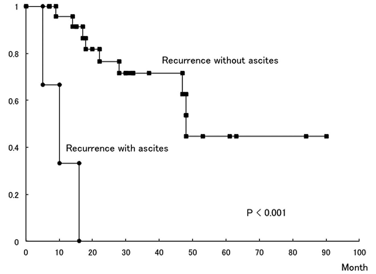
While the most frequent site of recurrence was
intraperitoneal dissemination, local pelvic peritoneal recurrence
(26.2%) and solitary recurrences in the lymph nodes (19.0%) and the
liver (16.7%) were relatively frequent (Table III). Secondary debulking surgery
(SDS) such as resection of the relapsed abdominal/pelvic mass or
lymph nodes, or partial hepatectomy was performed in as many as 44%
of patients with recurrence (Table
II). Conversely, recurrence was associated with ascites
(carcinomatous peritonitis) in just 4 of the relapsed 34 patients,
but all 4 patients died. The median survival time after recurrence
was 28 months: 7.5 and 30 months with and without ascites,
respectively (P<0.001) (Fig.
1).
| Table IIISites of recurrence. |
Table III
Sites of recurrence.
| Recurrent
sitea | No. of patients
(%) |
|---|
| Intraperitoneal
(with ascites) | 4 (9.5) |
| Intrapertitoneal
(without ascites) | 8 (19.0) |
| Pelvic cavity | 11 (26.2) |
| Lymph nodes | 8 (19.0) |
| Liver | 7 (16.7) |
| Lung | 3 (7.1) |
| Increase in CA-125
alone | 1 (2.5) |
Determination of factors predicting poor
outcome in fallopian tube malignancies
Univariate analysis by histological type, FIGO
stage, age, CA-125 level, operative procedure, completion rate,
recurrent site, presence or absence of SDS performance, and time to
recurrence was performed to assess predictive factors related to
the prognosis of FTM. Prognosis was significantly correlated solely
with whether SDS could be performed (Table IV).
| Table IVDetermination of factors predicting
poor outcome in fallopian tube malignancy. |
Table IV
Determination of factors predicting
poor outcome in fallopian tube malignancy.
| Factors | No. of
patients | Death events from
this disease | P-value |
|---|
| Histology |
| Carcinoma of the
fallopian tube | 60 | 12 | |
| Carcinosarcoma of
the fallopian tube | 8 | 2 | ns |
| Stage |
| I/II | 19 | 2 | |
| III/IV | 49 | 12 | ns |
| Age, years |
| ≥60 | 33 | 8 | |
| <60 | 35 | 6 | ns |
| CA-125 value |
| ≥330 | 33 | 7 | |
| <330 | 35 | 7 | ns |
| Completion of
surgery |
| Complete or
optimal | 44 | 10 | |
| Suboptimal | 24 | 4 | ns |
| Staging
laparotomy |
| Yes | 34 | 7 | |
| No | 34 | 7 | ns |
| Recurrent
sites |
|
Intraperitoneal | 12 | 6 | |
| Others | 30 | 8 | ns |
| Ascites in
recurrence |
| Yes | 4 | 4 | |
| No | 30 | 10 | ns |
| Secondary debulking
surgery |
| Yes | 15 | 1 | |
| No | 19 | 13 | <0.01 |
| Time to
recurrence |
| ≥6 months | 30 | 13 | |
| <6 months | 2 | 1 | ns |
Discussion
This study revealed that, although intraperitoneal
recurrence occurred in fallopian tube carcinoma and carcinosarcoma,
recurrence presented as carcinomatous peritonitis was noted in only
4 of the 34 patients with relapsed disease; isolated metastases to
the abdominal/pelvic cavity or lymph nodes and sole metastasis to
the liver were relatively frequent. Therefore, it appears that
post-recurrence survival was prolonged since in many patients SDS
such as excision of the relapsed abdominal/pelvic mass or the
relapsed lymph node or partial hepatic resection was indicated.
This fact was also supported by the univariate analysis which
revealed that prognosis was significantly correlated solely with
whether SDS was performed. Previous studies have shown that FTM
easily infiltrates surrounding organs and progresses aggressively
(1), and that lymph node metastasis
occurs more frequently than that in ovarian carcinoma (9). The present study, however, showed that
only 4 of the 34 patients with recurrent disease developed
carcinomatous peritonitis, demonstrating for the first time that
carcinomatous peritonitis is uncommon. It is already known in cases
of ovarian carcinoma that the prognosis of a solitary recurrence in
the liver is significantly improved by partial hepatic resection
(10). Thus, the results of the
present study suggest that SDS should be positively introduced to
recurrent FTMs unless patients develop carcinomatous
peritonitis.
With regard to the initial site of recurrence, FTM
shows contrasting findings with those for ovarian carcinoma
(Table III). While the present
results demonstrated a high frequency of lymph nodes recurrence
(19.0%) and solitary liver metastasis (16.7%), Ushijima reported
that lymph node metastases (7.1%) and solitary liver metastases
(6.3%) occur infrequently in ovarian carcinoma (11). FTM is richly permeated with
lymphatic channels that drain into the para-aortic lymph nodes
through infundibulopelvic lymphatics (12), and indeed a more frequent rate of
lymph node metastasis than ovarian carcinoma has been reported
(9,11). In addition, Ajithkumar et
al(13) found that metastasis
to the retroperitoneal lymph nodes was observed at the initial
visit in ~50% of patients, irrespective of the stage of disease
advancement. In this study as well, metastasis to the
retroperitoneal lymph nodes was observed in 16 of the 34 patients
(47.1%) on whom staging surgery was conducted initially (data not
shown).
It has been reported that platinum-based
chemotherapy for advanced or recurrent fallopian tube carcinoma
resulted in CR in 75% of patients (14) and that an 82.2% response rate was
obtained in progressive fallopian tube carcinoma (9). In this study, the combination therapy
with platinum and a taxane was chosen as a postoperative
chemotherapeutic regimen for most patients, and CR to postoperative
chemotherapy was 82% in the present patients, although suboptimal
surgery accounted for ~35% of all patients. There was no difference
in the number of deaths between patients who received optimal
surgery and those who received suboptimal surgery, suggesting that
aggressive postoperative chemotherapy was mandatory and the
combination of platinum and a taxane was effective, even in
patients undergoing suboptimal surgery. The results of the present
study were compatible with those of Baekelandt et
al(15) and Gemignani et
al(16), in which an 87.5%
response rate and significant prolongation of PFS were observed.
These reports and this study suggest that combination therapy with
platinum and a taxane is effective against FTM. Given that the
response rate to chemotherapy in epithelial ovarian carcinoma was
found to be 56% (17) and that the
rate for ovarian carcinosarcoma was 20 to 68% (18,19),
the favorable effect of initial therapy in FTM is noteworthy. This
favorable therapeutic effect may be attributed to the histological
type of fallopian tube carcinoma, which is largely composed of
serous and endometrioid adenocarcinoma.
Within Latzko’s triad, atypical genital bleeding was
observed in ~30% of patients in this study. If an adnexal tumor is
detected in patients with atypical genital bleeding or a watery
vaginal discharge, FTM should be considered as a differential
diagnosis. Nevertheless, a definitive preoperative diagnosis of FTM
is difficult in many patients. It has been reported that FTM is
diagnosed preoperatively in 0 to 10% of patients (20–22).
Moreover, Meng et al(23)
reported that in 50% of patients, even intraoperative findings fail
to identify that the primary site is the fallopian tube. In this
study, preoperative diagnosis was feasible in 13.2% of patients,
probably by virtue of advances in imaging diagnosis. Transvaginal
ultrasonography has been reported to be more effective than
abdominal ultrasonography (24),
and the presence of a sausage-like mass and/or multilobular mass
with a ‘cog-and-wheel’ appearance is considered to be the basis for
suspecting a FTM (25–27). Diagnostic ability has improved with
the development of color Doppler and three-dimensional Doppler
transvaginal ultrasonography (28).
However, findings that clearly discriminate FTM have not been
identified, even with full use of CT and MRI. Although this
provides no clinical specificity, the CA-125 level is increased in
80% or more of patients with fallopian tube carcinoma (13,29).
Although it has been reported occasionally that the preoperative
CA-125 level is an independent predictive factor for the prognosis
of fallopian tube carcinoma (13,30)
and that this level correlates with responsiveness to chemotherapy
(31,32), such a correlation with the prognosis
and therapeutic effect was not observed in this study. More cases
need to be accumulated to establish the clinical significance of
the CA-125 level in fallopian tube carcinoma.
A retrospective multicenter study was conducted
recently to clarify the prognosis of fallopian tube carcinoma.
Moore et al(33) used
matched, case-controlled methods to compare the prognosis of
fallopian tube carcinoma in 96 patients and epithelial ovarian
carcinoma in 189 patients. Comparison of stage I/II with a median
observation period of 57 months for fallopian tube carcinoma and 42
months for ovarian carcinoma showed a 5-year survival rate of 95%
for fallopian tube carcinoma and 76% for ovarian carcinoma
(P=0.02); a similar comparison in stage III/IV disease with a
median observation period of 33 months for fallopian tube carcinoma
and 35 months for ovarian carcinoma, showed a 3-year survival rate
that was comparable (59%) for both diseases. Accordingly, Moore
et al(33) re-evaluated the
concept that therapy for fallopian tube carcinoma could be similar
to that for ovarian carcinoma. Conversely, Pectasides et
al(34) analyzed 64 patients
with fallopian tube carcinoma and reported that advanced stage and
residual tumor diameter correlated significantly with prognosis. In
stage III/IV fallopian tube carcinoma, while the 5-year survival in
patients whose residual tumors could be debulked to less than 1 cm
was 55%, the 5-year survival of patients with larger residual tumor
deposits was 21% (9). Thus, optimal
debulking and subsequent platinum/taxan therapy were concluded to
confer a favorable prognosis. In addition, a large population-based
tumor registry study in which 416 patients with fallopian tube
carcinoma were included showed that the 5-year survival rate was
95% for stage I, 75% for stage II, 69% for stage III, and 45% for
stage IV, respectively, indicating that the prognosis was better
than the 5-year survival rate for each stage of ovarian carcinoma
evaluated at around the same time (35). More recently, using SEER database,
the outcome of 1,576 women with fallopian tube carcinoma and 54,249
with epithelial ovarian carcinoma was compared (4). Five-year cancer-specific survival was
54 and 36% in women with stage III and IV fallopian tube carcinoma,
respectively, vs. only 30 and 14% in those with the same stage
epithelial ovarian cancer, respectively (4). However, initially relapsed locations
and time to recurrence were not clarified in the SEER database
(4). The present results suggest
that there are many isolated recurrences and few recurrences of
carcinomatous peritonitis for FTMs. Thus, because there are many
cases of FTMs for which recurrent tumors can be extracted by
surgery, even if the disease relapsed, prognosis would be improved.
Although a hypothesis for the origin of ovarian and peritoneal
carcinoma has been proposed, based on the concept of transport and
implantation of malignant cells from the fallopian tube to the
ovary and peritoneum (36), the
results of this study suggest that FTM has a different biological
behavior when compared to ovarian carcinoma.
The present study suggests that FTM frequently
results in solitary recurrence, and that aggressive recurrence
treatment including SDS would improve the prognosis of this
disease. However, several limitations must be acknowledged in this
study. While differentiation of fallopian tube and ovarian
carcinoma for women with advanced stage disease is sometimes
difficult (37), a central
pathology review was not performed as a retrospective nature.
Although clinical features of fallopian tube carcinoma are not
distinct from those of ovarian carcinoma as reported using
case-control analysis (38), this
study did not adopt a case-control method. Further study to
elucidate the molecular mechanisms involved in the development of
FTM and to improve decision-making for disease treatment is
warranted.
References
|
1
|
Pectasides D, Pectasides E and
Economopoulos T: Fallopian tube carcinoma: a review. Oncologist.
11:902–912. 2006. View Article : Google Scholar : PubMed/NCBI
|
|
2
|
Rosenblatt KA, Weiss NS and Schwartz SM:
Incidence of malignant fallopian tube tumors. Gynecol Oncol.
35:236–239. 1989. View Article : Google Scholar : PubMed/NCBI
|
|
3
|
Riska A, Leminen A and Pukkala E:
Sociodemographic determinations of incidence of primary fallopian
tube carcinoma, Finland 1953–97. Int J Cancer. 104:643–645.
2003.PubMed/NCBI
|
|
4
|
Wethington SL, Herzog TJ, Seshan VE, et
al: Improved survival for fallopian tube cancer: a comparison of
clinical characteristics and outcome for primary fallopian tube and
ovarian cancer. Cancer. 113:3298–3306. 2008. View Article : Google Scholar
|
|
5
|
Sedlis A: Primary carcinoma of the
fallopian tube. Obstet Gynecol Surv. 16:209–226. 1961. View Article : Google Scholar : PubMed/NCBI
|
|
6
|
World Health Organization Classification
of Tumours. Pathology & Genetics. Tumours of the Breast and
Female Genital Organs. IARC Press; Lyon: 2003
|
|
7
|
Hoskins WJ, McGuire WP, Brady MF, et al:
The effect of diameter of largest residual disease on survival
after primary cytoreductive surgery in patients with suboptimal
residual epithelial ovarian carcinoma. Am J Obstet Gynecol.
170:974–979. 1994. View Article : Google Scholar
|
|
8
|
Eisenhauer EA, Therasse P, Bogaerts J, et
al: New response evaluation criteria in solid tumours: revised
RECIST guideline (version 1.1). Eur J Cancer. 45:228–247. 2009.
View Article : Google Scholar
|
|
9
|
Gadducci A, Landoni F, Sartori E, et al:
Analysis of treatment failures and survival of patients with
fallopian tube carcinoma: a Cooperation Task Force (CTF) study.
Gynecol Oncol. 81:150–159. 2001. View Article : Google Scholar : PubMed/NCBI
|
|
10
|
Roh HJ, Kim DY, Joo WD, et al: Hepatic
resection as part of secondary cytoreductive surgery for recurrent
ovarian cancer involving the liver. Arch Gynecol Obstet.
284:1223–1229. 2011. View Article : Google Scholar : PubMed/NCBI
|
|
11
|
Ushijima K: Treatment of recurrent ovarian
cancer - at first relapse. J Oncol. 2010:4974292010. View Article : Google Scholar : PubMed/NCBI
|
|
12
|
Klein M, Rosen AC, Lahousen M, et al:
Lymphadenectomy in primary carcinoma of the fallopian tube. Cancer
Lett. 147:63–66. 1999. View Article : Google Scholar : PubMed/NCBI
|
|
13
|
Ajithkumar TV, Minimole AL, John MM and
Ashokkumar OS: Primary fallopian tube carcinoma. Obstet Gynecol
Surv. 60:247–252. 2005. View Article : Google Scholar
|
|
14
|
Peter WA III, Andersen WA, Hopkins MP, et
al: Prognostic features of carcinoma of the fallopian tube. Obstet
Gynecol. 71:757–762. 1988.PubMed/NCBI
|
|
15
|
Baekelandt M, Joruun Nesbakken A,
Kristensen GB, et al: Carcinoma of the fallopian tube. Cancer.
89:2076–2084. 2000. View Article : Google Scholar : PubMed/NCBI
|
|
16
|
Gemignani ML, Hensley ML, Cohen R, et al:
Paclitaxel-based chemotherapy in carcinoma of the fallopian tube.
Gynecol Oncol. 80:16–20. 2001. View Article : Google Scholar : PubMed/NCBI
|
|
17
|
Katsumata N, Yasuda M, Takahashi F, et al:
Dose-dense paclitaxel once a week in combination with carboplatin
every 3 weeks for advanced ovarian cancer: a phase 3, open-label,
randomised controlled trial. Lancet. 374:1331–1338. 2009.
View Article : Google Scholar
|
|
18
|
Anderson B, Turner DA and Benda J: Ovarian
sarcoma. Gynecol Oncol. 26:183–192. 1987. View Article : Google Scholar
|
|
19
|
Tate Thigpen J, Blessing JA, DeGeest K, et
al: Cisplatin as initial chemotherapy in ovarian carcinosarcima: a
Gynecologic Oncology Group study. Gynecol Oncol. 93:336–339.
2004.
|
|
20
|
Eddy GL, Copeland LJ and Gershenson DM:
Second-look laparotomy in fallopian tube carcinoma. Gynecol Oncol.
19:182–186. 1984. View Article : Google Scholar : PubMed/NCBI
|
|
21
|
Podratz KC, Podczaski ES, Gaffey TA, et
al: Primary carcinoma of the fallopian tube. Am J Obstet Gynecol.
154:1319–1326. 1986. View Article : Google Scholar : PubMed/NCBI
|
|
22
|
Huber-Buchholz MM, Buchholz NP and
Staehelin J: Analysis of 23 cases of primary carcinoma of the
fallopian tube over 50 years. J Obstet Gynaecol Res. 22:193–199.
1996.PubMed/NCBI
|
|
23
|
Meng ML, Gan-Gao, Scheng-Sun, et al:
Diagnosis of primary adenocarcinoma of the fallopian tube. J Cancer
Res Clin Oncol. 110:136–140. 1985. View Article : Google Scholar : PubMed/NCBI
|
|
24
|
Timor-Tritsch IE and Rottem S:
Transvaginal ultrasonographic study of the fallopian tube. Obstet
Gynecol. 70:424–428. 1987.PubMed/NCBI
|
|
25
|
Subramanyam BR, Raghavendra BN, Whalen CA,
et al: Ultrasonic features of fallopian tube carcinoma. J
Ultrasound Med. 3:391–393. 1984.PubMed/NCBI
|
|
26
|
Kol S, Gal D, Friedman M and Paldi E:
Preoperative diagnosis of fallopian tube carcinoma by transvaginal
sonography and CA-125. Gynecol Oncol. 37:129–131. 1990. View Article : Google Scholar : PubMed/NCBI
|
|
27
|
Ajjimakorn S, Bhamarapravati Y and
Israngura N: Ultrasound appearance of fallopian tube carcinoma. J
Clin Ultrasound. 16:516–518. 1988. View Article : Google Scholar : PubMed/NCBI
|
|
28
|
Kurjak A, Kupesic S, Sparac V and Kosuta
D: Three-dimensional ultrasonographic and power Doppler
characterization of ovarian lesions. Ultrasound Obstet Gynecol.
16:365–371. 2000. View Article : Google Scholar : PubMed/NCBI
|
|
29
|
McMurray EH, Jacobs AJ, Perez CA, et al:
Carcinoma of the fallopian tube. Management and sites of failure.
Cancer. 58:2070–2075. 1986. View Article : Google Scholar : PubMed/NCBI
|
|
30
|
Rosen AC, Klein M, Hafner E, et al:
Management and prognosis of primary fallopian tube carcinoma.
Austrian Cooperative Study Group for Fallopian Tube Carcinoma.
Gynecol Obstet Invest. 47:45–51. 1999. View Article : Google Scholar : PubMed/NCBI
|
|
31
|
Tokunaga T, Miyazaki K, Matsuyama S, et
al: Serial measurement of CA 125 in patients with primary carcinoma
of the fallopian tube. Gynecol Oncol. 36:335–337. 1990. View Article : Google Scholar : PubMed/NCBI
|
|
32
|
Hefler LA, Rosen AC, Graf AH, et al: The
clinical value of serum concentrations of cancer antigen 125 in
patients with primary fallopian tube carcinoma: a multicenter
study. Cancer. 89:1555–1560. 2000. View Article : Google Scholar : PubMed/NCBI
|
|
33
|
Moore KN, Moxley KM, Fader AN, et al:
Serous fallopian tube carcinoma: a retrospective,
multi-institutional case-control comparison to serous
adenocarcinoma of the ovary. Gynecol Oncol. 107:398–403. 2007.
View Article : Google Scholar : PubMed/NCBI
|
|
34
|
Pectasides D, Pectasides E, Papaxoinis G,
et al: Primary fallopian tube carcinoma: results of a retrospective
analysis of 64 patients. Gynecol Oncol. 115:97–101. 2009.
View Article : Google Scholar : PubMed/NCBI
|
|
35
|
Kosary C and Trimble EL: Treatment and
survival for women with fallopian tube carcinoma: a
population-based study. Gynecol Oncol. 86:190–191. 2002. View Article : Google Scholar
|
|
36
|
Kurman RJ and Shih IeM: Molecular
pathogenesis and extraovarian origin of epithelial ovarian cancer -
shifting the paradigm. Hum Pathol. 42:918–931. 2011. View Article : Google Scholar : PubMed/NCBI
|
|
37
|
Berek JS, Crum C and Friedlander M: Cancer
of the ovary, fallopian tube, and peritoneum. Int J Gynaecol
Obstet. 119:S118–S129. 2012. View Article : Google Scholar : PubMed/NCBI
|
|
38
|
Dunn MS, Manahan KJ and Geisler JP:
Primary carcinoma of the fallopian tube and epithelial ovarian
carcinoma: a case-control analysis. J Reprod Med. 53:691–694.
2008.PubMed/NCBI
|