Introduction
The stem and root bark of Ulmus davidiana
var. japonica (UJ) are Korean herbal medicines which contain
many biologically active compounds. Recent studies have shown that
UJ has an immunomodulating effect and vasorelaxing activity in
vitro and in vivo(1,2). The
major constituents of UJ include flavan-3-ols [(+)-catechin,
(+)-catechin 7-O-β-D-apiofuranoside, (+)-catechin
7-O-β-D-xylopyranoside, and (+)-catechin 7-O-β-D-glucopyranoside],
triterpene esters, lignan, trihydroxy fatty acid and
polysaccharides.
Catechin, one of the major components of the
flavan-3-ols, shows efficacy against many cancer types (3–5).
Catechin-7-O-glucoside is a flavan-3-ol glycoside formed from
catechin and is found in natural traditional drugs such as in the
roots of the Chinese peony or is found in foods such as Korean
plum-yew (6). However, the
molecular mechanism of its selective anticancer role is not clearly
understood.
Apoptosis plays a critical role in the development
and homeostasis of eukaryotic cells, and impairment of apoptotic
function has been associated with several types of human diseases,
including cancer and neurodegenerative disorders (7–9).
Apoptosis is mediated by the caspases, a conserved family of
aspartate-specific cysteine proteases, that can be activated by the
mitochondrial pathway (7).
Recently, increasing evidence has identified that the apoptotic
pathway is linked to the endoplasmic reticulum (ER) stress
(10). ER stress is induced by
autophagy, oxidative stress and calcium depletion (11–13).
The start of ER stress-induced apoptosis occurs through unfolded
protein response signaling and involves transcriptional activation
of the proapoptotic transcription factor CCAAT/enhancer-binding
protein (C/EBP) homologous protein (CHOP) (14). CHOP is a key component in ER
stress-mediated apoptosis. For example, CHOP acts to downregulate
anti-apoptotic B-cell lymphoma 2 (Bcl-2) protein (15).
Catechin-7-O-xyloside (C7Ox) is a pentose analog of
catechin-7-O-glucoside. The cytotoxic effects of C7Ox and the
mechanism by which C7Ox exerts its cytotoxic effect remain largely
unknown in cancer cells including non-small cell lung cancer
(NSCLC) cells. In the present study, we examined the anticancer
effects and molecular mechanisms of C7Ox in H1299 cancer cells. Our
results suggest that C7Ox induces apoptosis via the loss of
mitochondrial membrane potential and caspase-6 activation, and that
the ER stress pathway is important in C7Ox-induced apoptotic cell
death in human lung tumor H1299 cells.
Materials and methods
Compound preparation and the biochemical
reagents
U. davidiana var. japonica (UJ) powder
was ground to ultrafine particle size using an herbal medicine
pulverizer (Delsa™Nano; Beckman Coulter Inc., Brea, CA, USA).
Catechin-7-O-xyloside (C7Ox) was obtained through purification of
an ethanol extract of UJ by using a Sep-Pak cartridge (Waters,
Milford, MA, USA). The water-soluble tetrazolium salt (WST)-8 cell
proliferation assay kit was obtained from Dojindo Laboratories
(Kumamoto, Japan). Lactate dehydrogenase (LDH) cytotoxicity assay
and caspase-6 colorimetric assay kits were purchased from Cayman
Chemical Co. (Ann Arbor, MI, USA) and Abcam (Cambridge, MA, USA),
respectively. The Annexin V/PI apoptosis detection kit was from BD
Biosciences (Bedford, MA, USA). Primary antibodies for
cleaved-caspase-6, cleaved-poly(ADP-ribose) polymerase (PARP), CHOP
and tubulin, and the secondary antibodies were obtained from Cell
Signaling Technology (Beverly, MA, USA).
LC-MS/MS analysis
The extract was dissolved in ethanol at a
concentration of 10 mg/ml and diluted with 50% ethanol to a final
concentration of 2 mg/ml, and 2 μl was analyzed using liquid
chromatography followed by tandem mass spectrometry (LC-MS/MS).
LC-MS/MS was performed using an LTQ Orbitrap XL ion trap mass
spectrometer (Thermo Fisher Scientific, Waltham, MA, USA) equipped
with a heated electrospray ionization source. Separation by ultra
HPLC (UHPLC) was performed on a Thermo Accela LC system by using an
Acquity BEH C18 column (1.7 μm, 2.1 × 150 mm; Waters). Mobile phase
A contained water and mobile phase B contained acetonitrile; both
contained 0.1% formic acid. Gradient elution at a flow rate of 0.4
ml/min was carried out as follows: 0–1 min with 1–5% B (linear
gradient) and 1–10 min with 5–20% B (linear gradient). Full-scan
mass spectra were obtained in the negative ion modes at a range
m/z 100–1000. To identify the structures of the compounds,
the data obtained from tandem mass spectrometry (MS/MS) analysis
were compared with those from an MS/MS spectral library search
(16).
Cell culture
The non-small cell lung cancer (NSCLC) cell line
H1299 was purchased from the American Type Culture Collection
(ATCC; Manassas, VA, USA). Cells were grown in RPMI-1640 medium
containing 10% fetal bovine serum (FBS), 100 U/ml penicillin and
100 μg/ml streptomycin (Gibco-BRL/Life Technologies) in a 5%
CO2 incubator at 37°C.
Determination of cytotoxicity and plasma
membrane integrity
Cell cytotoxicity was assessed by measuring the
optical density at 450 nm with a microplate reader (SpectraMax
190®; Molecular Devices Corp., Sunnyvale, CA, USA) 2 h
after the addition of
2-(2-methoxy-4-nitrophenyl)-3-(4-nitrophenyl)-5-(2,4-disulfophenyl)-2
H-tetrazolium (WST-8) reagent solution according to the
manufacturer’s guidelines. Plasma membrane integrity was assessed
based on lactate dehydrogenase (LDH) leakage into the culture
medium from cells. LDH leakage was determined by measuring the
optical density at 490 nm.
Determination of apoptosis by
fluorescence-activated cell sorting (FACS) analysis
After C7Ox treatment, cells were harvested and
stained with propidium iodide (PI) and Annexin V (BD Biosciences)
for 15 min at room temperature in binding buffer and then analyzed
using flow cytometry (BD Biosciences). PI and Annexin V emissions
were detected in the FL-2 and FL-1 channels, respectively. For each
sample, data from 10,000 cells were recorded in list mode on
logarithmic scales. Data analysis was conducted using CellQuest
software (BD Biosciences).
Assessment of mitochondrial membrane
potential
Mitochondrial membrane potential was assessed using
the cationic dye JC-1
(5,5′,6,6′-tetrachloro-1,1′,3,3′-tetraethylbenzimidazol-carbocyanine
iodide) according to the manufacturer’s instructions (Molecular
Probes, Eugene, OR, USA). Images were collected using a Zeiss LSM
510 fluorescence photomicroscope (Carl Zeiss, Oberkochen, Germany).
Visualization of JC-1 monomers (green fluorescence) and JC-1
aggregates (red fluorescence) was carried out using filter sets for
fluorescein and rhodamine dyes, respectively, and analyzed using
ImageJ software.
Detection of caspase-6 activation
A caspase-6 activity assay was conducted using
substrates of the color reporter molecule
Val-Glu-Ile-Asp-p-nitroaniline (VEID-pNA), which is specific for
caspase-6. Briefly, H1299 cells were collected for sampling and
lysed on ice by using lysis buffer containing protease inhibitors.
Lysates were centrifuged at 13,000 rpm for 10 min at 4°C, and the
supernatant collected was used for the assay. Caspase-6 activity
was measured using a microplate reader at 405 nm.
Immunoblotting
Total cell lysate and total tissue protein from lung
cancer xenografts of mice were prepared according to a previous
study (17). Equal amounts of
protein were resolved using sodium dodecyl sulfate-polyacrylamide
gel electrophoresis (SDS-PAGE) and transferred to polyvinylidene
fluoride (PVDF) membranes (Bio-Rad Laboratories, Hercules, CA,
USA). Membranes were probed with antibodies against cleaved
caspase-6, poly(ADP-ribose) polymerase (PARP), CHOP, tubulin and
secondary antibodies were detected using Pierce ECL-Plus
chemiluminescence kit (Thermo Fisher Scientific).
Gene silencing using small interfering
RNA (siRNA) transfection
CHOP antisense oligonucleotides (GTCCTGTC
TTCAGATGAATT) were synthesized by Genolution Pharmaceuticals, Inc.
(Seoul, Korea). Irrelevant scrambled siRNA was used as a control.
Cells were transfected with the siRNAs using Lipofectamine reagent
(Invitrogen, Carlsbad, CA, USA) according to the manufacturer’s
instructions.
Xenograft experiment
For the tumor model, H1299 cells (1×10⁶)
were subcutaneously injected into the right flank of female BALB/c
nude mice (6 weeks of age) by using a 28-gauge needle. One week
later, after the appearance of implanted tumors, the mice were
randomly divided into 2 groups: C7Ox group and vehicle group (n=5
mice per group). C7Ox was dissolved in ethanol (EtOH) and diluted
with phosphate-buffered saline (PBS) (EtOH:PBS=2:8). C7Ox or
vehicle (ie, 20% EtOH in PBS) was intraperitoneally administered
once daily for 1 month. Tumor size was calculated by measuring the
length and width of the tumor with a caliper. Tumor volume was
calculated as follows: Tumor volume (mm3) = length ×
width2/2. Next, tumor tissues were collected and frozen
at −80°C until use.
RNA extraction and reverse
transcriptase-PCR (RT-PCR)
Total RNA from the tumor tissues was extracted using
TRIzol reagent (Invitrogen). cDNA was synthesized using the
ImProm-II™ reverse transcription system (Promega, Madison, WI, USA)
according to the manufacturer’s protocol. RT-PCR was performed
using a Solgent PCR detection kit following the manufacturer’s
instructions. Glyceraldehyde 3-phosphate dehydrogenase (GAPDH) was
used as an internal control. Primers used included the following:
CHOP forward, GACCT GCAAGAGGTCCTG and reverse, CTACTTCCCTGGTCA GGC;
GAPDH forward, 5′-GGGCTCATCTGAAGGGTGGT GCTA-3′ and reverse,
5′-GTGGACGCTGGGATGATGTTC TGG-3′. The following PCR cycle was used:
1 min of denaturation at 92°C, 30 sec of annealing at 58°C and 1
min of extension at 72°C. PCR was conducted for 28 cycles. PCR
products were run on a 0.8% agarose gel and visualized under UV
illumination.
Statistical analysis
The data are expressed as the means ± standard error
(SE) of at least three independent experiments. Student’s t-test
was used to assess differences between the two groups. The level of
significance was set at P<0.01.
Results
C7Ox purification from ultrafine U.
davidiana var. japonica (UJ) ethanol extract and LC-MS/MS
analysis
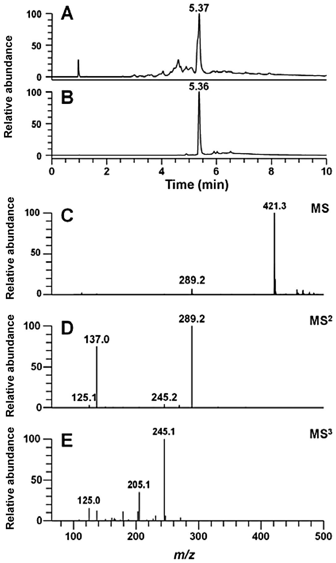
The 50-mg ethanol extract was loaded on Sep-Pak
cartridges and eluted stepwise using 0, 10, 20, 30, 40 and 50%
water-ethanol solvent (15 ml each). The 30% fraction was collected
and concentrated for the next experiment. The purity of the
isolated C7Ox was found to be 87% using ultra high-pressure liquid
chromatography (UHPLC) at 280 nm (Fig.
1B). The ultrafine UJ ethanol extract was characterized for its
major constituent compounds by simultaneous estimation using
negative ion-mode tandem mass analysis (MS2 and
MS3). The major peak was identified as C7Ox
([M-H]−, m/z (mass-to-charge ratio) of 421.3) at
RT at 5.37 min (Fig. 1). The sugar
moiety was defined using MS2 fragment ion analysis.
Indeed, in the MS2 spectrum, neutral losses of
m/z 132 indicated the loss of a pentose. The aglycone
structures of MS3 spectra were identified as catechin by
MS/MS spectrum matching (16).
C7Ox induces cell death and increases LDH
release
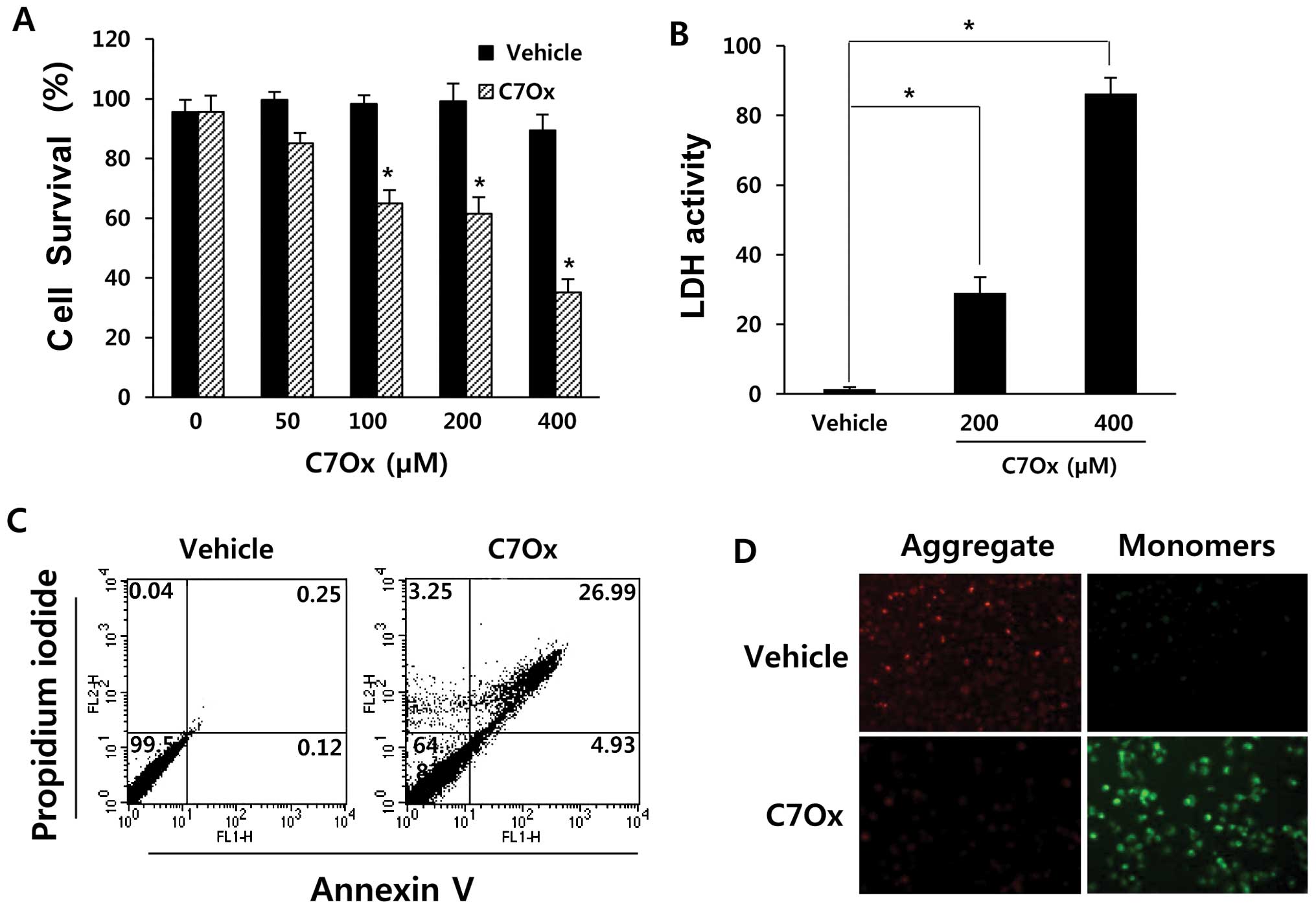
To investigate the cytotoxic effect of C7Ox on
cancer cells, we used the WST assay to monitor the survival rate of
C7Ox-exposed H1299 cells. Treatment of H1299 cells with C7Ox for 48
h caused dose-dependent decreases in cell survival (P<0.01;
Fig. 2A). Next, we investigated
whether C7Ox induces LDH release by measuring the activity of LDH
released from the cytosol of damaged cells into the medium. As
shown in Fig. 2B, LDH release into
the culture medium was significantly elevated after C7Ox treatment
in H1299 cells when compared with that in the vehicle-treated cells
(P<0.01). These results indicated that C7Ox-treated cells
underwent postapoptotic necrosis.
C7Ox causes H1299 cell apoptosis
To determine whether this agent induces typical
cancer cell apoptosis as well as necrosis, we treated H1299 cells
with 400 μM C7Ox for 30 h. After treatment, the cells were
harvested and apoptotic cells were examined by FACS analysis using
Annexin V/PI staining. As shown in Fig.
2C, C7Ox treatment increased the early apoptotic population
(stained by Annexin V only; quadrant 4) from 0.12 to 4.93% as well
as the necrotic population (stained by PI only; quadrant 2) from
0.04 to 3.25% or the late apoptotic population (stained by Annexiv
V + PI; quadrant 1) from 0.25 to 26.99%. These results indicated
that the proportion of apoptotic cells as well as necrotic cells
was significantly increased after C7Ox treatment.
C7Ox induces mitochondrial membrane
potential collapse
Loss of mitochondrial membrane potential has been
linked to the initiation and activation of the apoptotic process
(18). To evaluate whether C7Ox
triggers mitochondrial injury, the JC-1 probe was used to evaluate
the changes in mitochondrial membrane potential during C7Ox
treatment. In healthy cells, the dye accumulates in the
mitochondria as aggregates with red fluorescence, whereas in
apoptotic or dead cells, the dye remains in the cytosol as monomers
with green fluorescence. As shown in Fig. 2D, C7Ox-treated cells exhibited a
reduction in red emission intensity and a concomitant increase in
green emission intensity when compared to vehicle-treated cells.
These results indicated that C7Ox treatment induced H1299 cell
apoptosis by disrupting the mitochondrial membrane potential.
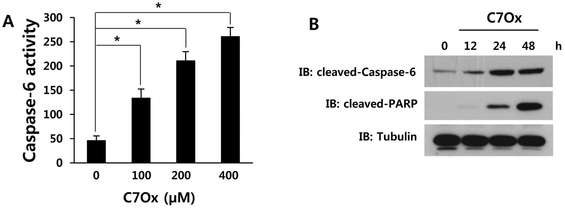
C7Ox induces proteolytic caspase-6
activation and the cleavage of PARP
Caspase activation and degradation of caspase
substrates are key markers of apoptosis (7). We confirmed the onset of apoptosis by
examining the protease activity using a colorimetric substrate
(VEID-pNA) specific for caspase-6. As shown in Fig. 3A, C7Ox activated caspase-6 in a
dose-dependent manner (P<0.01). We also examined whether the
expression levels of apoptosis-related proteins were affected by
C7Ox treatment at various time-points. Western blotting showed that
C7Ox significantly increased the cleavages of caspase-6 and PARP
proteins in H1299 cells (Fig.
3B).
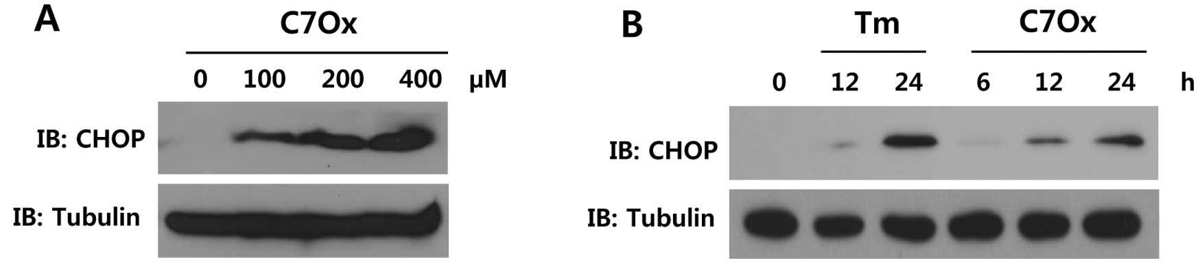
C7Ox triggers CHOP expression
Apoptotic cell death can be triggered by ER stress,
and several pathways have been directly implicated in ER
stress-induced apoptosis (10). One
of the major components of the ER stress-mediated apoptotic pathway
is CHOP expression (19). To
investigate whether C7Ox treatment causes ER stress, CHOP
expression was measured. Our results showed that C7Ox induced an
increase in CHOP expression in a dose- and time-dependent manner
(Fig. 4A and B).
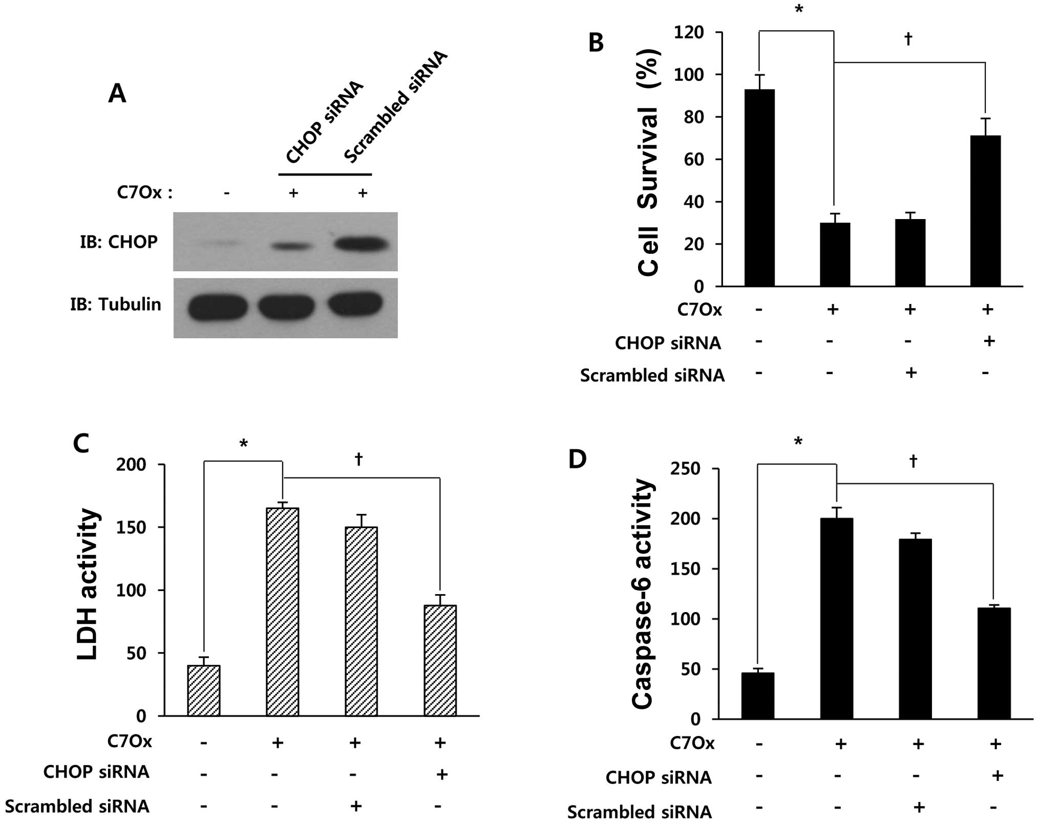
Inhibition of CHOP attenuates
C7Ox-induced cell death, LDH release and caspase-6 activation
Next, to evaluate the role of CHOP in C7Ox-induced
cytotoxicity and apoptosis, we examined the effect of the knockdown
of CHOP expression on cell viability, LDH release and caspase-6
activity. As shown in Fig. 5A, C7Ox
treatment increased CHOP expression, and CHOP siRNA, but not
scrambled siRNA, significantly downregulated CHOP expression
providing evidence for the specificity of siRNA inhibition. C7Ox
treatment significantly reduced cell viability, increased LDH
release and caspase-6 activity, as expected (P<0.01; Fig. 5B–D). However, downregulation of CHOP
expression by CHOP siRNA significantly attenuated C7Ox
treatment-induced cell death, LDH release and caspase-6 activation
(P<0.01; Fig. 5B–D). C7Ox
treatment-induced apoptosis, measured by Annexin V/PI staining, was
also attenuated by CHOP siRNA (data not shown). These results
suggest that ER stress is responsible for C7Ox-induced apoptotic
cell death and caspase-6 activation.
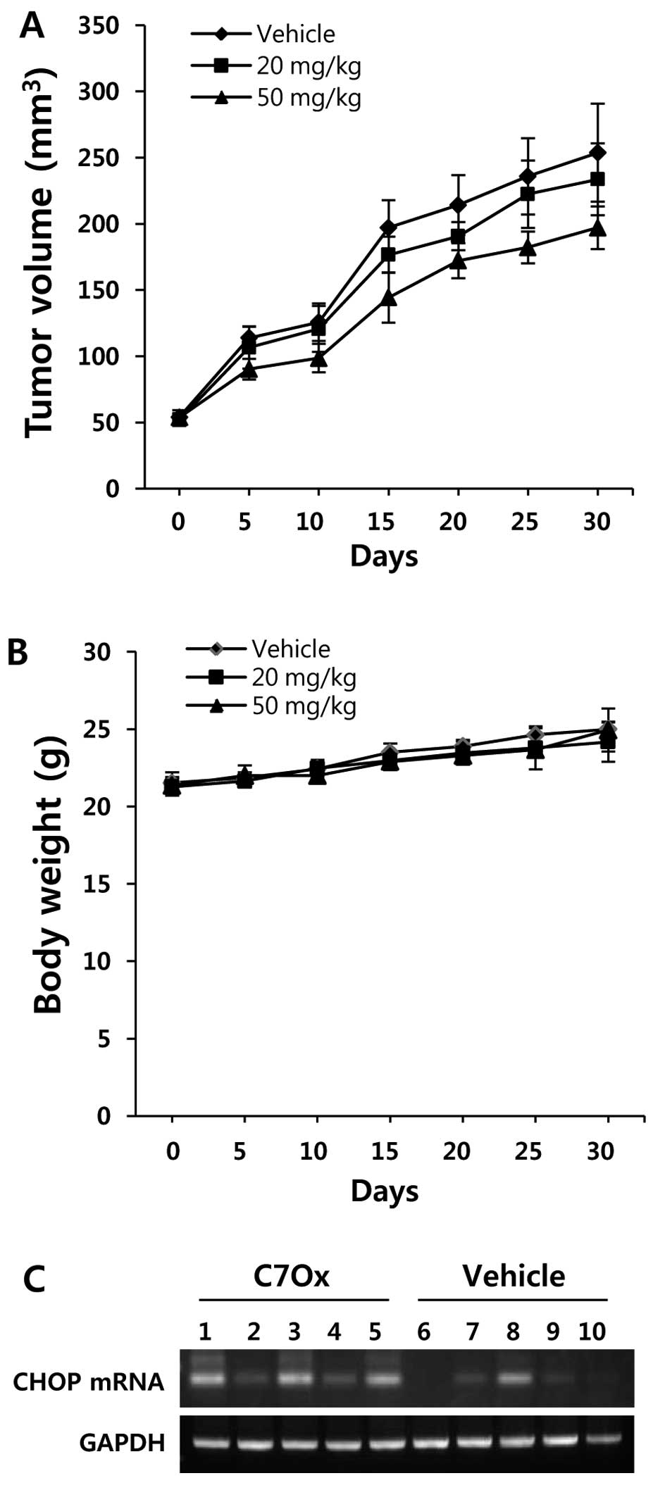
Inhibition of tumor growth in a lung
carcinoma xenograft model by C7Ox treatment
To further evaluate the anticancer activity of C7Ox
in vivo, we treated BALB/c nude mice bearing subcutaneous
H1299 cell-derived tumors with either C7Ox (20 mg or 50 mg/kg) or
vehicle. Tumor growth was inhibited by C7Ox as compared to the
vehicle (Fig. 6A) and there was no
apparent change in body weight in the C7Ox-injected animals
(Fig. 6B). We also measured the
mRNA level of CHOP within the tumor tissue. CHOP mRNA levels were
significantly upregulated in the tissues obtained from the
C7Ox-treated group when compared with levels obtained from the
vehicle-treated group (Fig. 6C).
The cleaved caspase-6 protein expression was also upregulated in
the tissues obtained from the C7Ox-treated group (data not shown).
Our results suggest that C7Ox may be a potential antitumor agent
for NSCLC and that ER stress signals are involved in the anticancer
activity of C7Ox.
Discussion
We determined whether catechin-7-O-xyloside (C7Ox),
the major active constituent purified from the ethanol extract of
U. davidiana var. japonica (UJ), demonstrates
anticancer effects and examined its working mechanisms. Although
some catechins have antitumor effects (5,20), the
effects of catechin derivatives such as catechin-7-O-glucoside or
C7Ox on tumors remain unclear. In the present study, we showed that
C7Ox induced apoptosis in human H1299 NSCLC cells. We also gained
insights into the signaling mechanisms underlying C7Ox-induced
apoptosis in this cell line. We monitored the dose response of
C7Ox-induced cell death and the elevated release of LDH into the
culture medium as indices of cytoplasmic membrane damage and loss
of membrane integrity in H1299 cells. The cell viability assay,
detection of LDH release, Annexin V/PI staining, caspase-6 activity
assay, and western blotting for caspase-6 and PARP showed that C7Ox
induced apoptotic cell death as well as post-apoptotic (necrotic)
cell death in NSCLC.
Since changes in mitochondrial membrane potential
have been directly associated with apoptosis, and a decrease in
mitochondrial membrane potential results in the release of
cytochrome c from impaired mitochondria into the cytosol,
resulting in apoptosis (8), we
evaluated changes in the mitochondrial membrane potential in NSCLC
cells during C7Ox treatment. The decrease in mitochondrial membrane
potential, observed using JC-1 dye in the present study, indicated
that C7Ox induced apoptosis via a mitochondrial-dependent
pathway.
The ER is significantly involved in protein
synthesis, maturation and calcium storage in mammalian cells
(21). Perturbation of the ER
function leads to ER stress, and prolonged ER stress can activate
apoptotic pathways in damaged cells (13,15).
Therefore, pharmacological interventions that promote cancer cell
death through ER stress are attractive options for anticancer
therapy (22,23). The transcription factor C/EBP
homologous protein (CHOP), an ER stress marker protein, is induced
by ER stress and is involved in ER stress-induced apoptosis
(19). Treatment of cells with C7Ox
was found to cause a dose- and time-dependent increase in the
levels of CHOP, indicating that C7Ox can induce ER stress in cancer
cells. We also examined the contribution of ER stress to
C7Ox-induced apoptotic cell death by using an siRNA that targets
CHOP. We found that CHOP siRNA, but not control siRNA,
significantly reduced C7Ox-induced cell death, LDH release and
caspase-6 activation. CHOP siRNA also suppressed C7Ox-induced
apoptosis as assessed by Annexin V/PI staining (data not shown).
These findings suggest that C7Ox-induced apoptosis, post-apoptotic
necrosis, and caspase-6 activation in human lung cancer cells
involve the ER stress pathway.
At a clinically feasible concentration (50 mg/kg),
C7Ox significantly delayed H1299 tumor growth in a nude mouse
xenograft model, supporting clinical application for anticancer
therapy. Notably, CHOP mRNA levels were significantly higher in
samples taken from xenografts treated with C7Ox than in samples
taken from the vehicle-treated groups. Caspase-6 protein expression
was also higher in xenografts treated with C7Ox (data not shown).
Collectively, these data indicated that C7Ox inhibited human lung
tumor growth in the mouse xenograft model by triggering ER
stress-mediated and caspase-6-mediated apoptosis.
In summary, we demonstrated that C7Ox, a derivative
of catechin, induced tumor cell death via both apoptosis and
necrosis in vitro and in vivo. Our findings also
suggest that C7Ox triggers ER stress signals and that ER stress
molecules such as CHOP contribute to C7Ox-induced apoptotic cell
death and caspase-6 activation. Many components from Ulmus
davidiana var. japonica are relatively non-toxic. C7Ox
may, therefore, be a potential chemotherapeutic candidate for
treating human cancers, including lung cancer.
Acknowledgements
The present study was supported by the Basic Science
Research Program through the National Research Foundation of Korea
(NRF) funded by the Ministry of Education, Science and Technology
(2012R1A1A3003467). J.W.Y. was supported by RP-Grant 2010 of Ewha
Womans University.
References
|
1
|
Lee EH, Park CW and Jung YJ:
Anti-inflammatory and immune-modulating effect of Ulmus
davidiana var. japonica Nakai extract on a macrophage
cell line and immune cells in the mouse small intestine. J
Ethnopharmacol. 146:608–613. 2013.PubMed/NCBI
|
|
2
|
Cho EJ, Park MS, Kim SS, et al:
Vasorelaxing activity of Ulmus davidiana ethanol extracts in
rats: activation of endothelial nitric oxide synthase. Korean J
Physiol Pharmacol. 15:339–344. 2011.
|
|
3
|
Li JJ, Gu QH, Li M, Yang HP, Cao LM and Hu
CP: Role of Ku70 and Bax in epigallocatechin-3-gallate-induced
apoptosis of A549 cells in vivo. Oncol Lett. 5:101–106.
2013.PubMed/NCBI
|
|
4
|
Hsu YC and Liou YM: The anti-cancer
effects of (−)-epigallocatechin-3-gallate on the signaling pathways
associated with membrane receptors in MCF-7 cells. J Cell Physiol.
226:2721–2730. 2011.
|
|
5
|
Singh BN, Shankar S and Srivastava RK:
Green tea catechin, epigallocatechin-3-gallate (EGCG): mechanisms,
perspectives and clinical applications. Biochem Pharmacol.
82:1807–1821. 2011. View Article : Google Scholar : PubMed/NCBI
|
|
6
|
Yoon KD, Jeong DG, Hwang YH, Ryu JM and
Kim J: Inhibitors of osteoclast differentiation from
cephalotaxus koreana. J Nat Prod. 70:2029–2032. 2007.
View Article : Google Scholar : PubMed/NCBI
|
|
7
|
Elmore S: Apoptosis: a review of
programmed cell death. Toxicol Pathol. 35:495–516. 2007. View Article : Google Scholar : PubMed/NCBI
|
|
8
|
Lowe SW and Lin AW: Apoptosis in cancer.
Carcinogenesis. 21:485–495. 2000. View Article : Google Scholar
|
|
9
|
Mattson MP: Apoptosis in neurodegenerative
disorders. Nat Rev Mol Cell Biol. 1:120–129. 2000. View Article : Google Scholar
|
|
10
|
Gorman AM, Healy SJ, Jäger R and Samali A:
Stress management at the ER: regulators of ER stress-induced
apoptosis. Pharmacol Ther. 134:306–316. 2012. View Article : Google Scholar : PubMed/NCBI
|
|
11
|
Kim SJ, Hong EH, Lee BR, et al:
α-Mangostin reduced ER stress-mediated tumor growth through
autophagy activation. Immune Netw. 12:253–260. 2012.
|
|
12
|
Bhandary B, Marahatta A, Kim HR and Chae
HJ: An involvement of oxidative stress in endoplasmic reticulum
stress and its associated diseases. Int J Mol Sci. 14:434–456.
2012. View Article : Google Scholar : PubMed/NCBI
|
|
13
|
Mekahli D, Bultynck G, Parys JB, De Smedt
H and Missiaen L: Endoplasmic-reticulum calcium depletion and
disease. Cold Spring Harb Perspect Biol. 3:1–30. 2011. View Article : Google Scholar
|
|
14
|
Zinszner H, Kuroda M, Wang X, et al: CHOP
is implicated in programmed cell death in response to impaired
function of the endoplasmic reticulum. Genes Dev. 12:982–995. 1998.
View Article : Google Scholar : PubMed/NCBI
|
|
15
|
Xu C, Bailly-Maitre B and Reed JC:
Endoplasmic reticulum stress: cell life and death decisions. J Clin
Invest. 115:2656–2664. 2005. View
Article : Google Scholar : PubMed/NCBI
|
|
16
|
Lee JS, Kim DH, Liu KH, Oh TK and Lee CH:
Identification of flavonoids using liquid chromatography with
electrospray ionization and ion trap tandem mass spectrometry with
an MS/MS library. Rapid Commun Mass Spectrom. 19:3539–3548. 2005.
View Article : Google Scholar : PubMed/NCBI
|
|
17
|
McIntyre A, Patiar S, Wigfield S, et al:
Carbonic anhydrase IX promotes tumor growth and necrosis in vivo
and inhibition enhances anti-VEGF therapy. Clin Cancer Res.
18:3100–3111. 2012. View Article : Google Scholar : PubMed/NCBI
|
|
18
|
Tsujimoto Y and Shimizu S: Role of the
mitochondrial membrane permeability transition in cell death.
Apoptosis. 12:835–840. 2007. View Article : Google Scholar : PubMed/NCBI
|
|
19
|
Szegezdi E, Logue SE, Gorman AM and Samali
A: Mediators of endoplasmic reticulum stress-induced apoptosis.
EMBO Rep. 7:880–885. 2006. View Article : Google Scholar : PubMed/NCBI
|
|
20
|
Bharrhan S, Koul A, Chopra K and Rishi P:
Catechin suppresses an array of signalling molecules and modulates
alcohol-induced endotoxin mediated liver injury in a rat model.
PLoS One. 6:e206352011. View Article : Google Scholar : PubMed/NCBI
|
|
21
|
Chakrabarti A, Chen AW and Varner JD: A
review of the mammalian unfolded protein response. Biotechnol
Bioeng. 108:2777–2793. 2011. View Article : Google Scholar : PubMed/NCBI
|
|
22
|
Rosati E, Sabatini R, Rampino G, De Falco
F, Di Ianni M, Falzetti F, Fettucciari K, Bartoli A, Screpanti I
and Marconi P: Novel targets for endoplasmic reticulum
stress-induced apoptosis in B-CLL. Blood. 116:2713–2723. 2010.
View Article : Google Scholar : PubMed/NCBI
|
|
23
|
Luo B and Lee AS: The critical roles of
endoplasmic reticulum chaperones and unfolded protein response in
tumorigenesis and anticancer therapies. Oncogene. 32:805–818. 2013.
View Article : Google Scholar : PubMed/NCBI
|