Introduction
Hepatocellular carcinoma (HCC) is the fifth most
common cancer in the world and the third most common cause of
cancer-related death (1–3). In Japan, most HCC cases are due to
chronic hepatitis C virus (HCV) infection (3). Curative therapies for HCC consist of
liver transplantation, surgical resection (SR) and radiofrequency
ablation (RFA) (1–3). The clinical outcome of HCC patients
undergoing these therapies has improved substantially in recent
years due to treatment advances. However, HCC often recurs even
after curative therapies, leading to high mortality, and the
pattern of HCC recurrence is frequently ectopic as well as local.
The identification of predictive factors and effective management
of HCC recurrence are essential for improving survival, even after
curative treatment (1–5).
99mTc-labeled diethylene triamine
pentaacetate-galactosyl human serum albumin (99mTc-GSA)
is a radiopharmaceutical that binds specifically to the hepatic
asialoglycoprotein receptor (ASGP-R). Expression of ASGP-R has been
reported to be decreased in patients with chronic liver damage and
thus it has been widely used to assess liver functional reserve in
various pathological and pharmacological states (6–8). In
clinical field practice, receptor index (uptake ratio of the liver
to the liver plus heart at 15 min; LHL15) and blood clearance index
(uptake ratio of the heart at 15 min to that at 3 min; HH15)
characteristics are frequently used for this purpose (6,7,9,10).
On the other hand, indocyanine green retention at 15 min (ICG15) is
an easy and convenient method for obtaining parameters to determine
the appropriate and safe extent of liver resection (11). However, in patients with jaundice or
when a porto-systemic shunt is present, the results of ICG15 are
not reliable. In addition, discrepancies between ICG clearance and
the extent of liver fibrosis are occasionally noted in such cases
(8,12). ICG mainly reflected hepatic blood
flow, while GSA was associated with both the amount of functional
hepatocytes and blood flow (6,7,9–11).
Recently, Yoshizumi et al demonstrated the
clinical significance of blood appearance corrected hepatic uptake
ratio (LHL15 to HH15 ratio; GSA index) as an index of liver
functional reserve in patients treated with living donor liver
transplantation (13). However, to
the best of our knowledge, there have been no reports regarding GSA
index on clinical outcome in HCV-related HCC patients treated with
SR. Furthermore, although there has been a substantial drive to
noninvasive assessment of liver fibrosis particularly for the
grading of severity of chronic hepatitis C (CHC), the relationship
between GSA index and the extent of liver fibrosis in patients with
CHC is unclear. The aims of the present analysis were thus to
examine the relationship between preoperative GSA index calculated
from 99mTc-GSA scintigraphy and background liver
fibrosis in non-tumor parts obtained from extracted surgical
specimens and to investigate whether the preoperative GSA index can
be a useful predictor in HCV-related HCC patients treated with
SR.
Patients and methods
Patients
Between March 2004 and April 2014, a total of 213
treatment-naïve HCV-related HCC patients in whom preoperative
99mTc-GSA scintigraphy was performed received SR at our
institution with curative intent and they were thus analyzed.
Curative surgery was defined as resection of all tumors detectable
using imaging modalities. HCV-related HCC was defined as HCC
positive for HCV antibody and negative for hepatitis B surface
antigen. A diagnosis of diabetes mellitus was based on past medical
history or 75-g oral glucose tolerance test results (14). We examined predictive factors
associated with overall survival (OS) and recurrence-free survival
(RFS) after SR in univariate and multivariate analyses.
Written informed consent was obtained from all
patients prior to SR, and the study protocol complied with all of
the provisions of the Declaration of Helsinki. The present study
was approved by the Ethics Committee of Osaka Red Cross Hospital,
Japan. The present study comprised a retrospective analysis of
patient records registered in our database, and all treatments were
conducted in an open-label manner.
99mTc-GSA scintigraphy and
calculated scores
Three milligrams of Tc-GSA (185 MBq; Nihon
Medi-Physics, Nishinomiya, Japan) was injected as a bolus into an
antecubital vein. Dynamic imaging was performed in the supine
position under a gamma camera with a large-field-of view. Digital
images were acquired at a rate of 30 sec/frame. Static anterior
abdominal images were obtained at 5, 10, 15, 20, 25 and 30 min
after injection of Tc-GSA (15).
LHL15 was calculated by dividing the radioactivity of the region of
interest (ROI) of the liver by the radioactivity of the ROI of the
liver and the heart 15 min after injection, and HH15 was calculated
by dividing the radioactivity of the ROI of the heart 15 min after
injection by that 3 min after injection (6). LHL15 to HH15 ratio (GSA index) was
also calculated.
The aspartate aminotransferase (AST) to platelet
ratio index (APRI) score was calculated using Wai’s formula:
(AST/upper limit of normal)/platelet count (expressed as platelets
× 109/l) × 100 (16).
The FIB-4 index was calculated using Sterling’s formula as: age
(years) × AST (IU/l)/platelet count (x109/l) × alanine
aminotransferase (ALT) (IU/l)½ (17).
HCC diagnosis
HCC was diagnosed using abdominal ultrasound and
dynamic CT scans (hyperattenuation during the arterial phase in all
or some part of the tumor and hypoattenuation in the portal-venous
phase) and/or magnetic resonance imaging (MRI), based mainly on the
recommendations of the American Association for the Study of Liver
Diseases (18). Arterial- and
portal-phase dynamic CT images were obtained at ~30 and 120 sec,
respectively, after the injection of the contrast material. HCC
stage was determined using the Liver Cancer Study Group of Japan
staging system (19). All HCC was
confirmed pathologically except for 19 cases with complete necrosis
due to the preoperative transcatheter arterial chemoembolization
(TACE).
Hepatectomy and surgical procedure
All surgical procedures were performed by one of
four surgeons with at least 10 years experience of SR. Anatomical
SR was defined as a resection in which tumors are completely
removed anatomically on the basis of Couinaud’s classification
(segmentectomy, sectionectomy, and hemihepatectomy or extended
hemihepatectomy). Non-anatomical partial SR was carried out as a
limited resection or tumor enucleation. Anatomical SR was performed
in 100 patients (46.9%) and non-anatomical SR was performed in 113
patients (53.1%) in the present study. Conventional open
hepatectomy was performed in 166 patients (77.9%) and laparoscopic
hepatectomy was performed in 47 patients (22.1%) in the present
study.
Histological evaluation of extracted
liver specimens
All extracted liver specimens were reviewed by a
single pathologist in our hospital. Background liver fibrosis was
staged as F0–F4: F0, no fibrosis; F1, portal fibrosis without
septa; F2, portal fibrosis and a few septa; F3, numerous septa
without cirrhosis; and F4, cirrhosis. The degree of differentiation
of HCC in each resected specimen was determined as
well-differentiated HCC, moderately differentiated HCC, poorly
differentiated HCC or combined type of HCC and cholangiocellular
carcinoma (CCC) (20).
Follow-up
Follow-up after each therapy consisted of periodic
blood tests and monitoring of tumor markers, including
α-fetoprotein (AFP) and des-γ-carboxy prothrombin (DCP), using
chemiluminescent enzyme immunoassays (Lumipulse PIVKAII Eisai,
Eisai, Tokyo, Japan). Dynamic CT scans and/or MRI were obtained
every 2–4 months after each therapy. Chest CT, whole abdominal CT,
brain MRI, and bone scintigraphy were performed when extrahepatic
HCC recurrence was suspected. When HCC recurred, the most
appropriate therapy for HCC recurrence was performed considering
tumor status, liver function or performance status of patients.
Statistical analysis
Data were analyzed using univariate and multivariate
analyses. Continuous variables were compared between groups by the
Mann-Whitney U test. Receiver operating characteristic (ROC) curve
analysis was performed for calculating the area under the ROC
(AUROC) for GSA index, ICG15, APRI, FIB-4 index, AST to ALT ratio,
serum albumin, total bilirubin, platelet count and prothrombin time
(PT) selecting the optimal cut-off value that maximized the sum of
sensitivity and specificity for cirrhosis (F4). Time to recurrence
was defined as the interval between initial therapy and first
confirmed recurrence. For analysis of RFS, follow-up ended at the
time of first recurrence; other patients were censored at their
last follow-up visit or the time of death from any cause without
recurrence. For analysis of OS, follow-up ended at the time of
death from any cause, and the remaining patients were censored at
the last follow-up visit. The cumulative OS and RFS rates were
calculated using the Kaplan-Meier method, and tested using the
log-rank test. Factors with a P-value <0.05 in univariate
analysis were subjected to multivariate analysis using the Cox
proportional hazards model. These statistical methods were used to
estimate the interval from initial treatment. Data were analyzed
using SPSS software (SPSS, Inc., Chicago, IL, USA) for Microsoft
Windows. Data are expressed as means ± standard deviation (SD).
Values of P<0.05 were considered to indicate a statistically
significant result.
Results
Baseline characteristics
The baseline characteristics of the analyzed
subjects (n=213) are shown in Table
I. There were 153 males and 60 females with the mean (± SD) age
of 69.9±7.9 years. The median observation periods were 2.8 years
(range, 0.1–10.5 years). The mean maximum tumor size was 4.1±2.3
cm. HH15 ranged from 0.452 to 0.897. LHL15 ranged from 0.669 to
0.982. Thus, the mean value of the GSA index was 1.41±0.28. As for
histological findings, in terms of the degree of liver fibrosis in
the non-tumor portion, F4 was observed in 132 patients, F3 in 34,
F2 in 19, F1 in 27 and F0 in 1, whereas in terms of HCC histology,
well-differentiated HCC was observed in 19 patients, moderately
differentiated HCC in 100, poorly differentiated HCC in 73,
combined type of HCC and CCC in 2 and complete necrosis due to
preoperative TACE in 19.
 | Table IBaseline characteristics of the
patients with HCV-related hepatocellular carcinoma (N=213). |
Table I
Baseline characteristics of the
patients with HCV-related hepatocellular carcinoma (N=213).
| Variables | N=213 |
|---|
| Age (years) | 69.9±7.9 |
| Gender,
male/female | 153/60 |
| Body mass index
(kg/m2) | 22.8±3.6 |
| Diabetes mellitus,
yes/no | 47/166 |
| HCC stage,
I/II/III/IV | 18/113/63/19 |
| Maximum tumor size
(cm) | 4.1±2.3 |
| Tumor number,
single/multiple | 123/90 |
| AST (IU/l) | 61.0±36.7 |
| ALT (IU/l) | 56.0±41.6 |
| ALP (IU/l) | 346.6±151.9 |
| GGT (IU/l) | 97.0±110.8 |
| LHL15 | 0.898±0.058 |
| HH15 | 0.657±0.094 |
| GSA index | 1.41±0.28 |
| Serum albumin
(g/dl) | 3.8±0.5 |
| Total bilirubin
(mg/dl) | 0.9±0.5 |
| Prothrombin time
(%)a | 88.6±14.8 |
| Platelets
(x104/mm3) | 12.8±5.7 |
| AFP (ng/ml) | 2,180±11,580 |
| DCP
(mAU/ml)b | 3,484±14,865 |
| Histological
findings (extracted surgical specimen) | |
| Background liver
fibrosis, F4/3/2/1/0 | 132/34/19/27/1 |
|
Tumor-differentiation | |
|
Well/moderate/poor/combined/necrosis | 19/100/73/2/19 |
Comparison of area under receiver
operating curves for GSA index and serum markers for cirrhosis
We evaluated the correlation between the GSA index
and serum markers including ICG15, FIB-4 index, APRI, AST to ALT
ratio, platelet count, serum albumin, total bilirubin and PT and
cirrhosis (F4). Receiver operating curves of the serum markers used
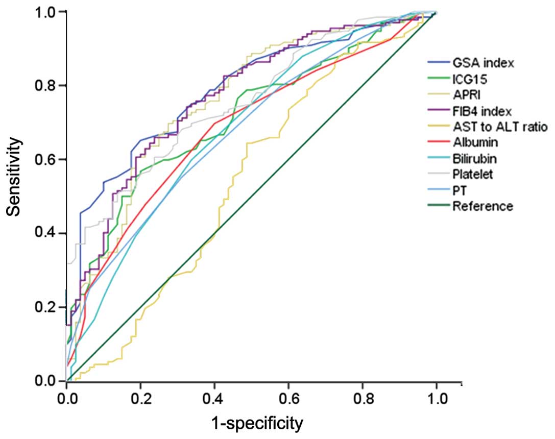
for predicting cirrhosis are demonstrated in Fig. 1. GSA index, ICG15, FIB-4 index, APRI
and platelet count exhibited reliable discriminative ability for
predicting cirrhosis. Among these, the GSA index yielded the
highest AUROC with a level of 0.786 at an optimal cut-off value of
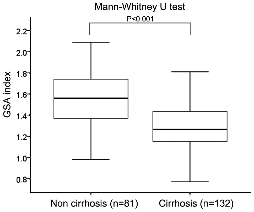
1.37 (sensitivity, 65.9%; specificity, 79.0%) (Table II). The GSA index in patients with
cirrhosis (F4, n=132) was significantly lower than that in those
with non-cirrhosis (F0–3, n=81) (P<0.001, Mann-Whitney U test)
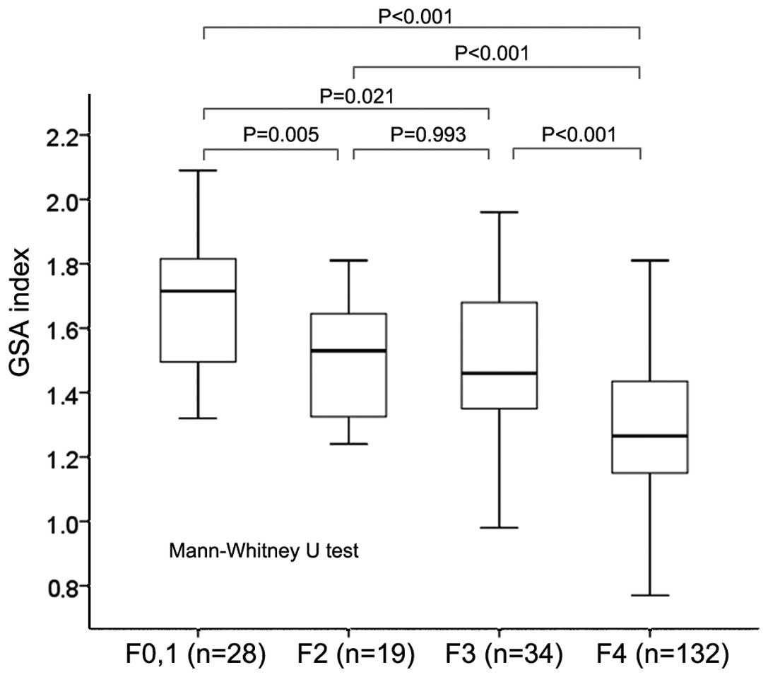
(Fig. 2). Between patients with F0
or 1 (n=28) and F4 (P<0.001), F2 (n=19) and F4 (P<0.001), F3
(n=34) and F4 (P<0.001), F0 or 1 and F2 (P=0.005) and F0 or 1
and F3 (P=0.021), significant differences were observed in terms of
the GSA index (Fig. 3).
| Table IIComparison of the area under receiver
operating curves (AUROCs) for the GSA index, ICG15, APRI, FIB-4
index, AST to ALT ratio, serum albumin, total bilirubin, platelet
count and prothrombin time for cirrhosis. |
Table II
Comparison of the area under receiver
operating curves (AUROCs) for the GSA index, ICG15, APRI, FIB-4
index, AST to ALT ratio, serum albumin, total bilirubin, platelet
count and prothrombin time for cirrhosis.
| Variables | AUROC | 95% CI | P-value |
|---|
| GSA index | 0.786 | 0.724–0.847 | <0.001 |
| ICG15 | 0.713 | 0.644–0.782 | <0.001 |
| APRI | 0.761 | 0.693–0.828 | <0.001 |
| FIB-4 index | 0.771 | 0.706–0.835 | <0.001 |
| AST to ALT
ratio | 0.542 | 0.458–0.627 | 0.304 |
| Serum albumin | 0.683 | 0.610–0.755 | <0.001 |
| Bilirubin | 0.681 | 0.606–0.756 | <0.001 |
| Platelet | 0.755 | 0.692–0.819 | <0.001 |
| Prothrombin
time | 0.676 | 0.603–0.749 | <0.001 |
Cumulative OS and RFS rates according to
GSA index
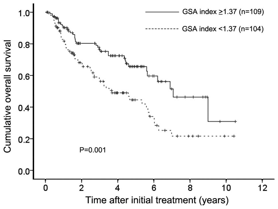
The 1-, 3- and 5-year cumulative OS rates in
patients with GSA index ≥1.37 (optimal cut-off value) (n=109) were
91.3, 76.5 and 65.6%, respectively, and the corresponding
cumulative OS rates in patients with GSA index <1.37 (n=104)
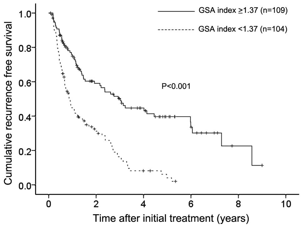
were 82.8, 57.6 and 44.5%, respectively (P=0.001) (Fig. 4). The 1-, 3- and 5-year cumulative
RFS rates in patients with GSA index ≥1.37 were 74.9, 50.2 and
39.7%, respectively, and the corresponding cumulative RFS rates in
patients with GSA index <1.37 were 44.9, 16.3 and 4.1%,
respectively (P<0.001) (Fig.
5).
Univariate and multivariate analyses of
factors contributing to OS
Univariate analysis identified the following factors
as significantly associated with OS for all cases (n=213): tumor
number (P=0.001); maximum tumor size ≥3.5 cm (P=0.010); microscopic
vascular invasion (MVI) (P=0.020); AST ≥50 IU/l (P=0.025); alkaline
phosphatase (ALP) ≥320 IU/l (P=0.008); GSA index ≥1.37 (P=0.001);
serum albumin ≥3.9 g/dl (P=0.005); total bilirubin ≥1.0 mg/dl
(P=0.002); AFP ≥100 ng/ml (P<0.001); and DCP ≥100 mAU/ ml
(P=0.001) (Table III). The hazard
ratios (HRs) and 95% confidence intervals (CIs) calculated using
multivariate analysis for the 10 factors with P<0.05 in
univariate analysis are detailed in Table III. Only the DCP value was found
to be a significant predictor linked to OS in the multivariate
analysis (P=0.005).
| Table IIIUnivariate and multivariate analysis
of factors contributing to overall survival. |
Table III
Univariate and multivariate analysis
of factors contributing to overall survival.
| Variables | n | Univariate
analysis | Multivariate
analysis |
|---|
|
|---|
| Hazard ratio (95%
CI) | P-valuea |
|---|
| Gender, male vs.
female | 153/60 | 0.639 | | |
| Age (years), ≥70
vs. <70 | 119/94 | 0.296 | | |
| Tumor number,
single vs. multiple | 123/90 | 0.001 | 0.715
(0.452–1.131) | 0.152 |
| Maximum tumor size
(cm), ≥3.5 vs. <3.5 | 109/104 | 0.010 | 0.906
(0.554–1.483) | 0.696 |
| Microscopic
vascular invasion, yes vs. no | 72/141 | 0.020 | 0.660
(0.414–1.052) | 0.081 |
| AST (IU/l), ≥50 vs.
<50 | 109/104 | 0.025 | 0.824
(0.523–1.300) | 0.406 |
| ALT (IU/l), ≥50 vs.
<50 | 91/122 | 0.459 | | |
| ALP (IU/l), ≥320
vs. <320 | 109/104 | 0.008 | 0.906
(0.566–1.452) | 0.683 |
| GGT (IU/l), ≥70 vs.
<70 | 104/109 | 0.482 | | |
| GSA index ≥1.37,
yes vs. no | 109/104 | 0.001 | 1.594
(0.957–2.658) | 0.074 |
| Serum albumin level
(g/dl), ≥3.9 vs. <3.9 | 112/101 | 0.005 | 1.642
(0.996–2.705) | 0.052 |
| Total bilirubin
(mg/dl), ≥1.0 vs. <1.0 | 67/146 | 0.002 | 0.647
(0.409–1.024) | 0.063 |
| Platelet count
(x104/mm3), ≥12 vs. <12 | 105/108 | 0.475 | | |
| Prothrombin time
(%), ≥88 vs. <88b | 105/107 | 0.113 | | |
| Diabetes mellitus,
yes vs. no | 47/166 | 0.475 | | |
| Body mass index
(kg/m2), ≥23 vs. <23 | 99/114 | 0.562 | | |
| Serum AFP (ng/ml),
≥100 vs. <100 | 62/151 | <0.001 | 0.623
(0.385–1.007) | 0.053 |
| DCP (mAU/ml), ≥100
vs. <100c | 129/81 | 0.001 | 0.451
(0.259–0.788) | 0.005 |
Univariate and multivariate analyses of
factors contributing to RFS
Univariate analysis identified the following factors
as significantly associated with RFS for all cases: tumor number
(P<0.001); MVI (P=0.002); AST ≥50 IU/l (P=0.010); ALP ≥320 IU/l
(P=0.008); GSA index ≥1.37 (P<0.001); serum albumin ≥3.9 g/dl
(P=0.016); total bilirubin ≥1.0 mg/dl (P=0.001); and PT ≥88%
(P=0.023) (Table IV). The HRs and
95% CIs calculated using multivariate analysis for the eight
factors with P<0.05 in univariate analysis are detailed in
Table IV. Tumor number (P=0.002),
MVI (P=0.002), ALP ≥320 IU/l (P=0.039) and GSA index (P<0.001)
were found to be significant prognostic factors linked to RFS.
| Table IVUnivariate and multivariate analyses
of the factors contributing to recurrence-free survival. |
Table IV
Univariate and multivariate analyses
of the factors contributing to recurrence-free survival.
| Variables | n | Univariate
analysis | Multivariate
analysis |
|---|
|
|---|
| Hazard ratio (95%
CI) | P-valuea |
|---|
| Gender, male vs.
female | 153/60 | 0.733 | | |
| Age (years), ≥70
vs. <70 | 119/94 | 0.560 | | |
| Tumor number,
single vs. multiple | 123/90 | <0.001 | 0.581
(0.410–0.823) | 0.002 |
| Maximum tumor size
(cm), ≥3.5 vs. <3.5 | 109/104 | 0.251 | | |
| Microscopic
vascular invasion, yes vs. no | 72/141 | 0.002 | 0.567
(0.395–0.815) | 0.002 |
| AST (IU/l), ≥50 vs.
<50 | 109/104 | 0.010 | 0.945
(0.663–1.345) | 0.753 |
| ALT (IU/l), ≥50 vs.
<50 | 91/122 | 0.141 | | |
| ALP (IU/l), ≥320
vs. <320 | 109/104 | <0.001 | 0.683
(0.475–0.982) | 0.039 |
| GGT (IU/l), ≥70 vs.
<70 | 104/109 | 0.483 | | |
| GSA index ≥1.37,
yes vs. no | 109/104 | <0.001 | 2.379
(1.594–3.550) | <0.001 |
| Serum albumin level
(g/dl), ≥3.9 vs. <3.9 | 112/101 | 0.016 | 1.056
(0.730–1.529) | 0.771 |
| Total bilirubin
(mg/dl), ≥1.0 vs. <1.0 | 67/146 | 0.001 | 0.840
(0.587–1.203) | 0.342 |
| Platelet count
(x104/mm3), ≥12 vs. <12 | 105/108 | 0.050 | | |
| Prothrombin time
(%), ≥88 vs. <88b | 105/107 | 0.023 | 1.064
(0.751–1.508) | 0.727 |
| Diabetes mellitus,
yes vs. no | 47/166 | 0.664 | | |
| Body mass index
(kg/m2), ≥23 vs. <23 | 99/114 | 0.662 | | |
| Serum AFP (ng/ml),
≥100 vs. <100 | 62/151 | 0.201 | | |
| DCP (mAU/ml), ≥100
vs. <100c | 129/81 | 0.118 | | |
Causesofdeath
In patients with preoperative GSA index ≥1.37
(n=109), 35 patients (32.1%) died during the follow-up period. The
causes of death were HCC recurrence in 26 patients, liver failure
in 6 patients and miscellaneous causes in 3 patients, while in
patients with preoperative GSA index <1.37 (n=104), 54 patients
(51.9%) died during the follow-up period. The causes of death were
HCC recurrence in 34 patients, liver failure in 10 patients and
miscellaneous causes in 10 patients.
HCC recurrence
In patients with preoperative GSA index ≥1.37, 59
patients (54.1%) had HCC recurrences during the follow-up period.
Nineteen patients (17.4%) had late first confirmed HCC recurrence
(≥2 years after initial SR). The patterns of HCC recurrence after
initial treatment were: single HCC recurrence in the liver in 23
patients; multiple HCC recurrences in the liver in 27 patients;
multiple HCC recurrences in the liver with lung metastases in 2
patients; multiple bone metastases in 2 patients; multiple HCC
recurrences in the liver with lymph node metastases in 2 patients;
multiple HCC recurrences in the liver with peritoneal dissemination
in one patient; multiple HCC recurrence in the liver with right
atrium invasion in one patient; and local tumor progression
(recurrence in the SR site) in one patient. Treatment methods for
the first HCC recurrence were: SR in 7 patients; RFA in 23
patients; percutaneous ethanol injection (PEI) in one patient; TACE
in 19 patients; systemic chemotherapy such as sorafenib in 2
patients; radiation therapy in 2 patients and no specific treatment
in 5 patients.
In patients with preoperative GSA index <1.37, 87
patients (83.7%) had HCC recurrences during the follow-up period.
Twenty patients (19.2%) had late first confirmed HCC recurrence (≥2
years after initial SR). The patterns of HCC recurrence after
initial treatment were: single HCC recurrence in the liver in 37
patients; multiple HCC recurrences in the liver in 43 patients;
multiple HCC recurrences in the liver with lung metastases in 3
patients; multiple bone metastases in one patient; multiple lung
metastases in one patient; multiple HCC recurrences in the liver
with lymph node metastases in one patient; and local tumor
progression (recurrence in the SR site) in one patient. Treatment
methods for the first HCC recurrence were: SR in 3 patients; RFA in
38 patients; PEI in 2 patients; TACE in 29 patients; systemic
chemotherapy such as sorafenib in 4 patients; radiation therapy in
one patient and no specific treatment in 10 patients.
Discussion
To the best of our knowledge, this is the first
reported study to examine the relationship between preoperative GSA
index calculated from 99mTc-GSA scintigraphy and liver
fibrosis and clinical outcomes in HCV-related HCC patients treated
with SR. Although several noninvasive serum markers such as ICG15,
FIB-4 index and APRI are associated with clinical outcomes in
HCV-related HCC patients, no reports have assessed the impact of
preoperative GSA index on clinical outcomes in HCV-related HCC
patients treated with SR (21–24).
Hence, we conducted the current analysis.
In the present study, the GSA index yielded the
highest AUROC for cirrhosis and in multivariate analyses, GSA index
was an independent predictor (P<0.001) linked to RFS and it had
a marginal significance in terms of OS (P=0.074). Our results
suggest that the preoperative GSA index well reflects hepatic
functional reserve and is a useful predictor of clinical outcomes
in HCV-related HCC patients treated with SR. Yoshizumi et al
demonstrated that the 6-month survival probability was improved in
the group with a GSA index ≥1.3 in patients who underwent liver
transplantation, whereas our optimal cut-off value of the GSA index
according to ROC analysis was 1.37 (13). Our results were consistent with
their results. As mentioned earlier, ICG mainly reflected hepatic
blood flow, while GSA was related to the amount of functional
hepatocytes as well as blood flow. As shown in our results, the GSA
index can reflect the liver fibrosis more accurately than
ICG15.
On the other hand, FIB-4 index and APRI exhibited
highly discriminative ability for predicting cirrhosis in our
analysis. Several investigators demonstrated that FIB-4 and ARPI
are useful noninvasive serum markers for predicting liver fibrosis
in patients with CHC (25–28). In addition, a recent meta-analysis
regarding diagnostic accuracy of FIB-4 and APRI in patients with
chronic hepatitis B infection showed that the mean AUROCs of FIB-4
and APRI for predicting cirrhosis were 0.78 and 0.72, while our
data of FIB-4 and APRI were 0.771 and 0.761. Although the causes of
liver diseases were different between their data and ours, our
results were similar to their reports (29).
In our analysis, as demonstrated in Fig. 3, the GSA index had well
discriminative ability between various stages of liver fibrosis
except for the relationship between F2 and F3. The reason why the
GSA index did not show well discriminative ability between patients
with F2 and F3 is unclear, however, the small sample size in
patients with F2 (n=19) may be attributed to our current
results.
Liver biopsy, which has been considered as the
‘golden standard’ for assessing the extent of liver fibrosis,
carries some drawbacks: sampling error and interobserver
variability, which have raised questions on its value, whereas in
our present analyses, we investigated the impact of the
preoperative GSA index on cirrhosis using non-tumor parts of
extracted surgical specimens, which had sufficient amount of liver
specimens for exact evaluation of the degree of liver fibrosis
(30–32). Thus, our data are highly reliable
and this is a major strength of the present study.
The presence of MVI was a significant factor linked
to RFS and it had a tendency toward poorer OS in our multivariate
analyses. Postoperative factors as well as preoperative factors may
be essential for predicting survival. Indeed, Lim et al
reported that MVI is a better predictor of HCC recurrence and OS
after SR for HCC (33). On the
other hand, it is of interest that a higher ALP value was
significantly linked to higher HCC recurrence in multivariate
analysis. Cumulative evidence derived from Asian populations with
HCC revealed that a higher ALP level was associated with poor
outcomes, which is in line with the present study results (34).
We acknowledge several limitations to the present
study. First, the present study was a retrospective observational
study with heterogeneous HCC patients with various HCC stages.
Second, postoperative therapy such as interferon was not included
in our analysis, leading to bias. Third, subjects in whom
99mTc-GSA scintigraphy prior to surgery was not
performed were excluded from our analysis (data not shown) and
whether 99mTc-GSA scintigraphy was performed or not
before SR mainly depends on the decision of attending surgeons in
our hospital, also leading to bias. Thus, a well characterized
study will be needed in the future. However, the present study
results demonstrated that the preoperative GSA index well reflected
the extent of liver fibrosis and it is closely associated with
clinical outcomes in patients with HCV-related HCC treated with
SR.
In conclusion, the preoperative GSA index calculated
from 99mTc-GSA scintigraphy can be a useful predictor
for patients with HCV-related HCC treated with SR.
Acknowledgements
The authors would like to thank Haruko Takada for
data collection.
References
|
1
|
El-Serag HB: Epidemiology of viral
hepatitis and hepatocellular carcinoma. Gastroenterology.
142:1264.e1–1273.e1. 2012. View Article : Google Scholar
|
|
2
|
de Lope CR, Tremosini S, Forner A, Reig M
and Bruix J: Management of HCC. J Hepatol. 56(Suppl 1): S75–S87.
2012. View Article : Google Scholar : PubMed/NCBI
|
|
3
|
Osaki Y and Nishikawa H: Treatment for
hepatocellular carcinoma in Japan over the last three decades: our
experience and published work review. Hepatol Res. Jun
26–2014.(Epub ahead of print). View Article : Google Scholar
|
|
4
|
Zhou WP, Lai EC, Li AJ, Fu SY, Zhou JP,
Pan ZY, Lau WY and Wu MC: A prospective, randomized, controlled
trial of preoperative transarterial chemoembolization for
resectable large hepatocellular carcinoma. Ann Surg. 249:195–202.
2009. View Article : Google Scholar : PubMed/NCBI
|
|
5
|
Nishikawa H, Osaki Y, Kita R, Kimura T,
Inuzuka T, Takeda H, Nakajima J, Matsuda F, Sakamoto A, Henmi S,
Hatamaru K, Saito S and Nasu A: Transcatheter arterial infusion
chemotherapy prior to radiofrequency thermal ablation for single
hepatocellular carcinoma reduces the risk of intrahepatic distant
recurrence. Int J Oncol. 41:903–909. 2012.PubMed/NCBI
|
|
6
|
Kudo M, Todo A, Ikekubo K and Hino M:
Receptor index via hepatic asialoglycoprotein receptor imaging:
correlation with chronic hepatocellular damage. Am J Gastroenterol.
87:865–870. 1992.PubMed/NCBI
|
|
7
|
Matsuzaki S, Onda M, Tajiri T and Kim DY:
Hepatic lobar differences in progression of chronic liver disease:
correlation of asialoglycoprotein scintigraphy and hepatic
functional reserve. Hepatology. 25:828–832. 1997. View Article : Google Scholar : PubMed/NCBI
|
|
8
|
Kaibori M, Ha-Kawa SK, Maehara M, Ishizaki
M, Matsui K, Sawada S and Kwon AH: Usefulness of Tc-99m-GSA
scintigraphy for liver surgery. Ann Nucl Med. 25:593–602. 2011.
View Article : Google Scholar : PubMed/NCBI
|
|
9
|
Ogasawara G, Inoue Y, Itoh Y, Tagami S,
Matsunaga K and Miki K: Improved reproducibility of simple
quantitative indices from 99mTc-GSA liver functional
imaging. Ann Nucl Med. 27:487–491. 2013. View Article : Google Scholar : PubMed/NCBI
|
|
10
|
Harada K, Mizuguchi T, Katagiri Y,
Kawamoto M, Nakamura Y, Meguro M, Ota S, Sasaki S, Miyanishi K,
Sonoda T, Mori M, Shinomura Y, Kato J and Hirata K: Area between
the hepatic and heart curves of 99mTc-galactosyl-human
serum albumin scintigraphy represents liver function and disease
progression for preoperative evaluation in hepatocellular carcinoma
patients. J Hepatobiliary Pancreat Sci. 19:667–673. 2012.
View Article : Google Scholar
|
|
11
|
Kubota K, Makuuchi M, Kusaka K, Kobayashi
T, Miki K, Hasegawa K, Harihara Y and Takayama T: Measurement of
liver volume and hepatic functional reserve as a guide to
decision-making in resectional surgery for hepatic tumors.
Hepatology. 26:1176–1181. 1997.PubMed/NCBI
|
|
12
|
Gupta S, Chawla Y, Kaur J, Saxena R,
Duseja A, Dhiman RK and Choudhary NS: Indocyanine green clearance
test (using spectrophotometry) and its correlation with model for
end stage liver disease (MELD) score in Indian patients with
cirrhosis of liver. Trop Gastroenterol. 33:129–134. 2012.
View Article : Google Scholar : PubMed/NCBI
|
|
13
|
Yoshizumi T, Taketomi A, Uchiyama H,
Harada N, Kayashima H, Yamashita Y, Soejima Y, Shimada M and
Maehara Y: Graft size, donor age, and patient status are the
indicators of early graft function after living donor liver
transplantation. Liver Transpl. 14:1007–1013. 2008. View Article : Google Scholar : PubMed/NCBI
|
|
14
|
Alberti KG and Zimmet PZ: Definition,
diagnosis and classification of diabetes mellitus and its
complications. Part 1: diagnosis and classification of diabetes
mellitus provisional report of a WHO consultation. Diabet Med.
15:539–553. 1998. View Article : Google Scholar : PubMed/NCBI
|
|
15
|
Kinoshita K, Ukikusa M, Iwaisako K,
Arimoto A, Fujisawa N, Ozaki T, Tanaka H, Seo S, Naitoh M, Nomura
A, Inomoto T, Kitai T, Ino K, Higashiyama H, Hanafusa T and
Nakajima Y: Preoperative assessment of hepatic function: utility of
a new convenient two-compartment model analysis using galactosyl
human serum albumin scintigraphy. J Gastroenterol. 18:99–104.
2003.
|
|
16
|
Wai CT, Greenson JK, Fontana RJ,
Kalbfleisch JD, Marrero JA, Conjeevaram HS and Lok AS: A simple
noninvasive index can predict both significant fibrosis and
cirrhosis in patients with chronic hepatitis C. Hepatology.
38:518–526. 2003. View Article : Google Scholar : PubMed/NCBI
|
|
17
|
Sterling RK, Lissen E, Clumeck N, Sola R,
Correa MC, Montaner J, Sulkowski MS, Torriani FJ, Dieterich DT,
Thomas DL, Messinger D and Nelson M; APRICOT Clinical
Investigators. Development of a simple noninvasive index to predict
significant fibrosis in patients with HIV/HCV coinfection.
Hepatology. 43:1317–1325. 2006. View Article : Google Scholar : PubMed/NCBI
|
|
18
|
Bruix J and Sherman M; Practice Guidelines
Committee, American Association for the Study of Liver Diseases.
Management of hepatocellular carcinoma. Hepatology. 42:1208–1236.
2005. View Article : Google Scholar : PubMed/NCBI
|
|
19
|
No authors listed. The general rules for
the clinical and pathological study of primary liver cancer. Liver
Cancer Study Group of Japan. Jpn J Surg. 19:98–129. 1989.
View Article : Google Scholar : PubMed/NCBI
|
|
20
|
Utsunomiya T, Shimada M, Kudo M, Ichida T,
Matsui O, Izumi N, Matsuyama Y, Sakamoto M, Nakashima O, Ku Y,
Takayama T and Kokudo N; for the Liver Cancer Study Group of Japan.
A comparison of the surgical outcomes among patients with
HBV-positive, HCV-positive, and non-B non-C hepatocellular
carcinoma: a nationwide study of 11,950 patients. Ann Surg. Jul
28–2014.(Epub ahead of print). PubMed/NCBI
|
|
21
|
Shen SL, Fu SJ, Chen B, Kuang M, Li SQ,
Hua YP, Liang LJ, Guo P, Hao Y and Peng BG: Preoperative aspartate
aminotransferase to platelet ratio is an independent prognostic
factor for hepatitis B-induced hepatocellular carcinoma after
hepatic resection. Ann Surg Oncol. 21:3802–3809. 2014. View Article : Google Scholar : PubMed/NCBI
|
|
22
|
Makuuchi M: Surgical treatment for HCC -
special reference to anatomical resection. Int J Surg. 11(Suppl 1):
S47–S49. 2013. View Article : Google Scholar
|
|
23
|
Shindoh J, Hasegawa K, Inoue Y, Ishizawa
T, Nagata R, Aoki T, Sakamoto Y, Sugawara Y, Makuuchi M and Kokudo
N: Risk factors of post-operative recurrence and adequate surgical
approach to improve long-term outcomes of hepatocellular carcinoma.
HPB. 15:31–39. 2013. View Article : Google Scholar :
|
|
24
|
Angulo P, Bugianesi E, Bjornsson ES,
Charatcharoenwitthaya P, Mills PR, Barrera F, Haflidadottir S, Day
CP and George J: Simple noninvasive systems predict long-term
outcomes of patients with nonalcoholic fatty liver disease.
Gastroenterology. 145:782.e4–789.e4. 2013. View Article : Google Scholar
|
|
25
|
Lin ZH, Xin YN, Dong QJ, Wang Q, Jiang XJ,
Zhan SH, Sun Y and Xuan SY: Performance of the aspartate
aminotransferase-to-platelet ratio index for the staging of
hepatitis C-related fibrosis: an updated meta-analysis. Hepatology.
53:726–736. 2011. View Article : Google Scholar : PubMed/NCBI
|
|
26
|
Smith JO and Sterling RK: Systematic
review: non-invasive methods of fibrosis analysis in chronic
hepatitis C. Aliment Pharmacol Ther. 30:557–576. 2009. View Article : Google Scholar : PubMed/NCBI
|
|
27
|
Joo SK, Kim JH, Oh S, Kim BG, Lee KL, Kim
HY, Jung YJ, Woo HS, Moon MH, Chang MS and Kim W: Prospective
comparison of noninvasive fibrosis assessment to predict advanced
fibrosis or cirrhosis in Asian patients with hepatitis C. J Clin
Gastroenterol. Sep 8–2014.(Epub ahead of print). View Article : Google Scholar : PubMed/NCBI
|
|
28
|
Vergniol J, Boursier J, Coutzac C,
Bertrais S, Foucher J, Angel C, Chermak F, Hubert IF, Merrouche W,
Oberti F, de Lédinghen V and Calès P: Evolution of noninvasive
tests of liver fibrosis is associated with prognosis in patients
with chronic hepatitis C. Hepatology. 60:65–76. 2014. View Article : Google Scholar : PubMed/NCBI
|
|
29
|
Xiao G, Yang J and Yan L: Comparison of
diagnostic accuracy of APRI and FIB-4 for detecting liver fibrosis
in adult patients with chronic hepatitis B virus infection: a
systemic review and meta-analysis. Hepatology. Aug 18–2014.(Epub
ahead of print). View Article : Google Scholar
|
|
30
|
Nalbantoglu I and Brunt EM: Role of liver
biopsy in nonalcoholic fatty liver disease. World J Gastroenterol.
20:9026–9037. 2014.PubMed/NCBI
|
|
31
|
Castera L, Vilgrain V and Angulo P:
Noninvasive evaluation of NAFLD. Nat Rev Gastroenterol Hepatol.
10:666–675. 2013. View Article : Google Scholar : PubMed/NCBI
|
|
32
|
Castera L: Transient elastography and
other noninvasive tests to assess hepatic fibrosis in patients with
viral hepatitis. J Viral Hepat. 16:300–314. 2009. View Article : Google Scholar : PubMed/NCBI
|
|
33
|
Lim KC, Chow PK, Allen JC, Chia GS, Lim M,
Cheow PC, Chung AY, Ooi LL and Tan SB: Microvascular invasion is a
better predictor of tumor recurrence and overall survival following
surgical resection for hepatocellular carcinoma compared to the
Milan criteria. Ann Surg. 254:108–113. 2011. View Article : Google Scholar : PubMed/NCBI
|
|
34
|
Chen CH, Hu FC, Huang GT, Lee PH, Tsang
YM, Cheng AL, Chen DS, Wang JD and Sheu JC: Applicability of
staging systems for patients with hepatocellular carcinoma is
dependent on treatment method - analysis of 2,010 Taiwanese
patients. Eur J Cancer. 45:1630–1639. 2009. View Article : Google Scholar : PubMed/NCBI
|