Differential proteomic profiling of primary and recurrent chordomas

  • Authors:
    • Su Chen
    • Wei Xu
    • Jian Jiao
    • Dongjie Jiang
    • Jian Liu
    • Tenghui Chen
    • Zongmiao Wan
    • Leqin Xu
    • Zhenhua Zhou
    • Jianru Xiao
  • View Affiliations

  • Published online on: February 26, 2015     https://doi.org/10.3892/or.2015.3818
  • Pages: 2207-2218
Metrics: Total Views: 0 (Spandidos Publications: | PMC Statistics: )
Total PDF Downloads: 0 (Spandidos Publications: | PMC Statistics: )


Abstract

Chordomas are locally destructive tumors with high rates of recurrence and a poor prognosis. The mechanisms involved in chordoma recurrence remain largely unknown. In the present study, we examined the proteomic profile of a chordoma primary tumor (CSO) and a recurrent tumor (CSR) through mass spectrum in a chordoma patient who underwent surgery. Bioinformatic analysis of the profile showed that 359 proteins had a significant expression difference and 21 pathways had a striking alteration between the CSO and the CSR. The CSR showed a significant increase in carbohydrate metabolism. Immunohistochemistry (IHC) confirmed that the cancer stem cell marker activated leukocyte cell adhesion molecule (ALCAM or CD166) expression level was higher in the recurrent than that in the primary tumor. The present study analyzed the proteomic profile change between CSO and CSR and identified a new biomarker ALCAM in recurrent chordomas. This finding sheds light on unraveling the pathophysiology of chordoma recurrence and on exploring more effective prognostic biomarkers and targeted therapies against this devastating disease.

Introduction

Chordoma is a rare slow-growing neoplasm thought to arise from cellular remnants of the notochord. The incidence of chordoma is approximately 1 case in one million individuals and it accounts for ~1–4% of all tumor cases. However, these rare tumors present very significant treatment challenges (1). It is notable that 67% of the surgically managed patients suffer local recurrence, and the disease-free survival at 5 years is almost 0% (1,2).

Complete surgical resection followed by radiation therapy offers the best chance of long-term control for chordoma. However, incomplete resection of the primary tumor makes controlling the disease more difficult and increases the odds of recurrence. Tumors at certain sites such as skull base chordoma can hardly be removed completely due to the complicated peritumoral tissue structure which makes the tumor difficult to be exposed. Thus, recurrence of skull base chordoma is as high as 85%. Chordomas are relatively radioresistant as well, requiring high doses of radiation to be controlled. The proximity of chordomas to vital neurological structures such as the brain stem and nerves limits the dose of radiation that can safely be delivered. A strategy to control the recurrence of chordomas is vital for improving the survival rate of patients. It is imperative to explore chordoma recurrence mechanisms at the molecular level and to search for alternative therapeutic methods including chemotherapy to treat it. Recently, Zhou et al assessed the chordoma proteome in chordoma tumor tissues and identified ENO1, PKM2, and gp96 proteins as being upregulated in chordomas. They reported that the expression of these proteins was higher in recurrent than that in the primary chordomas (3). However, the molecular mechanisms involved in chordoma recurrence remain unstudied, and the detailed changes in proteomic profiling in the process of recurrence remain unclear.

We analyzed 3,296 proteins identified by mass spectrum in a chordoma patient original tumor (CSO) and recurrent tumor (CSR) tissue. Bioconductor’s Global Anova test was applied to compare the overall proteomic profiling changes between CSO and CSR which indicated there was a significant difference (P≤0.1). Bioconductor’s multtest found that 359 proteins exhibited the highest expression difference between CSO and CSR. KEGG database analysis of the 359 proteins revealed that 21 pathways had a significant change between CSO and CSR. Immunohistochemistry (IHC) further verified that cancer stem cell marker activated leukocyte cell adhesion molecule (ALCAM or CD166) was markedly upregulated in the CSR tissue.

Materials and methods

Tissue specimen processing

Chordoma specimens were obtained from a resected tumor following an institutional review board approved protocol. The histological composition of the samples was assessed by examining adjacent sections. Tumor samples were dissected and only tissue that was superfluous to that required for pathological evaluation was taken. The samples were immediately snap-frozen in liquid nitrogen and stored at −80°C. Approximately 800 μg of tissue samples was cut into small pieces with a scalpel and transferred into a mortar filled with liquid nitrogen. The tissue was ground to a fine powder with a pestle in the continuous presence of liquid nitrogen and transferred into a reaction tube with extraction buffer [2 M thiocarbamide, 7 M urea and 10 μM proteinase inhibitor (Roche Diagnostics, Indianapolis, IN, USA)] at 4°C. The solution was centrifuged at 16,000 × g at 4°C for 15 min, and the supernatant (~300–400 μl) was stored frozen at −200°C.

Proteome analysis

The protein concentration was determined by Amersham 2D quant kit (GE Healthcare Bio-Sciences, Piscataway, NJ, USA). The protein samples were further lysed to peptides and prepared for proteome analysis as described previously (4). Peptides were analyzed using strong cation (SCX)/reversed phase, upgrade performance liquid chromatography (Nano-RPLC)/ESI/MS/MS. Samples were analyzed using a LTq Orbitrap XL (Thermo Electron Corp., Bremen, Germany) mass spectrometer. MS/MS spectra data were searched against the Swiss-Prot Human (2009.02.10, 20331 sequences) database or IPI database using Bioworks Browser 3.3.1 SP1. The identified proteins were quantified by APEX software (5,6). To control the false-positive rate, finally the quantitative results by false-discovery rate (FDR) 1% or less (false-positive rate of 1% or less) as the standard filter.

Immunohistochemistry

The paraffin sections were dried in an oven at 65°C for 1 h. The paraffin sections were then dewaxed in xylene and rehydrated in a series of ethanol solutions. The endogenous peroxidase activity was blocked by a 10-min pre-incubation with 3% H2O2. The paraffin sections were preheated at 100°C in antigen retrieval solution containing EDTA (pH 8.0) for 30 min and blocked by non-immune goat serum at room temperature for 15 min to decrease unspecific staining. Incubation with mouse polyclonal anti-ALCAM (1:1,000 dilution) was performed overnight at 4°C. After being washed 3 times with 1X PBS buffer for 3 min, the sections were incubated with the secondary (link) antibody (biotinylated mouse-anti-human IgG) for 30 min at room temperature. After reacting with the streptavidin-biotin-peroxidase complex for 20 min, the immunoreactivity was determined by 3,3′-diamino-benzidine tetrahydrochloride and H2O2 at room temperature according to the manufacturer’s instructions. The positive reaction was manifested as brown (DAB) staining. The sections were counterstained in Mayer’s hematoxylin. The selected sections were scanned at ×400 magnification to visualize the localization and distribution of ALCAM.

Statistical analysis

Statistically significant proteins were identified by first performing a two-tailed Student’s t-test with the ‘multtest’ package in rat the respective time-points by comparing protein abundance between CSO and CSR. Multiple hypothesis testing was then implemented with the ‘rawp2adjp’ function in R by correcting the P-values according to Benjamini and Hochberg procedures (7) to control the FDRs to ≤1%. Proteins with a FDR ≤1%, peptide count ≥3, and fold-change ≥2 were identified as statistically significant. Throughout the present study, upregulation is defined as higher protein abundance measured in CSR relative to CSO and downregulation refers to fewer proteins measured with CSR. Moreover, a positive expression ratio represents upregulation and negative represents downregulation. Blast2Go (8) was used as a comprehensive bioinformatics tool for the functional annotation of the protein sequences in the present study such as determining gene ontology terms. The metabolic map at CSR was generated by first using the statistically significant proteins to identify the key pathways. Once the pathways were identified, all of the detected proteins in the same pathway were evaluated to determine whether they were upregulated or downregulated relative to the control. If ≥50% of the proteins in the pathway was regulated similarly in the same direction, then the pathway would be designated as upregulated or Downregulated according to the majority. The cellular pathways are displayed using the iPath 2.0 platform (9).

Results and Discussion

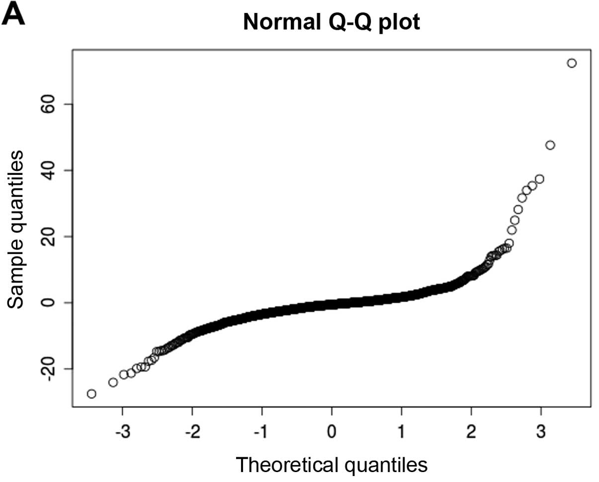
In order to obtain a comprehensive proteomic profile of CSO and CSR and to investigate the mechanism of recurrence, the proteome of patient CSO and CSR tumor tissue samples was analyzed with LC/MS. In total, 3,296 unique protein sequences were identified. Bioconductor’s Global Anova package was used to determine the significance of the protein expression change between CSO and CSR. A protein expression difference with P≤0.1±0.002 was defined as statistically significant (Table I). Furthermore, we applied Bioconductor’s multtest package to analyze the identified 3,296 proteins and found that a large number of proteins (359) showed significant changes in expression with BH ≤0.01 and P≤0.01 between CSO and CSR. These proteins were involved in central metabolism, genetic information transcription and other processes essential to cell functions. Of these 359 proteins, there were 244 Downregulated proteins (CSR/CSO value ≤0.1) and 115 upregulated proteins (CSR/CSO ≥9) (Fig. 1; Table III and IV). This analysis discovered many significant proteins which have never been reported before in recurrent chordomas.

Table I

Global test for differential gene expression.

Table I

Global test for differential gene expression.

ANOVASSQDFMS
Effect0.00207320732966.29007E-07
Error0.00065985131845.00493E-08
Test Result
 F.value12.56774798
 p.perm0.1
 p.approx0.002139239

[i] *p≤0.1.

Table III

Downregulated proteins (244) (CSR/CSO value ≤0.1).

Table III

Downregulated proteins (244) (CSR/CSO value ≤0.1).

Protein Protein_description
q7Z5L7Podocan GN=PODN
q7L5N1COP9 signalosome complex subunit 6 GN=COPS6
P31939Bifunctional purine biosynthesis protein PuRH GN=ATIC
O94808 Glucosamine-fructose-6-phosphate aminotransferase (isomerizing) 2 GN=GFPT2
q8TD55Pleckstrin homology domain-containing family O member 2 GN=PLEKHO2
O75400Pre-mRNA-processing factor 40 homolog A GN=PRPF40A
Q6KB66Keratin, type II cytoskeletal 80 GN=KRT80
q07157Tight junction protein ZO-1 GN=TJP1
P1015560 kDa SS-A/Ro ribonucleoprotein GN=TROVE2
P05156Complement factor I GN=CFI
q99983Osteomodulin GN=OMD
Q02790FK506-binding protein 4 GN=FKBP4
q9Y240C-type lectin domain family 11 member A GN=CLEC11A
q9H8Y8Golgi reassembly-stacking protein 2 GN=GORASP2
P27658Collagen α-1(VIII) chain GN=COL8A1
P27169Serum paraoxonase/arylesterase 1 GN=PON1
q99729Heterogeneous nuclear ribonucleoprotein A/B GN=HNRNPAB
q9H4A4Aminopeptidase B GN=RNPEP
P50570Dynamin-2 GN=DNM2
q14157 ubiquitin-associated protein 2-like GN=uBAP2L
P02788Lactotransferrin GN=LTF
q96S97Myeloid-associated differentiation marker GN=MYADM
O60841Eukaryotic translation initiation factor 5B GN=EIF5B
q96C19EF-hand domain-containing protein D2 GN=EFHD2
P50225Sulfotransferase 1A1 GN=SuLT1A1
A0AVT1Ubiquitin-like modifer-activating enzyme 6 GN=uBA6
A0MZ66Shootin-1 GN=KIAA1598
A1L4H1Scavenger receptor cysteine-rich domain-containing protein LOC284297
A8MWD9Small nuclear ribonucleoprotein G-like protein
O00154Cytosolic acyl coenzyme A thioester hydrolase GN=ACOT7
O00461Golgi integral membrane protein 4 GN=GOLIM4
O14791Apolipoprotein L1 GN=APOL1
O43592Exportin-T GN=XPOT
O43670Zinc fnger protein 207 GN=ZNF207
O43847Nardilysin GN=NRD1
O60240Perilipin GN=PLIN
O60684Importin subunit α-7 GN=KPNA6
O60687Sushi repeat-containing protein SRPX2 GN=SRPX2
O60831PRA1 family protein 2 GN=PRAF2
O75094Slit homolog 3 protein GN=SLIT3
O75110Probable phospholipid-transporting ATPase IIA GN=ATP9A
O75339Cartilage intermediate layer protein 1 GN=CILP
O75592Probable E3 ubiquitin-protein ligase MYCBP2 GN=MYCBP2
O76021Ribosomal L1 domain-containing protein 1
O94769Extracellular matrix protein 2 GN=ECM2
O94903Proline synthetase co-transcribed bacterial homolog protein GN=PROSC
O95302FK506-binding protein 9 GN=FKBP9
O95373Importin-7 GN=IPO7
O95425Supervillin GN=SVIL
O95433Activator of 90 kDa heat shock protein ATPase homolog 1 GN=AHSA1
O95757Heat shock 70 kDa protein 4L GN=HSPA4L
O95810Serum deprivation-response protein GN=SDPR
O95816BAG family molecular chaperone regulator 2 GN=BAG2
O95965Integrin β-like protein 1 GN=ITGBL1
O96005Cleft lip and palate transmembrane protein 1 GN=CLPTM1
P01614Ig κ chain V-II region cum
P01781Ig heavy chain V-III region GAL
P02724Glycophorin-A GN=GYPA
P02750Leucine-rich α-2-glycoprotein GN=LRG1
P04433Ig κ chain V–III region VG (fragment)
P05543Thyroxine-binding globulin GN=SERPINA7
P05546Heparin cofactor 2 GN=SERPIND1
P07093Glia-derived nexin GN=SERPINE2
P07358Complement component C8 β chain GN=C8B
P08174Complement decay-accelerating factor GN=CD55
P0825372 kDa type IV collagenase GN=MMP2
P08493Matrix Gla protein GN=MGP
P0870840S ribosomal protein S17 GN=RPS17
P10253Lysosomal α-glucosidase GN=GAA
P10451Osteopontin GN=SPP1
P10600Transforming growth factor β-3 GN=TGFB3
P11234Ras-related protein Ral-B GN=RALB
P12004Proliferating cell nuclear antigen GN=PCNA
P12107Collagen α-1(XI) chain GN=COL11A1
P15104Glutamine synthetase GN=GLuL
P19367Hexokinase-1 GN=HK1
P20036HLA class II histocompatibility antigen, DP α chain GN=HLA-DPA1
P20591Interferon-induced GTP-binding protein Mx1 GN=MX1
P20851C4b-binding protein β chain GN=C4BPB
P22102Trifunctional purine biosynthetic protein adenosine-3 GN=GART
P23193Transcription elongation factor A protein 1 GN=TCEA1
P23497Nuclear autoantigen Sp-100 GN=SP100
P2637360S ribosomal protein L13 GN=RPL13
P26599Polypyrimidine tract-binding protein 1 GN=PTBP1
P26639Threonyl-tRNA synthetase, cytoplasmic GN=TARS
P28300Protein-lysine 6-oxidase GN=LOX
P31153 S-adenosylmethionine synthetase isoform type-2 GN=MAT2A
P32321Deoxycytidylate deaminase GN=DCTD
P35542Serum amyloid A-4 protein GN=SAA4
P35625Metalloproteinase inhibitor 3 GN=TIMP3
P35858Insulin-like growth factor-binding protein complex acid labile chain GN=IGFALS
P36969Phospholzipid hydroperoxide glutathione peroxidase, mitochondrial GN=GPX4
P3902360S ribosomal protein L3 GN=RPL3
P41218Myeloid cell nuclear differentiation antigen GN=MNDA
P41240Tyrosine-protein kinase CSK GN=CSK
P45877Peptidyl-prolyl cis-trans isomerase C GN=PPIC
P46108Proto-oncogene C-crk GN=CRK
P46109Crk-like protein GN=CRKL
P4855626S proteasome non-ATPase regulatory subunit 8 GN=PSMD8
P49321Nuclear autoantigenic sperm protein GN=NASP
P49354Protein farnesyltransferase/geranylgeranyltransferase type-1 subunit α GN=FNTA
P49458Signal recognition particle 9 kDa protein GN=SRP9
P49591Seryl-tRNA synthetase, cytoplasmic GN=SARS
P50135Histamine N-methyltransferase GN=HNMT
P50479PDZ and LIM domain protein 4 GN=PDLIM4
P50583Bis (5′-nucleosyl)-tetraphosphatase (asymmetrical) GN=NuDT2
P51148Ras-related protein Rab-5C GN=RAB5C
P51812Ribosomal protein S6 kinase a-3 GN=RPS6KA3
P52788Spermine synthase GN=SMS
P55039 Developmentally-regulated GTP-binding protein 2 GN=DRG2
P55196Afadin GN=MLLT4
P55212Caspase-6 GN=CASP6
P60983Glia maturation factor β GN=GMFB
P61221ATP-binding cassette sub-family E member 1 GN=ABCE1
P61225Ras-related protein Rap-2b GN=RAP2B
P6131360S ribosomal protein L15 GN=RPL15
P61758Prefoldin subunit 3 GN=VBP1
P61970Nuclear transport factor 2 GN=NuTF2
P6219526S protease regulatory subunit 8 GN=PSMC5
P6226640S ribosomal protein S23 GN=RPS23
P6227740S ribosomal protein S13 GN=RPS13
P6228040S ribosomal protein S11 GN=RPS11
P62304Small nuclear ribonucleoprotein E GN=SNRPE
P62316Small nuclear ribonucleoprotein Sm D2 GN=SNRPD2
P6275060S ribosomal protein L23a GN=RPL23A
P6284740S ribosomal protein S24 GN=RPS24
P6285740S ribosomal protein S28 GN=RPS28
P6289960S ribosomal protein L31 GN=RPL31
P80217Interferon-induced 35 kDa protein GN=IFI35
P80303Nucleobindin-2 GN=NuCB2
P82987ADAMTS-like protein 3 GN=ADAMTSL3
P83110Probable serine protease HTRA3 GN=HTRA3
Q00341Vigilin GN=HDLBP
q03518Antigen peptide transporter 1 GN=TAP1
q04446 1,4-α-glucan-branching enzyme GN=GBE1
q06124Tyrosine-protein phosphatase non-receptor type 11 GN=PTPN11
q08J23tRNA (cytosine-5-)-methyltransferase NSuN2 GN=NSuN2
q12965Myosin-Ie GN=MYO1E
Q13123Protein Red GN=IK
q13315Serine-protein kinase ATM GN=ATM
q13838Spliceosome RNA helicase BAT1 GN=BAT1
q14011Cold-inducible RNA-binding protein GN=CIRBP
q14558Phosphoribosyl pyrophosphate synthetase-associated protein 1 GN=PRPSAP1
q14699Raftlin GN=RFTN1
q1500826S proteasome non-ATPase regulatory subunit 6 GN=PSMD6
q15121Astrocytic phosphoprotein PEA-15 GN=PEA15
q15181Inorganic pyrophosphatase GN=PPA1
q15465Sonic hedgehog protein GN=SHH
q15907Ras-related protein Rab-11B GN=RAB11B
Q3LXA3Dihydroxyacetone kinase GN=DAK
q3ZCW2Galectin-related protein GN=GRP
Q5KU26Collectin-12 GN=COLEC12
q5TC82Roquin GN=RC3H1
Q66K74 Microtubule-associated protein 1S GN=MAP1S
Q6ZVZ8Ankyrin repeat and SOCS box-containing protein 18 GN=ASB18
q7Z304MAM domain-containing protein 2 GN=MAMDC2
q7Z333Probable helicase senataxin GN=SETX
q86uE8 Serine/threonine-protein kinase tousled-like 2 GN=TLK2
q86W92Liprin-p-1 GN=PPFIBP1
q86X55Histone-arginine methyltransferase CARM1 GN=CARM1
q8IWE2Protein NOXP20 GN=FAM114A1
q8IWu6Extracellular sulfatase Sulf-1 GN=SuLF1
q8IXB1DnaJ homolog subfamily C member 10 GN=DNAJC10
q8IXM2uncharacterized potential DNA-binding protein C17orf49 GN=C17orf49
q8N129Protein canopy homolog 4 GN=CNPY4
q8N573Oxidation resistance protein 1 GN=OXR1
q8N6q3CD177 antigen GN=CD177
q8NB37Parkinson disease 7 domain-containing protein 1 GN=PDDC1
Q8TDX7 Serine/threonine-protein kinase Nek7 GN=NEK7
q8WWI1LIM domain only protein 7 GN=LMO7
q92673Sortilin-related receptor GN=SORL1
q92696Geranylgeranyl transferase type-2 subunit α GN=RABGGTA
q92882 Osteoclast-stimulating factor 1 GN=OSTF1
q93009ubiquitin carboxyl-terminal hydrolase 7 GN=uSP7
q96AT9Ribulose-phosphate 3-epimerase GN=RPE
q96C23Aldose 1-epimerase GN=GALM
q96CG8Collagen triple helix repeat-containing protein 1 GN=CTHRC1
Q96CV9Optineurin GN=OPTN
q96FW1ubiquitin thioesterase OTuB1 GN=OTuB1
q96GS4uncharacterized protein C17orf59 GN=C17orf59
q96HF1Secreted frizzled-related protein 2 GN=SFRP2
q96HN2Putative adenosylhomocysteinase 3 GN=AHCYL2
q96JB1Dynein heavy chain 8, axonemal GN=DNAH8
q96Jq2Calmin GN=CLMN
q96MM6Heat shock 70 kDa protein 12B GN=HSPA12B
q96N66Membrane-bound O-acyltransferase domain-containing protein 7 GN=MBOAT7
q96PX9Pleckstrin homology domain-containing family G member 4B GN=PLEKHG4B
q96RF0Sorting nexin-18 GN=SNX18
Q96RL7Vacuolar protein sorting-associated protein 13A GN=VPS13A
q99426Tubulin folding cofactor B GN=TBCB
q99538Legumain GN=LGMN
q99622Protein C10 GN=C12orf57
q99627COP9 signalosome complex subunit 8 GN=COPS8
Q9BRG1Vacuolar protein-sorting-associated protein 25 GN=VPS25
q9BuT13-hydroxybutyrate dehydrogenase type 2 GN=BDH2
Q9BVJ7Dual specifcity protein phosphatase 23 GN=DuSP23
q9BXJ0Complement C1q tumor necrosis factor-related protein 5 GN=C1qTNF5
q9BXP5Arsenite-resistance protein 2 GN=ARS2
q9BXS5AP-1 complex subunit mu-1 GN=AP1M1
q9BY32Inosine triphosphate pyrophosphatase GN=ITPA
q9H0W9Ester hydrolase C11orf54 GN=C11orf54
q9H2D6TRIO and F-actin-binding protein GN=TRIOBP
q9H488GDP-fucose protein O-fucosyltransferase 1 GN=POFuT1
Q9H6V9UPF0554 protein C2orf43 GN=C2orf43
q9HAB8 Phosphopantothenate-cysteine ligase GN=PPCS
q9HB40Retinoid-inducible serine carboxypeptidase GN=SCPEP1
Q9HCJ1Progressive ankylosis protein homolog GN=ANKH
q9NqR4Nitrilase homolog 2 GN=NIT2
q9NRN5Olfactomedin-like protein 3 GN=OLFML3
q9NS15Latent-transforming growth factor β-binding protein 3 GN=LTBP3
q9NZL9Methionine adenosyltransferase 2 subunit β GN=MAT2B
q9P258Protein RCC2 GN=RCC2
q9uBB6Neurochondrin GN=NCDN
q9uBR2Cathepsin Z GN=CTSZ
q9uBW8COP9 signalosome complex subunit 7α GN=COPS7A
q9uDY2Tight junction protein ZO-2 GN=TJP2
q9uEY8y-adducin GN=ADD3
q9uHL4 Dipeptidyl-peptidase 2 GN=DPP7
q9uHY7Enolase-phosphatase E1 GN=ENOPH1
q9uJC5SH3 domain-binding glutamic acid-rich-like protein 2 GN=SH3BGRL2
Q9UKU9 Angiopoietin-related protein 2 GN=ANGPTL2
q9uM19Hippocalcin-like protein 4 GN=HPCAL4
q9uM47Neurogenic locus notch homolog protein 3 GN=NOTCH3
Q9UM54Myosin-VI GN=MYO6
q9uMS0NFu1 iron-sulfur cluster scaffold homolog, mitochondrial GN=NFu1
q9uNF0Protein kinase C and casein kinase substrate in neurons protein 2 GN=PACSIN2
q9uNH6Sorting nexin-7 GN=SNX7
q9uPN7 Serine/threonine-protein phosphatase 6 regulatory subunit 1 GN=SAPS1
q9Y266Nuclear migration protein nudC GN=NuDC
q9Y287Integral membrane protein 2B GN=ITM2B
q9Y3C6Peptidyl-prolyl cis-trans isomerase-like 1 GN=PPIL1
q9Y4E8ubiquitin carboxyl-terminal hydrolase 15 GN=uSP15
Q9Y5K8V-type proton ATPase subunit D GN=ATP6V1D
q9Y5u9Immediate early response 3-interacting protein 1 GN=IER3IP1
q9Y5X1Sorting nexin-9 GN=SNX9
q9Y5X3Sorting nexin-5 GN=SNX5
Q9Y6K5 2′-5′-Oligoadenylate synthetase 3 GN=OAS3
q9Y6R7IgGFc-binding protein GN=FCGBP
O43765Small glutamine-rich tetratricopeptide repeat-containing protein α GN=SGTA
O60749Sorting nexin-2 GN=SNX2
q12765Secernin-1 GN=SCRN1
Q8N0U8Vitamin K epoxide reductase complex subunit 1-like protein 1 GN=VKORC1L1
Q9NYL4FK506-binding protein 11 GN=FKBP11

Table IV

Upregulated proteins (115).

Table IV

Upregulated proteins (115).

Protein Protein_description
O00148ATP-dependent RNA helicase DDX39 GN=DDX39
O00330Pyruvate dehydrogenase protein X component, mitochondrial GN=PDHX
O00629Importin subunit a-4 GN=KPNA4
O00748Carboxylesterase 2 GN=CES2
O14958Calsequestrin-2 GN=CASq2
O43676NADH dehydrogenase (ubiquinone) 1 β subcomplex subunit 3 GN=NDuFB3
O60784Target of Myb protein 1 GN=TOM1
O75112LIM domain-binding protein 3 GN=LDB3
O75128Protein cordon-bleu GN=COBL
O75298Reticulon-2 GN=RTN2
O75306NADH dehydrogenase (ubiquinone) iron-sulfur protein 2, mitochondrial GN=NDuFS2
O94826Mitochondrial import receptor subunit TOM70 GN=TOMM70A
O94906Pre-mRNA-processing factor 6 GN=PRPF6
O94925Glutaminase kidney isoform, mitochondrial GN=GLS
O95248 Myotubularin-related protein 5 GN=SBF1
O95299NADH dehydrogenase (ubiquinone) 1 α subcomplex subunit 10, mitochondrial GN=NDuFA10
P02585Troponin C, skeletal muscle GN=TNNC2
P05166Propionyl-CoA carboxylase β chain, mitochondrial GN=PCCB
P06732Creatine kinase M-type GN=CKM
P07451Carbonic anhydrase 3 GN=CA3
P08590Myosin light chain 3 GN=MYL3
P10916Myosin regulatory light chain 2, ventricular/cardiac muscle isoform GN=MYL2
P11217Glycogen phosphorylase, muscle form GN=PYGM
P11233Ras-related protein Ral-A GN=RALA
P13805Troponin T, slow skeletal muscle GN=TNNT1
P13807Glycogen (starch) synthase, muscle GN=GYS1
P14649Myosin light chain 6B GN=MYL6B
P19237Troponin I, slow skeletal muscle GN=TNNI1
P23327Sarcoplasmic reticulum histidine-rich calcium-binding protein GN=HRC
P28289Tropomodulin-1 GN=TMOD1
P29218Inositol monophosphatase GN=IMPA1
P30038 δ-1-pyrroline-5-carboxylate dehydrogenase, mitochondrial GN=ALDH4A1
P31513Dimethylaniline monooxygenase (N-oxide-forming) 3 GN=FMO3
P35080Proflin-2 GN=PFN2
P35609a-actinin-2 GN=ACTN2
P42704Leucine-rich PPR motif-containing protein, mitochondrial GN=LRPPRC
P45378Troponin T, fast skeletal muscle GN=TNNT3
P48788Troponin I, fast skeletal muscle GN=TNNI2
P50461Cysteine and glycine-rich protein 3 GN=CSRP3
P51553Isocitrate dehydrogenase (NAD) subunit γ, mitochondrial GN=IDH3G
P52179Myomesin-1 GN=MYOM1
P54296Myomesin-2 GN=MYOM2
P63316Troponin C, slow skeletal and cardiac muscles GN=TNNC1
q00872Myosin-binding protein C, slow-type GN=MYBPC1
q02045Myosin light chain 5 GN=MYL5
Q09013Myotonin-protein kinase GN=DMPK
q10589Bone marrow stromal antigen 2 GN=BST2
q13061Triadin GN=TRDN
q14118Dystroglycan GN=DAG1
q14324Myosin-binding protein C, fast-type GN=MYBPC2
q15111Inactive phospholipase C-like protein 1 GN=PLCL1
Q16630Cleavage and polyadenylation specifcity factor subunit 6 GN=CPSF6
q16775 Hydroxyacylglutathione hydrolase GN=HAGH
Q5BKX8PTRF/SDPR family protein
q5T1J5 Coiled-coil-helix-coiled-coil-helix domain-containing protein 9, mitochondrial GN=CHCHD9
Q5VTT5Myomesin-3 GN=MYOM3
Q5VXT5Synaptophysin-like protein 2 GN=SYPL2
Q5W0V3UPF0518 protein FAM160B1 GN=FAM160B1
q6ZMu5Tripartite motif-containing protein 72 GN=TRIM72
q702N8Xin actin-binding repeat-containing protein 1 GN=XIRP1
q86TD4Sarcalumenin GN=SRL
q86uW8Hyaluronan and proteoglycan link protein 4 GN=HAPLN4
Q86VU5 Catechol-O-methyltransferase domain-containing protein 1 GN=COMTD1
q8IWX7Protein unc-45 homolog B GN=uNC45B
q8IZL8Proline-, glutamic acid- and leucine-rich protein 1 GN=PELP1
q8N1G4Leucine-rich repeat-containing protein 47 GN=LRRC47
q8NE86Coiled-coil domain-containing protein 109A GN=CCDC109A
q8NF37 1-acylglycerophosphocholine O-acyltransferase 1 GN=LPCAT1
q8NFW1Collagen α-1(XXII) chain GN=COL22A1
q8NI60Chaperone activity of bc1 complex-like, mitochondrial GN=CABC1
q8WW22DnaJ homolog subfamily A member 4 GN=DNAJA4
q926296-sarcoglycan GN=SGCD
q96A32Myosin regulatory light chain 2, skeletal muscle isoform GN=MYLPF
q96EY8Cob(I)yrinic acid a,c-diamide adenosyltransferase, mitochondrial GN=MMAB
Q9BQS8FYVE and coiled-coil domain-containing protein 1 GN=FYCO1
q9BWD1Acetyl-CoA acetyltransferase, cytosolic GN=ACAT2
Q9GZV1Ankyrin repeat domain-containing protein 2 GN=ANKRD2
q9HC07Transmembrane protein 165 GN=TMEM165
q9NP98Myozenin-1 GN=MYOZ1
q9NPC6Myozenin-2 GN=MYOZ2
q9NTI5Sister chromatid cohesion protein PDS5 homolog B GN=PDS5B
q9NZq9Tropomodulin-4 GN=TMOD4
q9uBF9Myotilin GN=MYOT
Q9UKS6Protein kinase C and casein kinase substrate in neurons protein 3 GN=PACSIN3
q9Y235Probable C-)u-editing enzyme APOBEC-2 GN=APOBEC2
q9Y2J8Protein-arginine deiminase type-2 GN=PADI2
q9Y639Neuroplastin GN=NPTN
P12883Myosin-7 GN=MYH7
P31415Calsequestrin-1 GN=CASq1
P20929Nebulin GN=NEB
q8WZ42Titin GN=TTN
P05976Myosin light chain 1, skeletal muscle isoform GN=MYL1
P02144Myoglobin GN=MB
P11532Dystrophin GN=DMD
q14315Filamin-C GN=FLNC
q9uHq9NADH-cytochrome b5 reductase 1 GN=CYB5R1
q9NZ01Synaptic glycoprotein SC2 GN=GPSN2
q13740CD166 antigen GN=ALCAM
O95817BAG family molecular chaperone regulator 3 GN=BAG3
P25786Proteasome subunit α type-1 GN=PSMA1
q01130Splicing factor, arginine/serine-rich 2 GN=SFRS2
P12235ADP/ATP translocase 1 GN=SLC25A4
P13929(3-enolase GN=ENO3
O75923Dysferlin GN=DYSF
P53634 Dipeptidyl-peptidase 1 GN=CTSC
P23258Tubulin γ-1 chain GN=TuBG1
O75746Calcium-binding mitochondrial carrier protein Aralar1 GN=SLC25A12
O94919Endonuclease domain-containing 1 protein GN=ENDOD1
P24043Laminin subunit α-2 GN=LAMA2
P11216Glycogen phosphorylase, brain form GN=PYGB
P12829Myosin light chain 4 GN=MYL4
P55042GTP-binding protein RAD GN=RRAD
P62491Ras-related protein Rab-11A GN=RAB11A
q14BN4Sarcolemmal membrane-associated protein GN=SLMAP
Q96JG9Zinc fnger protein 469 GN=ZNF469

Among the top downregulated proteins, podocan is involved in negative regulation of cell migration and proliferation, concomitant with increased p21 expression which is a tumor-suppressor gene and can mediate cellular senescence (10). ZO-1 has been shown to be downregulated in poorly differentiated, highly invasive breast cancer cell lines (11), and downregulation of complement factor I (CFI) is regarded as a potential suppressive protein for gastric cancer identified by serum proteome analysis (12). Downregulation of osteomodulin (OMD) is referred to in the context of uterine serous papillary carcinoma. It was further disclosed that activation of OMD or/and PRELP gene expression or function can suppress cancer initiation and development (13). FK506-binding protein 4 (FKBP4) was reported to have cancer-specific methylation which usually inactivates this gene in breast cancer tissues (14). We are the first to report downregulation of these tumor-suppressing proteins in recurrent chordomas. The markedly decreased expression of these tumor-suppressing proteins suggests that recurrent chordomas are more aggressive than primary chordomas. While the top upregulated proteins included myosin-7 (MYH7) which is related to eukaryotic cell motility; CD166 (ALCAM) has been regarded as a potential cancer stem cell marker (15). Splicing factor, arginine/serine-rich 2 (SFRS2) demonstrated Wnt signaling-dependent activation which promotes cell migration (16); and Ras-related protein Rab-11A (RAB11A) can differentially modulate epidermal growth factor-induced proliferation and motility in immortal breast cells (17). These newly identified upregulated molecules in recurrent chordomas by our analysis may be possible biomarkers for diagnosis and/or targets for treating recurrent chordomas. Our study also provides valuable information for future studies on chordoma recurrence mechanisms which remain unelucidated.

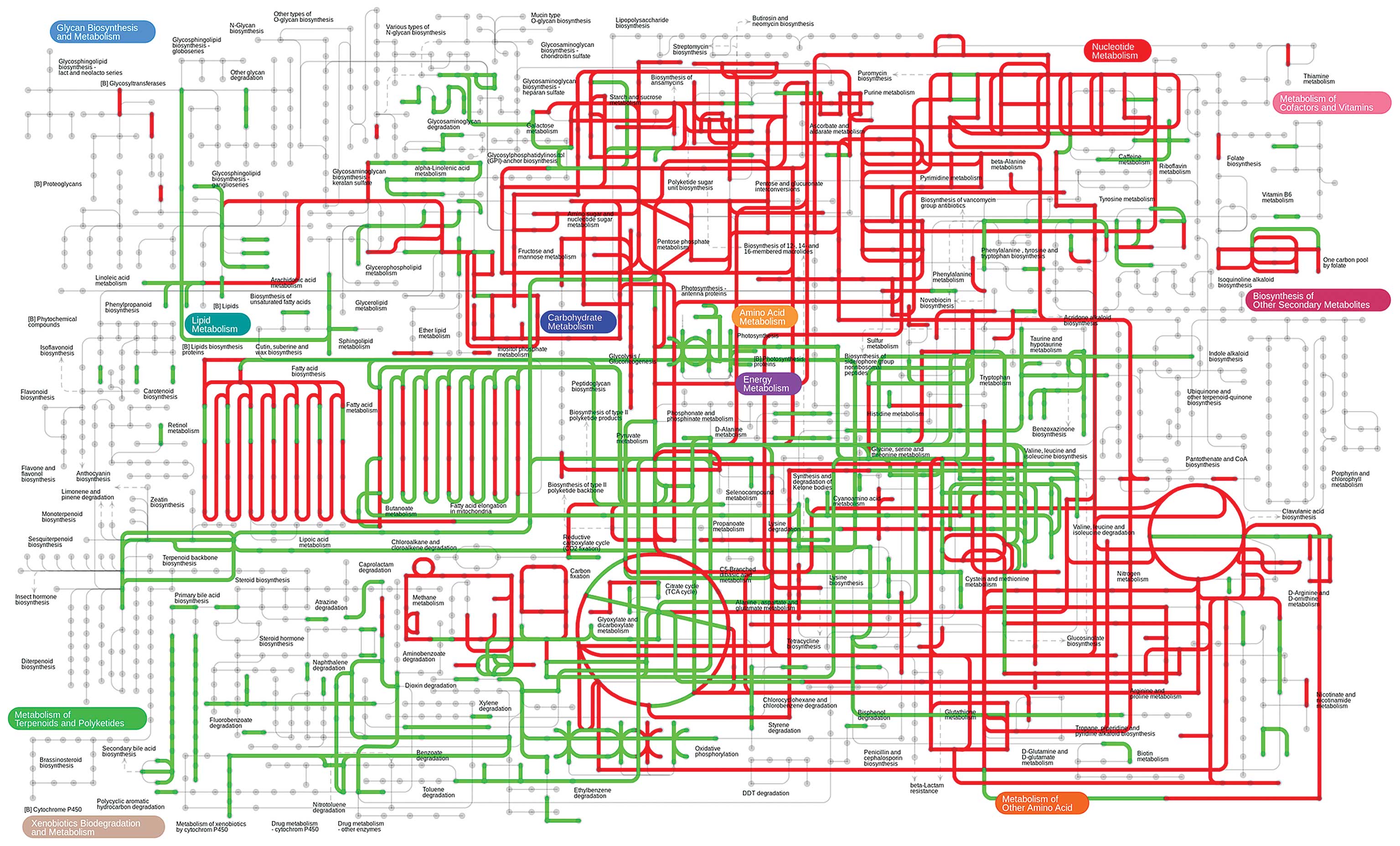
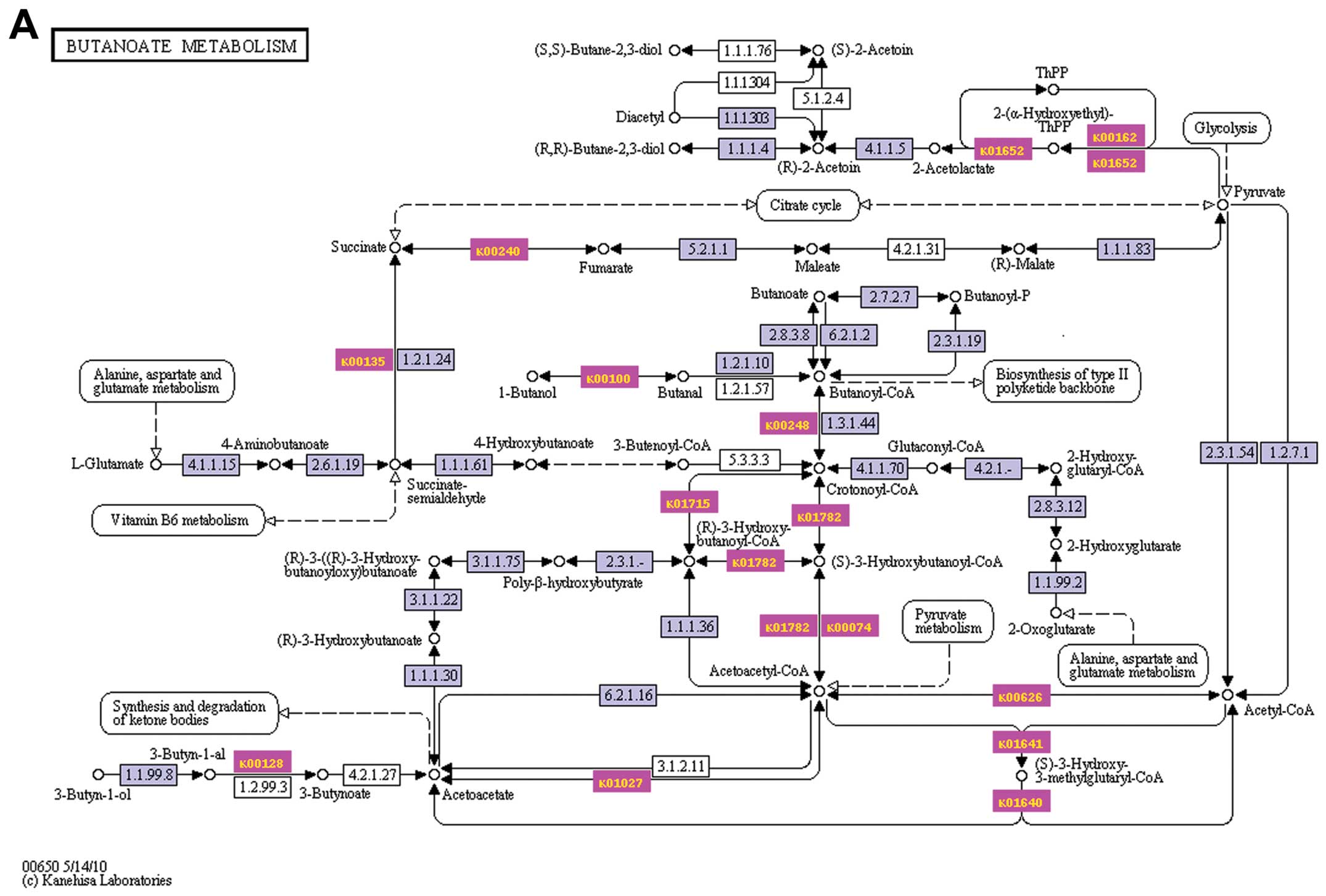
To investigate which signaling pathways have alterations in CSR, we searched the 359 identified proteins with significant change in the KEGG database and found that there were over 21 pathways with Seqs ≥3 between CSO and CSR as shown in Table II. Eight pathways were markedly upregulated in CSR compared with CSO (num upregulated protein ≥60%) and nine pathways were apparently downregulated in CSR compared with CSO (num downregulated protein ≥60%) (Fig. 2). Notably, most of the upregulated pathways (6 of 8) and cellular components are involved in carbohydrate metabolism indicating that carbohydrate metabolic activity was higher in recurrent chordomas than in primary chordomas (Fig. 2). Fig. 3 shows that the top 3 upregulated pathways including butanoate, inositol phosphate and glyoxylate and dicarboxylate metabolism are all involved in carbohydrate metabolism. Those 3 pathways were upregulated by 85.7, 75 and 73.3%, respectively, in the CSR compared with the CSO (Fig. 3). The glycolysis/gluconeogenesis pathway was also upregulated (Fig. 2). It has been reported that a high glycolytic rate has advantages for malignant cells (18). High glycolytic activity produces high levels of lactate and H+ ions which are transported outside the cell where they directly promote tumor aggressiveness through invasion and metastasis, two other hallmarks of cancer (19). Additionally, the genes and pathways that upregulate glycolysis are themselves anti-apoptotic (20). The increased carbohydrate metabolism in recurrent chordomas suggests that recurrent chordomas have a more aggressive phenotype and are resistant to therapy. The other 2 upregulated pathways in the recurrent chordomas are involved in energy metabolism and amino acid metabolism. In contrast to the upregulated pathways, the downregulated pathways participate in various biological processes and molecular functions including nucleotide metabolism, amino acid metabolism, carbohydrate metabolism, genetic information processing and translation, and biosynthesis of other secondary metabolites.

Table II

KEGG database identified 21 pathways with Seqs ≥3.

Table II

KEGG database identified 21 pathways with Seqs ≥3.

Pathways#Seqs#Enzs
Purine metabolism1011
Arginine and proline metabolism56
Starch and sucrose metabolism56
Pyruvate metabolism44
Glycolysis/gluconeogenesis44
Oxidative phosphorylation42
Alanine, aspartate and glutamate metabolism45
Cysteine and methionine metabolism45
Aminoacyl-tRNA biosynthesis33
Butanoate metabolism33
Methane metabolism33
Streptomycin biosynthesis33
Amino sugar and nucleotide sugar metabolism35
Propanoate metabolism34
Valine, leucine and isoleucine degradation33
β-alanine metabolism34
Drug metabolism - other enzymes32
Glyoxylate and dicarboxylate metabolism33
Phosphatidylinositol signaling system33
Inositol phosphate metabolism33
Pyrimidine metabolism33

To further determine the cellular function change between CSO and CRO, the statistically significant 359 proteins identified were classified according to gene ontology terms. The proteins were found to be involved in either a biological process, a cellular component and/or a molecular function (Fig. 4), indicating that there were diverse cellular components and catalytic activity change in recurrent chordomas compared to the original chordoma. Furthermore, when the identified proteins in the recurrent chordoma were mapped to the corresponding metabolic pathways, many key cellular pathways including amino acid, carbohydrate metabolism, energy, and nucleotide metabolism were found to be downregulated (Fig. 4).

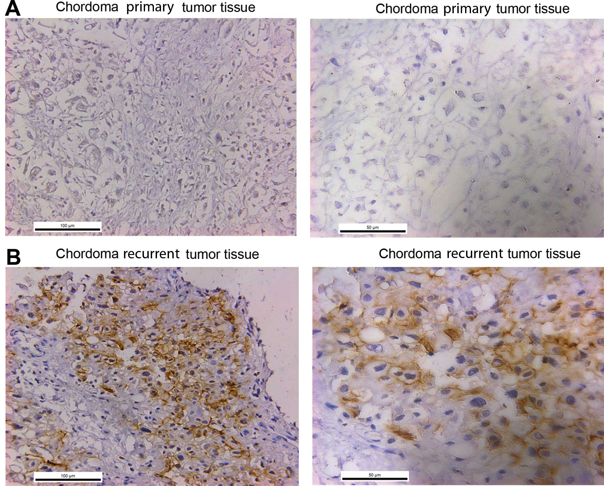
In order to confirm our results from the proteome, we examined tumor-related protein expression from the list of upregulated proteins through IHC in the chordoma patient primar y and CSR tissues. ALCAM (or CD166) is a 100–105 kDa type I transmembrane glycoprotein that is a member of the immunoglobulin superfamily of proteins (21). ALCAM has been reported as a cancer stem cell marker for non-small cell lung cancer (NSCLC) (15). Physiologically, it plays a role in the development of different tissues during embryogenesis. It is also expressed in various malignant lesions such as melanoma and esophageal, gynecologic, prostate, and pancreatic cancers, and its expression is associated with diverse outcomes in different tumors (2232). But the expression of ALCAM in chordomas has never been reported and the association between ALCAM and chordoma prognosis is not fully elucidated. In our study, we firstly detected ALCAM expression by using IHC in chordoma patient primary and CSR tissues. Fig. 5A clearly demonstrates that the primary chordoma tumor was negative for staining; however, the CSR had strong expression of ALCAM which suggests that ALCAM is a positive biomarker for recurrent chordomas and may play important roles for chordoma recurrence (Fig. 5B).

In conclusion, we analyzed the proteomic profile of a chordoma patient CSO and CSR and identified 359 proteins and 21 pathways with significant changes between CSO and CSR. Many of these molecular changes are reported in chordomas for the first time. Further investigation of the potential roles of these proteins in chordoma aggression is of interest. We also firstly found that the recurrent chordoma tumor showed enhanced carbohydrate metabolism, and the cancer stem cell marker ALCAM (CD166) expression level was increased markedly in CSR. The present study can serve as the basis for further research of recurrent chordomas.

Acknowledgments

The present study was supported by the National Nature Science Foundation for Distinguished Young Scholars of China (no. 81102036/H1624), the National Basic Research Program (no. 81272943), and the Nature Science Foundation of Shanghai (no. 12JC1411300).

References

1 

Stacchiotti S, Casali PG, Lo Vullo S, et al: Chordoma of the mobile spine and sacrum: a retrospective analysis of a series of patients surgically treated at two referral centers. Ann Surg Oncol. 17:211–219. 2010. View Article : Google Scholar

2 

DeLaney TF, Liebsch NJ, Pedlow FX, et al: Phase II study of high-dose photon/proton radiotherapy in the management of spine sarcomas. Int J Radiat Oncol Biol Phys. 74:732–739. 2009. View Article : Google Scholar :

3 

Zhou H, Chen CB, Lan J, et al: Differential proteomic profiling of chordomas and analysis of prognostic factors. J Surg Oncol. 102:720–727. 2010. View Article : Google Scholar : PubMed/NCBI

4 

Wisniewski JR, Zougman A, Nagaraj N and Mann M: universal sample preparation method for proteome analysis. Nat Methods. 6:359–362. 2009. View Article : Google Scholar : PubMed/NCBI

5 

Vogel C and Marcotte EM: Calculating absolute and relative protein abundance from mass spectrometry-based protein expression data. Nat Protoc. 3:1444–1451. 2008. View Article : Google Scholar : PubMed/NCBI

6 

Lu P, Vogel C, Wang R, Yao X and Marcotte EM: Absolute protein expression profiling estimates the relative contributions of transcriptional and translational regulation. Nat Biotechnol. 25:117–124. 2007. View Article : Google Scholar

7 

Benjamini Y and Hochberg Y: Controlling the false discovery rate - a practical and powerful approach to multiple testing. J R Stat Soc B. 57:289–300. 1995.

8 

Conesa A, Gotz S, Garcia-Gomez JM, Terol J, Talon M and Robles M: Blast2GO: a universal tool for annotation, visualization and analysis in functional genomics research. Bioinformatics. 21:3674–3676. 2005. View Article : Google Scholar : PubMed/NCBI

9 

Yamada T, Letunic I, Okuda S, Kanehisa M and Bork P: IPath2.0: interactive pathway explorer. Nucleic Acids Res. 39:W412–W415. 2011. View Article : Google Scholar : PubMed/NCBI

10 

Shimizu-Hirota R, Sasamura H, Kuroda M, Kobayashi E and Saruta T: Functional characterization of podocan, a member of a new class in the small leucine-rich repeat protein family. FEBS Lett. 563:69–74. 2004. View Article : Google Scholar : PubMed/NCBI

11 

Hoover KB, Liao SY and Bryant PJ: Loss of the tight junction MAGUK ZO-1 in breast cancer: relationship to glandular differentiation and loss of heterozygosity. Am J Pathol. 153:1767–1773. 1998. View Article : Google Scholar : PubMed/NCBI

12 

Liu W, Liu B, Xin L, Zhang Y, Chen X, Zhu Z and Lin Y: Downregulated expression of complement factor I: a potential suppressive protein for gastric cancer identified by serum proteome analysis. Clin Chim Acta. 377:119–126. 2007. View Article : Google Scholar

13 

Ohnuma SI: Cancer diagnosis and treatment. Google Patents. 2011

14 

Ostrow KL, Park HL, Hoque MO, et al: Pharmacologic unmasking of epigenetically silenced genes in breast cancer. Clin Cancer Res. 15:1184–1191. 2009. View Article : Google Scholar : PubMed/NCBI

15 

Tachezy M, Zander H and Wolters-Eisfeld G: Activated leukocyte cell adhesion molecule (CD166): an ‘inert’ cancer stem cell marker for non-small cell lung cancer? Stem Cells. 32:1429–1436. 2014. View Article : Google Scholar : PubMed/NCBI

16 

Ibrahim SA, Reed KR, Clarke AR, Pritchard DM and Jenkins JR: Sa1992 SFRS2 and CDC5L interaction: an insight into downstream events following APC deletion during colorectal carcinogenesis. Gastroenterology. 144:S353–S354. 2013.

17 

Palmieri D, Bouadis A, Ronchetti R, Merino MJ and Steeg PS: Rab11a differentially modulates epidermal growth factor-induced proliferation and motility in immortal breast cells. Breast Cancer Res Treat. 100:127–137. 2006. View Article : Google Scholar : PubMed/NCBI

18 

Klement RJ and Kammerer U: Is there a role for carbohydrate restriction in the treatment and prevention of cancer? Nutr Metab. 8:752011. View Article : Google Scholar

19 

Walenta S, Wetterling M, Lehrke M, Schwickert G, Sundfør K, Rofstad EK and Mueller-Klieser W: High lactate levels predict likelihood of metastases, tumor recurrence, and restricted patient survival in human cervical cancers. Cancer Res. 60:916–921. 2000.PubMed/NCBI

20 

Seyfried TN and Shelton LN: Cancer as a metabolic disease. Nutr Metab. 7:72010. View Article : Google Scholar

21 

Skonier JE, Bowen MA, Emswiler J, Aruffo A and Bajorath J: Recognition of diverse proteins by members of the immunoglobulin superfamily: delineation of the receptor binding site in the human CD6 ligand ALCAM. Biochemistry. 35:12287–12291. 1996. View Article : Google Scholar : PubMed/NCBI

22 

Weidle HU, Eggle D, Klostermann S and Swart GW: ALCAM/CD166: cancer-related issues. Cancer Genom Proteom. 7:231–243. 2010.

23 

Van Kilsdonk JW, Wilting RH, Bergers M, van Muijen GN, Schalkwijk J, van Kempen LL and Swart GW: Attenuation of melanoma invasion by a secreted variant of activated leukocyte cell adhesion molecule. Cancer Res. 68:3671–3679. 2008. View Article : Google Scholar : PubMed/NCBI

24 

Ofori-Acquah SF and King JA: Activated leukocyte cell adhesion molecule: a new paradox in cancer. Transl Res. 151:122–128. 2008. View Article : Google Scholar : PubMed/NCBI

25 

Ihnen M, Müller V, Wirtz RM, et al: Predictive impact of activated leukocyte cell adhesion molecule (ALCAM/CD166) in breast cancer. Breast Cancer Res Treat. 112:419–427. 2008. View Article : Google Scholar : PubMed/NCBI

26 

Mezzanzanica D, Fabbi M, Bagnoli M, et al: Subcellular localization of activated leukocyte cell adhesion molecule is a molecular predictor of survival in ovarian carcinoma patients. Clin Cancer Res. 14:1726–1733. 2008. View Article : Google Scholar : PubMed/NCBI

27 

Tachezy M, Zander H, Marx AH, Stahl PR, Gebauer F, Izbicki JR and Bockhornet M: ALCAM (CD166) expression and serum levels in pancreatic cancer. PLoS One. 7:e390182012. View Article : Google Scholar : PubMed/NCBI

28 

Tachezy M, Effenberger K, Zander H, et al: ALCAM (CD166) expression and serum levels are markers for poor survival of esophageal cancer patients. Int J Cancer. 131:396–405. 2012. View Article : Google Scholar

29 

Tachezy M, Zander H, Gebauer F, Marx A, Kaif JT, Izbicki JR and Bockhorn M: Activated leukocyte cell adhesion molecule (CD166) - its prognostic power for colorectal cancer patients. J Surg Res. 177:e15–e20. 2012. View Article : Google Scholar : PubMed/NCBI

30 

Tachezy M, Zander H, Marx AH, et al: ALCAM (CD166) expression as novel prognostic biomarker for pancreatic neuroendocrine tumor patients. J Surg Res. 170:226–232. 2011. View Article : Google Scholar : PubMed/NCBI

31 

Ishiguro F, Murakami H, Mizuno T, et al: Membranous expression of activated leukocyte cell adhesion molecule contributes to poor prognosis and malignant phenotypes of non-small-cell lung cancer. J Surg Res. 179:24–32. 2013. View Article : Google Scholar

32 

Simon R, Mirlacher M and Sauter G: Immunohistochemical analysis of tissue microarrays. Methods Mol Biol. 664:113–126. 2010. View Article : Google Scholar : PubMed/NCBI

Related Articles

Journal Cover

May-2015
Volume 33 Issue 5

Print ISSN: 1021-335X
Online ISSN:1791-2431

Sign up for eToc alerts

Recommend to Library

Copy and paste a formatted citation
x
Spandidos Publications style
Chen S, Xu W, Jiao J, Jiang D, Liu J, Chen T, Wan Z, Xu L, Zhou Z, Xiao J, Xiao J, et al: Differential proteomic profiling of primary and recurrent chordomas. Oncol Rep 33: 2207-2218, 2015
APA
Chen, S., Xu, W., Jiao, J., Jiang, D., Liu, J., Chen, T. ... Xiao, J. (2015). Differential proteomic profiling of primary and recurrent chordomas. Oncology Reports, 33, 2207-2218. https://doi.org/10.3892/or.2015.3818
MLA
Chen, S., Xu, W., Jiao, J., Jiang, D., Liu, J., Chen, T., Wan, Z., Xu, L., Zhou, Z., Xiao, J."Differential proteomic profiling of primary and recurrent chordomas". Oncology Reports 33.5 (2015): 2207-2218.
Chicago
Chen, S., Xu, W., Jiao, J., Jiang, D., Liu, J., Chen, T., Wan, Z., Xu, L., Zhou, Z., Xiao, J."Differential proteomic profiling of primary and recurrent chordomas". Oncology Reports 33, no. 5 (2015): 2207-2218. https://doi.org/10.3892/or.2015.3818