Introduction
The human epidermal growth factor receptor type 3
(HER3 or ErbB3) has recently attracted attention as a molecular
target for anticancer therapy (1–3). HER3
has no tyrosine kinase activity of its own and participates in
signaling by heterodimerization with other members of the HER
family (4). One of the mechanisms
behind acquired resistance of breast cancer patients to
HER2-targeting therapies may be due to the upregulation of HER3 and
hyperactivation of its signaling (5,6). In
addition, HER3 has been shown to be involved in resistance to
hormone therapy in breast cancer (7). Furthermore, HER3 expression in ovarian
cancer is associated with more aggressive disease; in lung cancer,
with resistance to tyrosine kinase-inhibiting therapy; and in
prostate cancer HER3 expression may drive tumor cell growth in
hormone refractory cancer (1).
Thus, there is a strong rationale for the development of
pharmaceuticals specifically targeting HER3. Besides monoclonal
antibodies, HER3-targeting antisense oligonucleotides and scaffold
proteins are under pre-clinical and early clinical development
(3,8,9).
The clinical implementation of anti-HER3 therapy is
a challenge. Treatment should be initiated at onset of HER3
overexpression to avoid overtreatment. As HER3 overexpression often
occurs as a response to therapy, the samples from primary tumors
would not be informative. Serial biopsies are undesirable due to
invasiveness of the procedure and HER3 overexpression may not be
synchronous in all the metastases that occur. The use of
radionuclide molecular imaging is a useful strategy since it is
non-invasive, repeatable and provides whole-body information.
Prerequisites for the successful application of
molecular imaging are high specificity and sensitivity. Although
radio-labeling of available therapeutic anti-HER3 monoclonal
antibodies is an approach to the development of imaging probes, it
is difficult to obtain a high sensitivity and specificity in this
manner. Anti-HER3 antibody (10)
and F(ab′)2 antibody fragments (11) were recently evaluated for positron
emission tomography (PET) imaging in a preclinical setting. Slow
clearance of antibodies from blood results in a high background
even several days after injection, reducing imaging contrast and
sensitivity. Additionally, bulky immunoglobulins with a molecular
weight of >45 kDa tend to accumulate non-specifically in tumors
due to an ‘enhanced permeability and retention’ (EPR) effect
(12). This may cause
false-positive findings, i.e., specificity of antibody-mediated
imaging is low. A possible solution is the use of smaller imaging
agents, which provide rapid extravasation and diffusion into the
extracellular space, rapid clearance of unbound tracer, and exclude
the EPR effect.
Previously, we demonstrated the feasibility of
radio-nuclide imaging of HER3 expression in vivo (murine
models) using a 99mTc-labeled HER3-antagonistic affibody
molecule 99mTc(CO)3-HEHEHE-Z08699 (13). Affibody molecules are small (7 kDa)
scaffold proteins that are selected using molecular display
techniques for high-affinity binding to different target proteins
(14). Affibody molecules have
demonstrated the ability of imaging of HER2 expression in breast
cancer patients (15,16).
HER3-targeting affibody molecules Z08698 and Z08699
have been selected and demonstrate high affinities to HER3 (50 and
21 pM, respectively) (9). Such high
affinities are important for the imaging of molecular targets with
a relatively low tumor expression. Z08698 and Z08699 were also
found to have high affinity to the murine counterpart of HER3,
mErbM3, rendering mice an adequate model for the evaluation of
imaging properties (13).
The aim of this study was the development of a HER3
imaging agent labeled with radiometals suitable for single photon
emission computed tomography (SPECT) and PET.
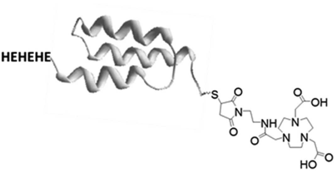
Engineering of a unique cysteine into affibody
molecules provides site-specific conjugation of the chelator,
resulting in well-defined products with reproducible
biodistribution properties. Findings regarding affibody molecules
suggested that placement of a cysteine at the C-terminus is
convenient, as this provides a maximum distance from a binding site
and minimizes possible interference of the chelator with the
molecular recognition (17,18). The use of the NOTA chelator
(2,2′,2″-(1,4,7-triazonane-1,4,7-triyl)triacetic acid) provides
stable labeling with 111 In for SPECT, and with
68Ga, 64Cu or 18F (using aluminum
monofluoride 18F-AlF chemistry) for PET. Previously, we
showed that placement of a HEHEHE-tag
(histidyl-glytamyl-histidyl-glytamyl-histidyl-glytamyl) at the
N-terminus permits immobilized metalion affinity chromatography
(IMAC) purification of affibody molecules and improves their
biodistribution by reducing hepatic uptake (13,19).
Bearing these considerations in mind, we generated two anti-HER3
affibody molecules with the HEHEHE-tag at the N-terminus and NOTA
chelator at the C-terminus (i.e., HEHEHE-Z-NOTA) (Fig. 1).
Investigations on factors influencing imaging using
HER2-targeting affibody molecules have demonstrated that low
picomolar affinity is essential for the imaging of targets with low
expression, such as HER3 (20).
Previous findings have also demonstrated that small changes in
amino acid composition can modify off-target interactions and
affect blood clearance rate and hepatic uptake (20). Thus high affinity is not the only
parameter to be considered for the selection of affibody molecules
for imaging, and evaluation of several variants with approximately
equal affinity may prove useful in selecting an agent with the most
suitable in vivo properties.
Findings obtained showed that targeting of HER3 and
other tyrosine kinases with an expression pattern similar to that
of HER3 (endogenous expression in healthy tissue, e.g., IGF-1R)
suggested that imaging contrast could be improved at later
time-points (i.e., at 24 h p.i.) (13,21).
Thus, 111In was used as a label whose half-life permits
studying biodistribution at the day of injection and during the
following day. The targeting properties of
111In-HEHEHE-Z08698-NOTA and
111In-HEHEHE-Z08699-NOTA were compared in vitro
and in vivo in nude mice bearing HER3-expressing BT-474
breast carcinoma xenografts.
Materials and methods
High-quality Milli-Q water (resistance >18 MΩ/cm)
was used to prepare the solutions. [111In]-indium
chloride was purchased from Covidien, Ltd. (Dublin, Ireland). Cells
used during the in vitro experiments were detached using
trypsin-EDTA solution (0.05% trypsin, 0.02% EDTA in buffer;
Biochrom AG, Berlin, Germany). Radioactivity was measured using an
automated gamma-counter with a 3-inch NaI(Tl) detector (1480
Wizard; Wallac Oy, Turku, Finland). The purity of radiolabeled
affibody molecules was determined by radio ITLC (150-771 Dark
Green, Tec-Control Chromatography strips from Biodex Medical
Systems) and cross-validated by sodium dodecyl sulfate
polyacrylamide gel electrophoresis (SDS-PAGE). The distribution of
radioactivity along the thin layer chromatography strips was
measured on a Cyclone Storage Phosphor System and analyzed using
the OptiQuant image analysis software (Perkin-Elmer, Waltham, MA,
USA).
For the in vitro and in vivo
experiments, the HER3-expressing BT-474 breast carcinoma cell line
[American Type Tissue Culture Collection (ATCC) via LGC Promochem,
Borås, Sweden] was used. Animal experiments were planned and
performed in accordance with national legislation on protection of
laboratory animals and were approved by the Ethics Committee for
Animal Research in Uppsala, Sweden.
Data on the cell uptake and biodistribution were
assessed by an unpaired, two-tailed t-test using GraphPad Prism
(version 4.00 for Windows GraphPad Software) to determine
significant differences (P<0.05).
Production, purification,
NOTA-conjugation and analysis of affibody molecules
Production of Z08698 and Z08699 was previously
described (9,13). In the present study, the DNA
sequences encoding Z08698 and Z08699 were amplified using
polymerase chain reaction (PCR) with primers incorporating a
HEHEHE-tag (N-terminal) and cysteine (C-terminal). Coupling of NOTA
chelator was performed as previously described (22). The NOTA-conjugated proteins were
subsequently purified using a 1200 series HPLC system in a Zorbax
C18 semi-preparative column (Agilent Technologies, Santa Clara, CA,
USA). The molecular masses of the purified proteins were confirmed
using a 6520 Accurate-Mass Q-TOF LC/MS (Agilent Technologies). The
purity of the NOTA-conjugated affibody molecules was determined
using an analytical column Zorbax 300B-C18 (Agilent Technologies)
on 1200 series RP-HPLC (Agilent Technologies). The secondary
structure content, thermal stability and refolding capacity was
analyzed using circular dichroism (CD) spectroscopy on a Jasco-810
(Jasco Inc., Easton, MD, USA). Affinities to human ErbB3-FC
(R&D Systems, Minneapolis, MN, USA) were determined on a
BiaCore 3000 system (GE Healthcare, Pittsburgh, PA, USA).
Labeling of HEHEHE-Z08698-NOTA and
HEHEHE-Z08699-NOTA with 111In
For labeling, 111In-HEHEHE-Z08698-NOTA or
111In-HEHEHE-Z08699-NOTA (40 μg, 6 nmol, in 100
μl 0.2 M ammonium acetate, pH 5.5) was incubated with 54
μl 111In-chloride solution (40 MBq) at 85°C for
40 min. The reaction mixture was obtained and analyzed by
radio-ITLC (instant thin-layer chromatography) eluted with 0.2 M
citric acid, pH 2.0. To cross-validate the ITLC, an SDS-PAGE
analysis (200 V constant, NuPAGE 4–16% Bis-Tris gel; Invitrogen AB)
was performed. To ensure high radiochemical purity, the conjugates
were purified using disposable NAP-5 columns (GE Healthcare)
according to the manufacturer’s instructions.
An EDTA-challenge was performed to evaluate the
labeling stability of conjugates [500-fold molar excess of the
tetrasodium salt of ethylenediaminetetraacetic acid (EDTA) solution
in water, 2 h at room temperature]. The samples were analyzed using
radio-ITLC. To evaluate stability of the conjugates in serum,
labeled conjugates were incubated at 37°C in fetal bovine serum (5
μg in 200 μl PBS diluted with 200 μl serum).
At 4- and 24-h incubation, the samples were analyzed using
SDS-PAGE. For the control, labeled conjugates were incubated under
the same conditions in PBS. No release of free 111In or
transchelation to serum proteins was detected (data not shown).
Real-time ligand-binding kinetics, and
KD determination for 111In-HEHEHE-Z08698-NOTA
and 111In-HEHEHE-Z08699-NOTA on BT-474 cells
The binding of 111In-HEHEHE-Z08698-NOTA
and 111In-HEHEHE-Z08699-noTA on BT-474 cells was
measured in real-time at room temperature using LigandTracer Yellow
(Ridgeview Instruments AB, Vänge, Sweden) as previously described
(23). The radioligand
concentrations of 0.2 and 2 nM were used. These concentrations were
selected to receive a clear increase in signal by addition of the
second concentration. Uptake was monitored for 105 min and
retention for 960 min. Interaction analysis and calculation of
equilibrium dissociation constant (KD) was performed
with TracerDrawer software (Ridgeview Instruments AB).
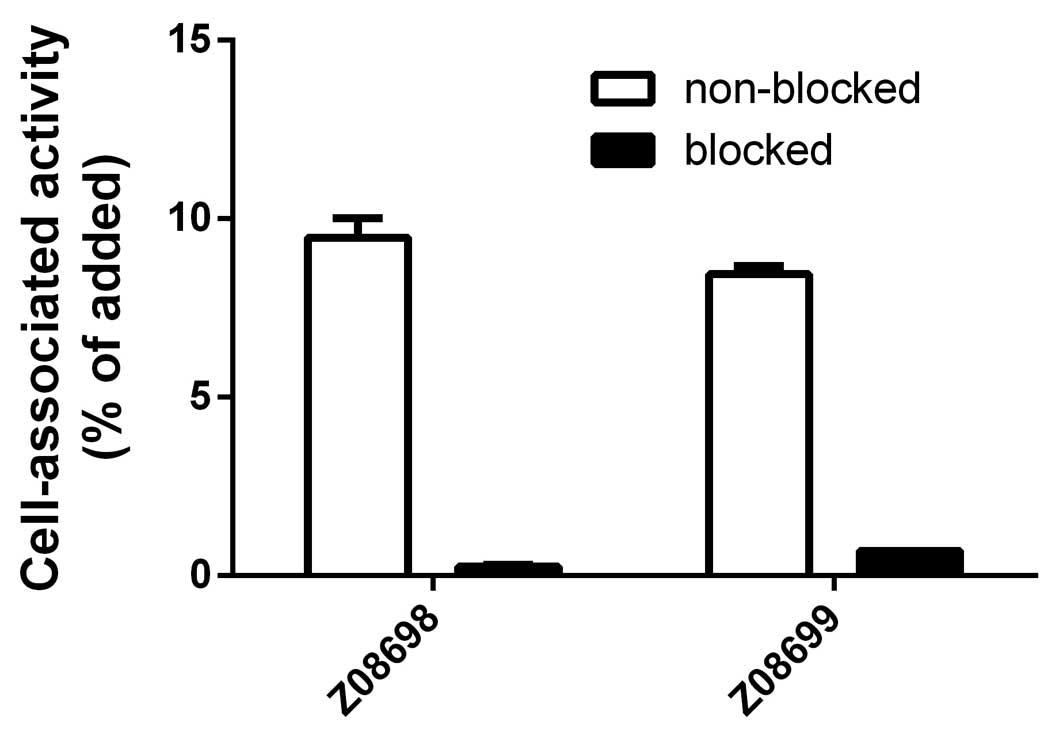
In vitro specificity of
111In-HEHEHE-Z08698-NOTA and
111In-HEHEHE-Z08699-NOTA to HER3-expressing cells
In vitro experiments were performed in
triplicate. For experiments 1×10⁶ BT-474 cells/dish were
seeded the day prior to the experiment.
An in vitro specificity test was performed as
previously described (24).
Briefly, a solution of radiolabeled affibody molecules (at 0.1 nM)
was added to the cell plates. For blocking, 5 nM of non-labeled
affibody molecule was added 15 min before the radiolabeled
conjugates to saturate the receptors in half of the dishes. The
cells were incubated for 1 h at 37°C. Thereafter, the medium was
collected and the cells were detached by trypsin-EDTA solution. The
samples were measured on radioactivity content to enable
calculation of the fraction of cell-bound radioactivity.
In vivo studies
To evaluate targeting of HER3-expressing tumors
in vivo, mice bearing BT-474 breast carcinoma xenografts
were used. Ten million BT-474 cells were implanted in 50% Matrigel
in Balb/c nu/nu female mice pre-implanted with estradiol pellets
(0.025 mg/day, 20 days; Innovative Research of America, Sarasota,
FL, USA) and used 3 weeks after implantation. Tumor size at
initiation of the experiment was 0.6±0.3 g. Mice (3–4 per group)
were intravenously injected with
111In-HEHEHE-Z08698-NOTA or
111In-HEHEHE-Z08699-NOTA (1 μg/30 kBq in 100
μl PBS). The animals were sacrificed at 1, 4, 8 and 24 h
p.i. by injection of a lethal dose of anesthesia [20 μl of
Ketalar Rompun per gram body weight: Ketalar (50 mg/ml; Pfizer), 10
mg/ml; Rompun, (20 mg/ml; Bayer] followed by heart puncture and
exsanguination with a syringe rinsed with heparin (5000 IE/ml; Leo
Pharma). Tumors and samples of blood, lung, liver, spleen, stomach,
small intestines, kidney, salivary gland, muscle and bone were
collected, weighed and their radioactivity was measured. The data
were corrected for background. Tissue uptake was calculated as
%IA/g (percent of injected radioactivity per gram).
To evaluate if uptake in tumors and
mErbB3-expressing organs and tissues was saturable, labeled
affibody conjugates were intravenously injected in a group of mice
with injected protein dose adjusted by dilution with non-labeled
affibody molecule to 70 μg per mouse. Biodistribution was
performed 4 h p.i. as described above.
Imaging studies
The study was performed to obtain visual
confirmation of the biodistribution data. Tumor-bearing mice were
injected with 0.8 MBq 111In-HEHEHE-Z08698-NOTA (1
μg) and 111In-HEHEHE-Z08699-NOTA (1 and 70
μg). At 4 h p.i., the animals were euthanized and the
urinary bladder was excised post-mortem to improve image quality.
Static planar imaging of the three mice was performed
simultaneously using a GE Infinia gamma camera equipped with a MEGP
(medium energy general purpose) collimator. Static images (20 min)
of the three animals were obtained with a zoom factor of 3 in a
256×256 matrix.
Statistical analysis
Data are presented as mean ± SD.
Results
Production, purification and analysis of
conjugates
The affibody molecules were expressed in E.
coli and purified using heat-induced precipitation of host
proteins. Correct size and sufficient purity for conjugation was
confirmed using SDS-PAGE (data not shown). Mass-spectrometry
analysis revealed that the two binders had the expected molecular
weight after conjugation to the NOTA chelator at the C-terminal
cysteine using maleimide chemistry [HEHEHE-Z08698-NOTA-8149.55
(8149) and HEHEHE-Z08699-NOTA-8112.39 (8112) Da]. Analysis using
RP-HPLC showed purities of 95.6 and 96.6% for HEHEHE-Z08698-NOTA
and HEHEHE-Z08699-NOTA, respectively. Circular dichroism (CD)
spectra verified expected α-helical secondary structure content,
indicating that correct folding into a three-helical bundle, and
variable temperature measurement (VTM) at 221 nm revealed melting
temperatures (Tm) of 65°C and 63°C for Z08698 and
Z08699, respectively. CD measurements following VTM treatment
showed no loss of helical content, demonstrating complete refolding
upon lowering the temperature after heat-induced denaturation.
According to the SPR analysis, dissociation constants for binding
to HER3 were 45 and 77 pM for HEHEHE-Z08698-NOTA and
HEHEHE-Z08699-NOTA, respectively.
Labeling and in vitro stability
The average yield of
111In-HEHEHE-Z08698-NOTA was 95±3%, and that of
111In-HEHEHE-Z08699-NOTA was 96.2±0.5%. Simple
purification using disposable size-exclusion columns provided a
radiochemical purity of >98%. A specific activity of 8
GBq/μmol was obtained routinely during labeling. The two
conjugates were stable under EDTA-challenge for 2 h and in fetal
bovine serum for 24 h at 37°C.
In vitro receptor binding
An in vitro cell binding assay showed that
the two affibody molecules bound to HER3-positive BT-474 cells.
Moreover, saturation of the receptors by pre-incubation with
non-labeled affibody molecules significantly decreased the binding
of the radiolabeled conjugates, demonstrating that the interactions
were specific (Fig. 2).
According to the analysis of real-time
ligand-binding kinetics data, the calculated KD values
for 111In-HEHEHE-Z08698-NOTA and
111In-HEHEHE-Z08699-NOTA were in the picomolar range
(5.4±0.4 and 4.2±0.4 pM, respectively) (data not shown).
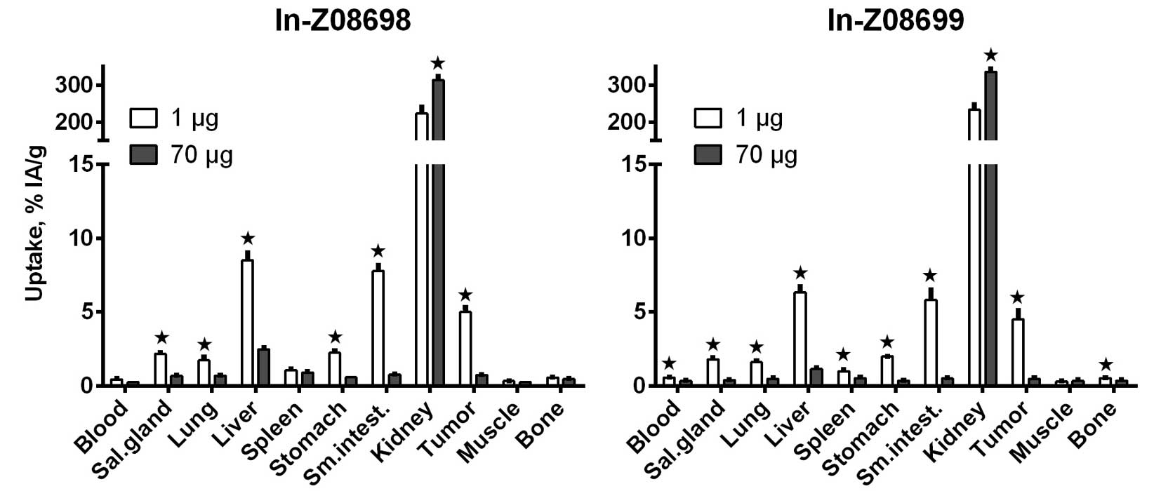
In vivo studies
The two conjugates demonstrated specific binding
in vivo to HER3 (BT-474 xenografts) and to mErbB3 (Fig. 3). Uptake of radioactivity in BT-474
xenografts and the mErbB3-expressing organs and tissues was
significantly reduced when 70 μg of affibody molecule was
injected compared to 1 μg. Reduction in radioactivity uptake
in liver was more pronounced for the Z08699 variant. At the same
time, significantly increased uptake was identified in kidneys for
the two conjugates.
Biodistribution over time of
111In-HEHEHE-Z08698-NOTA and
111In-HEHEHE-Z08699-NOTA in BT-474 xenograft-bearing
mice is presented in Tables I and
II. The overall pattern of
radioactivity distribution in tumors and normal organs was similar
for the two conjugates. The tumor uptake of radioactivity exceeded
radioactivity concentration in blood at 1 h p.i. and the
tumor-to-blood ratio increased over time (Table I). The uptake of radioactivity in
tumors was higher than that in organs with mErbB3 expression
(salivary glands, lungs and stomach). On the other hand, the uptake
of radioactivity in liver and small intestine tissue was higher
than that in tumors. The uptake of radioactivity in tissues without
mErbB3 expression (spleen, muscle and bone) was low. The main
excretion pathway was renal for the two conjugates with substantial
retention of radioactivity in kidneys. Differences in the
biodistribution pattern between 111In-HEHEHE-Z08698-NOTA
and 111In-HEHEHE-Z08699-NOTA were observed.
111In-HEHEHE-Z08698-NOTA demonstrated more rapid blood
clearance, better radioactivity retention in tumors, higher
tumor-to-blood ratio at 24 h p.i., but higher uptake of
radioactivity in liver. The tumor-to-muscle ratios were in the
range of 15–20 and the tumor-to-bone ratios, in the range of 6–12
(Table II). Tumor-to-liver ratios
were <1, and the tumor-to-intestine ratio was >1 for
111In-HEHEHE-Z08699-NOTA at 24 h p.i. (Table II).
 | Table IBiodistribution of
111In-HEHEHE-Z08698-NOTA and
111In-HEHEHE-Z08699-NOTA in BT-474 tumor-bearing Balb/c
nu/nu mice. |
Table I
Biodistribution of
111In-HEHEHE-Z08698-NOTA and
111In-HEHEHE-Z08699-NOTA in BT-474 tumor-bearing Balb/c
nu/nu mice.
| Organ |
111In-HEHEHE-Z08698-NOTA
|
111In-HEHEHE-Z08699-NOTA
|
|---|
| 1 h | 4 h | 8 h | 24 h | 1 h | 4 h | 8 h | 24 h |
|---|
| Blood | 0.95±0.04 | 0.4±0.1 | 0.34±0.05 | 0.24±0.02 | 1.10±0.09b | 0.56±0.06 | 0.42±0.02b | 0.20±0.02 |
| Salivary gland | 2.97±0.08 | 2.2±0.2 | 1.9±0.2 | 1.4±0.5 | 2.5±0.1 | 1.8±0.2 | 1.5±0.4 | 0.9±0.3 |
| Lung | 2.1±0.5 | 1.7±0.5 | 1.1±0.2 | 0.73±0.04 | 2.3±0.2 | 1.6±0.3 | 1.2±0.1 | 0.8±0.3 |
| Liver | 7.7±0.8a | 8±1a | 7.5±0.4a | 4.9±0.2a | 6.0±0.4 | 6.3±0.7 | 4.7±0.6 | 3.0±0.1 |
| Spleen | 1.0±0.2a | 1.1±0.2 | 1.0±0.1 | 1.01±0.05a | 0.91±0.02 | 1.0±0.2 | 0.92±0.05 | 0.71±0.04 |
| Stomach | 3.0±0.4 | 2.2±0.3 | 1.8±0.2a | 1.51±0.09a | 2.9±0.6 | 1.99±0.07 | 1.3±0.1 | 0.92±0.01 |
| Small
intestine | 12±5 | 7.8±0.8a | 5.2±0.9 | 5±2 | 9±2 | 6±1 | 5±2 | 2.1±0.7 |
| Kidney | 187±9 | 223±33 | 182±24 | 188±24 | 194±14 | 233±28 | 244±23b | 195±11 |
| Tumor | 5.1±0.4 | 5.0±0.6 | 4.0±0.2 | 3.7±0.2a | 4.7±0.8 | 5±1 | 3.9±0.4 | 2.4±0.2 |
| Muscle | 0.37±0.06 | 0.32±0.08 | 0.21±0.05 | 0.3±0.2 | 0.34±0.06 | 0.29±0.06 | 0.3±0.1 | 0.19±0.08 |
| Bone | 0.61±0.05 | 0.57±0.07 | 0.34±0.04 | 0.37±0.07 | 0.7±0.2 | 0.53±0.07 | 0.53±0.06 | 0.37±0.04 |
| Table IITumor-to-organ ratios of
111In-HEHEHE-Z08698-NOTA and
111In-HEHEHE-Z08699-NOTA in BT-474 tumor-bearing Balb/c
nu/nu mice. |
Table II
Tumor-to-organ ratios of
111In-HEHEHE-Z08698-NOTA and
111In-HEHEHE-Z08699-NOTA in BT-474 tumor-bearing Balb/c
nu/nu mice.
| Organ |
111In-HEHEHE-Z08698-NOTA
|
111In-HEHEHE-Z08699-NOTA
|
|---|
| 1 h | 4 h | 8 h | 24 h | 1 h | 4 h | 8 h | 24 h |
|---|
| Blood | 5.3±0.4 | 12±3a | 12±2 | 15.5±0.7a | 4.3±0.8 | 8±1 | 9±1 | 11.8±0.1 |
| Salivary gland | 1.7±0.2 | 2.3±0.3 | 2.1±0.1 | 3±1 | 1.9±0.4 | 2.5±0.7 | 2.8±0.9 | 3.0±1.0 |
| Lung | 2.5±0.6 | 3.0±0.4 | 3.6±0.7 | 5.1±0.4 | 2.0±0.3 | 2.9±0.8 | 3.2±0.4 | 3±1 |
| Liver | 0.66±0.06 | 0.59±0.06 | 0.53±0.01 | 0.75±0.07 | 0.8±0.2 | 0.7±0.2 | 0.8±0.1b | 0.80±0.08 |
| Spleen | 5.1±0.6 | 4.7±0.5 | 4.1±0.4 | 3.7±0.3 | 5.2±0.8 | 4.5±0.4 | 4.3±0.5 | 3.4±0.2 |
| Stomach | 1.7±0.2 | 2.3±0.2 | 2.3±0.3 | 2.45±0.08 | 1.7±0.4 | 2.3±0.6 | 3.0±0.6 | 2.6±0.3 |
| Small
intestine | 0.5±0.2 | 0.65±0.09 | 0.77±0.10 | 0.8±0.3 | 0.5±0.2 | 0.8±0.2 | 0.8±0.1 | 1.2±0.3 |
| Muscle | 14±2 | 16±4 | 20±6 | 16±8 | 14±4 | 16±7 | 15±6 | 14±4 |
| Bone | 8±1 | 8.9±0.5 | 12±2a | 10±3 | 7±1 | 9±2 | 7.5±0.9 | 6.3±0.2 |
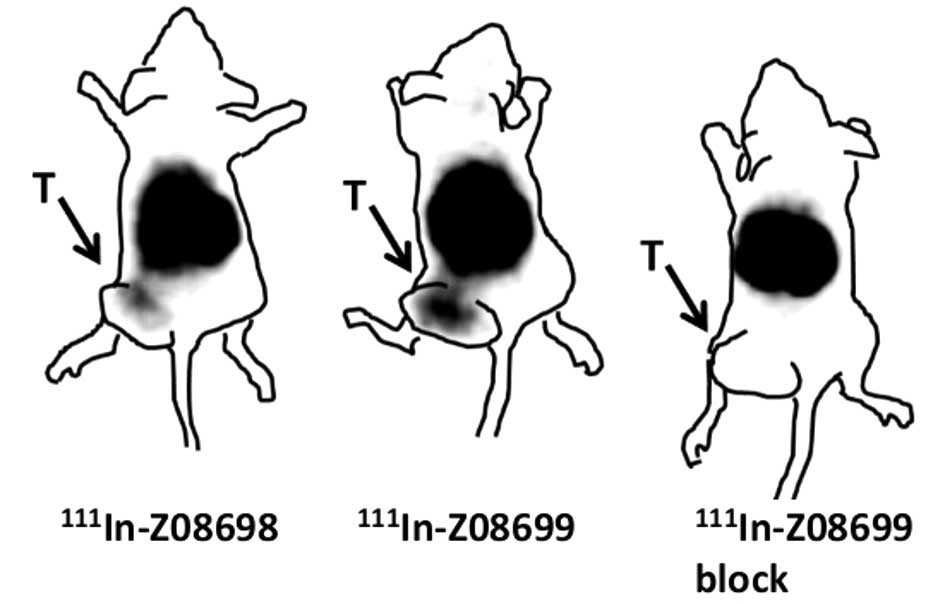
Gamma-camera images (Fig. 4) confirmed the major findings of the
biodistribution study. The two conjugates visualized
HER3-expressing xenografts at 4 h p.i. It was clearly demonstrated
that co-injection of 70 μg of non-labeled Z08699 decreased
the uptake of radioactivity in the xenograft. In agreement with the
biodistribution data, the radioactivity uptake in kidneys dominated
the images and radioactivity uptake in liver was at least on the
same level as in the tumors. It was also confirmed that blood
clearance occurred more rapidly for
111In-HEHEHE-Z08698-NOTA, resulting in lower background
radioactivity.
Discussion
Elucidation of the role of HER3 in cancer resistance
to different types of treatments has resulted in the development of
HER3-targeting therapies. Imaging may be a valuable clinical tool
for the stratification of patients for such therapies, making
treatment more personalized. Previously, we demonstrated the
feasibility of imaging of HER3-expressing xenografts using a
99mTc-labeled anti-HER3 HEHEHE-Z08698 affibody molecule
(13). That investigation initiated
a series of studies focused on the development of affibody-based
HER3-imaging agents suitable for PET and SPECT applications. To
achieve that aim, a NOTA chelator, site-specifically conjugated to
a C-terminal cysteine of the affibody molecule was selected. In a
new HEHEHE-Z-NOTA format tested for two variants, Z08698 and
Z08699, the affibody molecules retained their low picomolar
affinity to HER3. However, the altered format was found to have
some effect on the mutual affinities. The dissociation constant of
HEHEHE-Z08698-NOTA (45 pM) was lower than that of
HEHEHE-Z08699-NOTA (77 pM), while the opposite relationship was
observed for the parental affibody molecules Z08698 (50 pM) and
Z08699 (21 pM). The two conjugates retained a capacity for
high-fidelity refolding after heating, which is essential as
labeling using macrocyclic chelators requires relatively harsh
temperature conditions. Labeling of conjugates was performed at
elevated temperatures (85°C) that provided stable labeling of
111In-HEHEHE-Z08698-NOTA and
111In-HEHEHE-Z08699-NOTA. However, the two conjugates
demonstrated preserved binding specificity to HER3-expressing
BT-474 breast cancer cells after labeling.
The two conjugates demonstrated saturable binding to
HER3-expressing breast cancer xenografts in vivo, which
suggests target-specific uptake. Of note, saturable uptake was also
demonstrated in mErbB3-expressing murine tissues (Fig. 3). Expression of a molecular target
in normal tissues may appreciably affect imaging, and
cross-reactivity of affibody molecules with the murine counterpart
makes the animal model adequate for assessment of imaging
properties.
Uptake of conjugates in tumor xenografts did not
differ significantly between 111In-HEHEHE-Z08698-NOTA
and 111In-HEHEHE-Z08699-NOTA at earlier time points (1–8
h p.i.). However, 111In-HEHEHE-Z08698-NOTA had improved
tumor retention of radioactivity at 24 h p.i. The main difference
between the conjugates was in their uptake in normal organs and
tissues. It is apparent that 111 In-HEHEHE-Z08698-NOTA
cleared more rapidly from blood but had a higher uptake in liver
and spleen. Specifically, the two phenomena may be connected and
elevated hepatic uptake may be a reason for more rapid conjugate
elimination from blood. Furthermore, the HER3-binding site of the
tested conjugates may be involved in off-target interactions and
thereby influence their biodistribution profiles. The only
differences between these affibody molecules in the binding sites
were that Q11 and W25 in Z08698 were substituted by N11 and Y25 in
Z08699. Previous findings on HER2-targeting affibody molecules
(20) have shown that substitution
of two amino acids in the binding site by two more lipophilic ones
can cause higher hepatic uptake. Overall,
111In-HEHEHE-Z08698-NOTA provided higher tumor-to-blood
ratio, and should be considered as superior.
In a previous study, we evaluated targeting of
BT-474 xenografts using HEHEHE-Z08699 labeled with
99mTc(CO)3 (4 h p.i.) (13). A comparison with
111In-HEHEHE-Z08699-NOTA suggests that the combination
of 111In as a label and NOTA as a chelator provided a
marked increase in tumor uptake (5±1 vs. 1.7±0.7 %IA/g, for
111In-HEHEHE-Z08699-NOTA and
99mTc(CO)3-HEHEHE-Z08699, respectively).
111In-HEHEHE-Z08699-NOTA provided ~2-fold higher
tumor-to-stomach and tumor-to-lung ratios, the latter of which is
important in the context of breast cancer as lung metastases are
extremely common in this disease. This finding suggests that the
HEHEHE-containing NOTA-conjugated affibody molecule is a preferable
platform for further development of HER3-imaging agents.
Few studies are available on the development of
imaging agents visualizing HER3 in vivo. One potential
agent, 111In-DTPA-HRG provided a tumor uptake of 2.1±0.4
%IA/g, and a tumor-to-blood ratio of 7 at 2 days p.i. in mice
bearing HER3-positive xenografts (25). An anti-HER3 antibody
(89Zr-RG7116) demonstrated the highest tumor uptake and
imaging contrast at 6 days p.i. (10). Another PET imaging probe based on a
F(ab′)2 antibody fragment was presented in an abstract (11). This probe demonstrated comparable
contrast to the anti-HER3 affibody molecules shown in the present
study, although the time point was not specified (11).
111In-HEHEHE-Z08698-NOTA has reasonable
imaging contrast (tumor-to-blood ratio of 12±3, tumor-to-muscle
ratio of 16±4, tumor-to-lung ratio of 2.3±0.3, and tumor-to-bone
ratio of 8.9±0.5) already at 4 h p.i. This indicates that this
variant labeled with short-lived 68Ga (T½ =67.6 min) and
18F (T½ =110 min) may be useful for imaging using PET.
The labeling chemistry should be re-optimized, and imaging
properties have to be re-evaluated for these conjugates. However,
the current study provides a good rationale for future studies.
The suggested imaging agent may enable a
non-invasive and repeatable visualization of HER3 expression in
metastatic cancer. Detection of HER3 overexpression onset suggests
including anti-HER3 therapy to the treatment of disseminated
cancer. Use of HER3-imaging agents in combination with
18F-FDG PET may initially detect metastases and provide
their anatomical location prior to imaging of HER3 using affibody
molecules, as has been described for HER2-imaging affibody
molecules.
In conclusion, 111In-HEHEHE-Z08698-NOTA
is a promising imaging agent for visualization of HER3-expression
in cancer metastasis using SPECT. A good imaging contrast at 4 h
after injection indicates that this conjugate may be used for
patient stratification for anti-HER3 therapy.
Acknowledgments
The present study was supported by grants from the
Swedish Cancer Society (Cancerfonden) and Swedish Research Council
(Vetenskapsrådet).
References
|
1
|
Baselga J and Swain SM: Novel anticancer
targets: Revisiting ERBB2 and discovering ERBB3. Nat Rev Cancer.
9:463–475. 2009. View
Article : Google Scholar : PubMed/NCBI
|
|
2
|
Lipton A, Goodman L, Leitzel K, Cook J,
Sperinde J, Haddad M, Köstler WJ, Huang W, Weidler JM, Ali S, et
al: HER3, p95HER2, and HER2 protein expression levels define
multiple subtypes of HER2-positive metastatic breast cancer. Breast
Cancer Res Treat. 141:43–53. 2013. View Article : Google Scholar : PubMed/NCBI
|
|
3
|
Aurisicchio L, Marra E, Roscilli G,
Mancini R and Ciliberto G: The promise of anti-ErbB3 monoclonals as
new cancer therapeutics. Oncotarget. 3:744–758. 2012.PubMed/NCBI
|
|
4
|
Yarden Y and Sliwkowski MX: Untangling the
ErbB signalling network. Nat Rev Mol Cell Biol. 2:127–137. 2001.
View Article : Google Scholar : PubMed/NCBI
|
|
5
|
Sergina NV, Rausch M, Wang D, Blair J,
Hann B, Shokat KM and Moasser MM: Escape from HER-family tyrosine
kinase inhibitor therapy by the kinase-inactive HER3. Nature.
445:437–441. 2007. View Article : Google Scholar : PubMed/NCBI
|
|
6
|
Kruser TJ and Wheeler DL: Mechanisms of
resistance to HER family targeting antibodies. Exp Cell Res.
316:1083–1100. 2010. View Article : Google Scholar : PubMed/NCBI
|
|
7
|
Hamburger AW: The role of ErbB3 and its
binding partners in breast cancer progression and resistance to
hormone and tyrosine kinase directed therapies. J Mammary Gland
Biol Neoplasia. 13:225–233. 2008. View Article : Google Scholar : PubMed/NCBI
|
|
8
|
Wu Y, Zhang Y, Wang M, Li Q, Qu Z, Shi V,
Kraft P, Kim S, Gao Y, Pak J, et al: Downregulation of HER3 by a
novel antisense oligonucleotide, EZN-3920, improves the antitumor
activity of EGFR and HER2 tyrosine kinase inhibitors in animal
models. Mol Cancer Ther. 12:427–437. 2013. View Article : Google Scholar : PubMed/NCBI
|
|
9
|
Malm M, Kronqvist N, Lindberg H,
Gudmundsdotter L, Bass T, Frejd FY, Höidén-Guthenberg I, Varasteh
Z, Orlova A, Tolmachev V, et al: Inhibiting HER3-mediated tumor
cell growth with affibody molecules engineered to low picomolar
affinity by position-directed error-prone PCR-like diversification.
PLoS One. 8:e627912013. View Article : Google Scholar : PubMed/NCBI
|
|
10
|
Terwisscha van Scheltinga AG, Lub-de Hooge
MN, Abiraj K, Schröder CP, Pot L, Bossenmaier B, Thomas M,
Hölzlwimmer G, Friess T, Kosterink JG, et al: ImmunoPET and
biodistribution with human epidermal growth factor receptor 3
targeting antibody 89Zr-RG7116. MAbs. 6:1051–1058. 2014.
View Article : Google Scholar : PubMed/NCBI
|
|
11
|
Wehrenberg-Klee E, Turker NS, Chang B,
Heidari P and Mahmood U: Development of a HER3 PET probe for breast
cancer imaging. J Nucl Med. 55(Suppl 1): s5502014.
|
|
12
|
Wester HJ and Kessler H: Molecular
targeting with peptides or peptide-polymer conjugates: Just a
question of size? J Nucl Med. 46:1940–1945. 2005.PubMed/NCBI
|
|
13
|
Orlova A, Malm M, Rosestedt M, Varasteh Z,
Andersson K, Selvaraju RK, Altai M, Honarvar H, Strand J, Ståhl S,
et al: Imaging of HER3-expressing xenografts in mice using a
99mTc(CO)3-HEHEHE-ZHER3:08699
affibody molecule. Eur J Nucl Med Mol Imaging. 41:1450–1459. 2014.
View Article : Google Scholar : PubMed/NCBI
|
|
14
|
Löfblom J, Feldwisch J, Tolmachev V,
Carlsson J, Ståhl S and Frejd FY: Affibody molecules: Engineered
proteins for therapeutic, diagnostic and biotechnological
applications. FEBS Lett. 584:2670–2680. 2010. View Article : Google Scholar : PubMed/NCBI
|
|
15
|
Baum RP, Prasad V, Müller D, Schuchardt C,
Orlova A, Wennborg A, Tolmachev V and Feldwisch J: Molecular
imaging of HER2-expressing malignant tumors in breast cancer
patients using synthetic 111In- or
68Ga-labeled affibody molecules. J Nucl Med. 51:892–897.
2010. View Article : Google Scholar : PubMed/NCBI
|
|
16
|
Sörensen J, Sandberg D, Sandström M,
Wennborg A, Feldwisch J, Tolmachev V, Åström G, Lubberink M,
Garske-Román U, Carlsson J, et al: First-in-human molecular imaging
of HER2 expression in breast cancer metastases using the
111In-ABY-025 affibody molecule. J Nucl Med. 55:730–735.
2014. View Article : Google Scholar
|
|
17
|
Tolmachev V, Altai M, Sandström M, Perols
A, Karlström AE, Boschetti F and Orlova A: Evaluation of a
maleimido derivative of NOTA for site-specific labeling of affibody
molecules. Bioconjug Chem. 22:894–902. 2011. View Article : Google Scholar : PubMed/NCBI
|
|
18
|
Ahlgren S, Orlova A, Rosik D, Sandström M,
Sjöberg A, Baastrup B, Widmark O, Fant G, Feldwisch J and Tolmachev
V: Evaluation of maleimide derivative of DOTA for site-specific
labeling of recombinant affibody molecules. Bioconjug Chem.
19:235–243. 2008. View Article : Google Scholar : PubMed/NCBI
|
|
19
|
Hofstrom C, Orlova A, Altai M, Wangsell F,
Graslund T and Tolmachev V: Use of a HEHEHE purification tag
instead of a hexahistidine tag improves biodistribution of affibody
molecules site-specifically labeled with 99mTc,
111In, and 125I. J Med Chem. 54:3817–3826.
2011. View Article : Google Scholar : PubMed/NCBI
|
|
20
|
Tolmachev V, Tran TA, Rosik D, Sjöberg A,
Abrahmsén L and Orlova A: Tumor targeting using affibody molecules:
Interplay of affinity, target expression level, and binding site
composition. J Nucl Med. 53:953–960. 2012. View Article : Google Scholar : PubMed/NCBI
|
|
21
|
Orlova A, Hofström C, Strand J, Varasteh
Z, Sandstrom M, Andersson K, Tolmachev V and Gräslund T:
[99mTc(CO)3]+-(HE)3-ZIGF1R:4551,
a new Affibody conjugate for visualization of insulin-like growth
factor-1 receptor expression in malignant tumours. Eur J Nucl Med
Mol Imaging. 40:439–449. 2013. View Article : Google Scholar
|
|
22
|
Heskamp S, Laverman P, Rosik D, Boschetti
F, van der Graaf WT, Oyen WJ, van Laarhoven HW, Tolmachev V and
Boerman OC: Imaging of human epidermal growth factor receptor type
2 expression with 18F-labeled affibody molecule
ZHER2:2395 in a mouse model for ovarian cancer. J Nucl
Med. 53:146–153. 2012. View Article : Google Scholar
|
|
23
|
Björke H and Andersson K: Measuring the
affinity of a radio-ligand with its receptor using a rotating cell
dish with in situ reference area. Appl Radiat Isot. 64:32–37. 2006.
View Article : Google Scholar
|
|
24
|
Wållberg H and Orlova A: Slow
internalization of anti-HER2 synthetic affibody monomer
111In-DOTA-ZHER2:342-pep2: implications for
development of labeled tracers. Cancer Biother Radiopharm.
23:435–442. 2008. View Article : Google Scholar
|
|
25
|
Razumienko EJ, Scollard DA and Reilly RM:
Small-animal SPECT/CT of HER2 and HER3 expression in tumor
xenografts in athymic mice using trastuzumab Fab-heregulin
bispecific radioimmunoconjugates. J Nucl Med. 53:1943–1950. 2012.
View Article : Google Scholar : PubMed/NCBI
|