Introduction
Breast cancer is the most common cancer in women
both in developed or less developed world and is estimated that
over 508,000 women die due to it each year according to WHO
statistics (Global Health Estimates, WHO 2013) (1,2),
breast cancer is recognized as the fifth most frequent cause of
cancer-associated mortality in women worldwide (3). With advances in treatment of breast
cancer such as cytotoxic chemotherapy, antihormonal therapy or
radiation therapy, the outcomes of patients have improved
significantly in recent years. However, recurrent metastatic breast
cancer is still incurable and only 3% of patients with metastatic
disease achieve a complete response for >5 years (4). Metastatic breast cancer is a
complicated and multi-step malignant tumor involving a biological
process that has many stages, and steps, and multiple genes and 90%
of cancer-associated deaths are related to cancer cells metastasis.
Growing evidence has demonstrated that the change of intercellular
adhesion molecule play an important role in the metastasis of
breast cancer (5).
Intercellular adhesion molecule-1 (ICAM-1) is a cell
surface glycoprotein belonging to immunoglobulin superfamily widely
expressed on leukocytes, fibroblasts, keratinocytes, endothelial or
epithelial cells and can be upregulated in response to various of
inflammatory mediators (6).
Previous studies have shown that ICAM-1 plays an important role in
tumor cell adhesion, and metastasis progression (7). However, the exact role of ICAM-1
during the process of breast cancer is not well known and there are
various reports on whether ICAM-1 can facilitate breast cancer
progression. Some argue that ICAM-1 is low expressed in breast
cancer tissues than that of benign breast cells or normal breast
tissues and in this way escape the lysis by CTLs or NK cells, but
others argue that ICAM-1 was overexpressed in breast cancer and
promoted the development of tumors (8,9). In
previous work carried out on the BCSC-1 gene (breast cancer
candidate inhibiting protein 1) it was regarded as a melanoma tumor
suppressor (10,11), we found that the migration and
invasion ability of breast cancer cell were reduced significantly
after transfected with the BCSC-1 gene, then we found that ICAM-1
was affected most significantly by qRT-PCR method. However, the
exact role of ICAM-1 in breast cancer remains unclear and further
investigation is required to understand the role and molecular
mechanisms of ICAM-1 in breast cancer. To this end, we first stably
silenced ICAM-1 expression in the breast cancer MCF-7 cells by
infection with lentivirus, then we examined the effects of ICAM-1
on MCF-7 cell metastasis in vitro. Previous research has
indicated that MMP-14 which belong to the gelatinase MMP subfamily
was over expressed on different tumor cell surfaces including
breast cancer and has a critical role for tumor invasion or
metastasis (12,13), so the changes of MMP-14 in MCF-7
cells after infected by ICAM-1 lentivirus was also assessed in this
study.
Materials and methods
Cells and cell culture
Human breast cancer cells MCF-7 and human embryonic
kidney HEK293T cells were cultured in DMEM (Invitrogen, Carlsbad,
CA, USA) media containing 10% fetal bovine serum (FBS) (Gibco), 100
IU/ml penicillin and 100 µg/ml streptomycin. All cells were
kept at 37°C and in 5% carbon dioxide humidified atmosphere.
Tissue microarrays and clinical tissue
specimens for immunohistochemistry
Tissue microarray contain 69 cases of breast cancer
tissues and 3 cases of normal breast tissues was purchased from
Alenabio Co. (Xi'an, China). For further confirmation, we also
gathered 35 cases of pathologically and clinically confirmed breast
cancer from breast surgery centre of Weifang People's Hospital. The
breast cancer tissues and adjacent non-tumor tissues were obtained
before the patients received any treatment. All patients above were
required to sign an informed consent and our experiments were
approved by the institutional ethics committee. The SP
immunohistochemical staining was performed according to the
manufacturer's instructions (Bioss, Beijing, China). Briefly,
tissue microarray of breast cancer was put into citrate buffer (pH
6.0) at 100°C for 30 min for antigen retrieved, sequential
incubations with mouse anti-human ICAM-1 antibody (Cell Signaling
Technology, Inc., Danvers, MA, USA) at 1:500 dilution, secondary
antibody and finally chromogenic DAB substrate (ZSGB-BIO, Beijing,
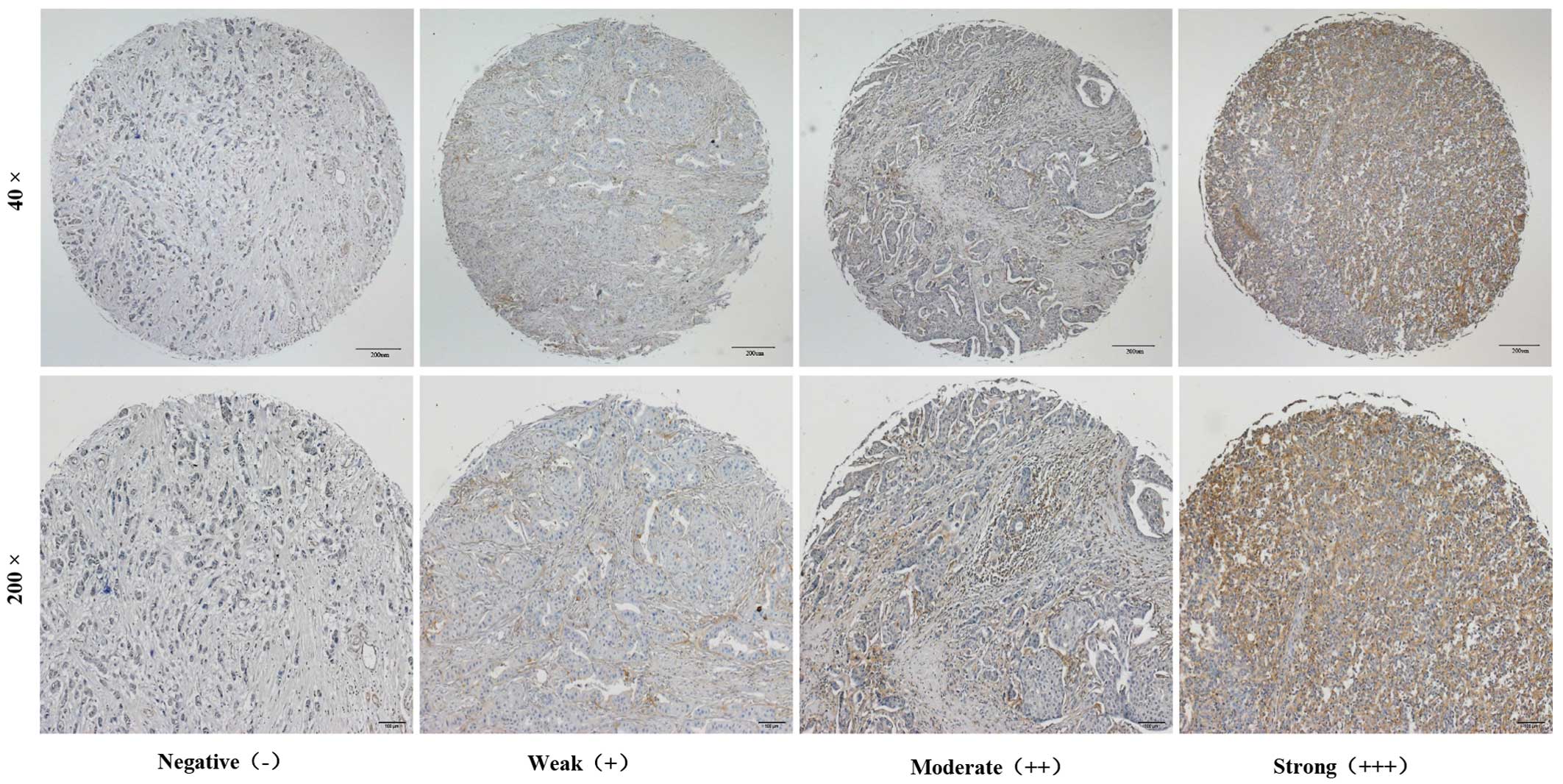
China). The degree of immunostaining of the tissue microarray was
viewed and scored separately by three independent investigators.
Cytoplasmic and membranous staining intensity were categorized as
follows: absent staining, 0; weak, 1; moderate, 2; and strong, 3.
The percentage of positive stained cells were categorized as: no
staining, 0; 0–24%, 1; 25–49%, 2; 50–74%, 3; and 75–100%, 4. The
total score was calculated by multiplying the staining intensity
and percentage of positive stained cells. The final score <2 was
negative (−); 3–4 was weak (+); 6–8 was moderate (++) and 9–12 was
strong (+++).
Construction of ICAM-1 shRNA lentivirus
and MCF-7 cell infection
Three shRNA sequences targeting human ICAM-1 and a
negative control sequence (Table I)
were designed and cloned into the pLKO.1-SP6-PGK-GFP vector. For
lentivirus packaging, HEK293T cells were transfected with ICAM-1
shRNA or control shRNA together with two helper plasmids (psPAX2
and pMD2.G) using Lipofectamine™ 2000 (Invitrogen) according to the
manufacturer's instructions. Four days after transfection, the
supernatant of HEK293T cells that containing packaged lentivirus
was collected and passed through 0.45-µm filters. To infect
MCF-7 cells, lentivirus particles were added to the culture medium
at a multiplicity of infection (MOI) of 5. The efficiency of
infection was determined by GFP percentage by flow cytometry (BD
FACSCalibur; BD Biosciences, Baltimore, MD, USA) four days after
lentivirus infection.
 | Table ITarget genes and sequences of
lentiviral shRNAs. |
Table I
Target genes and sequences of
lentiviral shRNAs.
| Target gene | Name | Target sequences | Site |
|---|
| Human ICAM-1 | Lv-sh1 |
5′-aagaaccttaccctacgctgc-3′ | 385–405 |
| Human ICAM-1 | Lv-sh2 |
5′-gcctcagcacgtacctcta-3′ | 940–962 |
| Human ICAM-1 | Lv-sh3 |
5′-aacgtgattctgacgaagccaga-3′ | 1490–1508 |
| Negative control | Lv-shNC |
5′-cctaaggttaagtcgccctc-3′ | Nonsense |
RNA extraction and quantitative real-time
PCR (qRT-PCR)
Total RNA of MCF-7 cells were extracted with TRIzol
solution (Takara Bio, Inc., Otsu, Japan) at 24 h after infection
and reverse transcribed to cDNA using a PrimerScript RT reagent kit
(Takara Bio, Inc.) by the manufacturer's instructions. β-actin was
used as a normalization control. Primers were chemically
synthesized by BioSune Biotechnology (Jinan, China). All primer
pairs used for Real-time PCR were designed according to the
sequences from GenBank (Table II).
Real-time PCR was performed using SYBR Premix Ex Taq (Takara Bio,
Inc.) on an CFX96 real-time PCR detection system (Bio-Rad,
Berkeley, CA, USA). The PCR conditions were as follows:
predegeneration at 94°C for 1 min, then 35 cycles of denaturation
at 94°C for 30 sec, renaturation at 55°C for 30 sec, extension at
72°C for 45 sec. The relative mRNA expression level was determined
by cycle threshold (Ct) normalized with β-actin using the 2−ΔΔCt
formula. (CT, cycle threshold) where ΔCT = CT (target gene) − CT
(β-actin).
| Table IIExpression of ICAM-1 in breast
cancers. |
Table II
Expression of ICAM-1 in breast
cancers.
| Sample | N | ICAM-1
immunostaining
|
χ2-value | P-value |
|---|
Negative (−)
| Weak (+)
| Moderate (++)
| Strong (+++)
|
|---|
| N | % | N | % | N | % | N | % |
|---|
| Breast cancers | 104 | 6 | 5.77 | 26 | 25 | 30 | 28.85 | 42 | 40.38 | | |
| Adjacent non-tumor
tissues | 38 | 18 | 47.37 | 11 | 28.94 | 6 | 15.79 | 3 | 7.89 | 39.81 | <0.001 |
Sequences of sense and antisense primers used for
qRT-PCR were as follows: ICAM-1-forward: 5′-ATGCCCA
GACATCTGTGTCC-3′, ICAM-1-reverse: 5′-GGGGTCTCTATGCCCAACAA-3′;
β-actin-forward: 5′-CCTAGAAGCATTTGCGGTGG-3′, β-actin-reverse:
5′-GAGCTACGAGCTGCCTGACG-3′.
Western blot analysis
Four days after lentivirus infection, MCF-7 cells
were washed by PBS and harvested, the protein concentration of each
group were determined using BCA assay kit (Beyotime Institute of
Biotechnology, Haimen, China). Subsequently, 40 µg protein
per lane was separated by 10% SDS-PAGE gel and transferred to NC
membranes. The NC membranes (Beyotime Institute of Biotechnology)
were blocked by 5% skim milk for 1 h and incubated with primary
antibodies to ICAM-1 (Signaling Technology, Inc., 1:1000), MMP-14
(Abcam, Cambridge, MA, USA, 1:1000) and β-actin (Beyotime Institute
of Biotechnology, 1:500) overnight, then incubated with their
respective secondary antibody for 2 h. All NC membranes were
detected by chemiluminescence (ECL, Sangon Biotech, Shanghai,
China) for 5 min and exposed to X-ray film for 5–10 min.
Wound-healing assay
For wound-healing assay, MCF-7 cells were plated
into 6-well plates at a density of 1×10⁵/ml and grown to
form a confluent monolayer, wounds were made with sterile pipette
tips and photographs of the wounds were taken at indicated time
points (0, 24 and 48 h).
Cell migration and invasion assay
MCF-7 cells (5×10⁵) were plated in 300
µl of serum-free medium and placed into cell chambers
(8-µm pore size, Corning) coated with (Invasion) or without
(migration) Matrigel (BD Biosciences), then incubated for 24 h.
Cells in the upper filters (inside the chambers) were removed by a
sterile swab, the migrated or invaded cells in the lower filters
(outside the chambers) were fixed by methanol for 2 h, and stained
with crystal violet. The number of migrated or invaded cells in
five random optical fields were counted under a microscope.
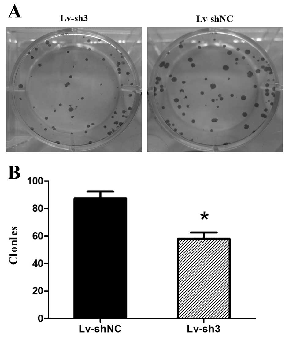
Colony formation assay
Four days after lentivirus infection, 300 MCF-7
cells of Lv-sh3 group and NS group were seeded into different wells
of 6-well plates, respectively, and cultured at 37°C in an
incubator for ~14 days until most single colonies had >50 cells.
All the colonies were washed by PBS and fixed with 4% methanol,
then stained by Giemsa. Images were captured by light
microscopy.
Statistical analysis
All the above experiments were performed three times
(biological replicates), data are presented as mean ± standard
deviation (mean ± SD) and analyzed by SPSS 10.0 software.
χ2 test or Student's t test were applied for statistical
analysis and P-value <0.05 was considered as statistical
significant in all cases.
Results
ICAM-1 is highly expressed in breast
cancer
Previous studies indicated that the expression of
ICAM-1 is involved in several carcinomas (6,14,15).
To investigate the function of ICAM-1 in breast cancer, we
evaluated the expression of ICAM-1 in breast cancer tissue
microarrays and clinical tissue specimens using immunohistochemical
staining. Of the 104 breast cancer samples, 6 (5.77%) were
negative, 26 (25%) were weakly positive, 30 (28.85%) were
moderately positive and 42 (40.38%) were strongly positive. In
normal breast tissue samples or adjacent non-tumor tissues, the
results were: 18 (47.37%) negative, 11 (28.94%) weakly positive, 6
(15.79%) moderately positive and 3 (7.89) strongly positive. The
ICAM-1 expression level in breast cancer was significantly higher
than the normal breast tissue or adjacent non-tumor tissues. These
results suggest that ICAM-1 is highly expressed in breast cancer
and might be involved in the pathogenesis of breast cancers
(Fig. 1 and Table II).
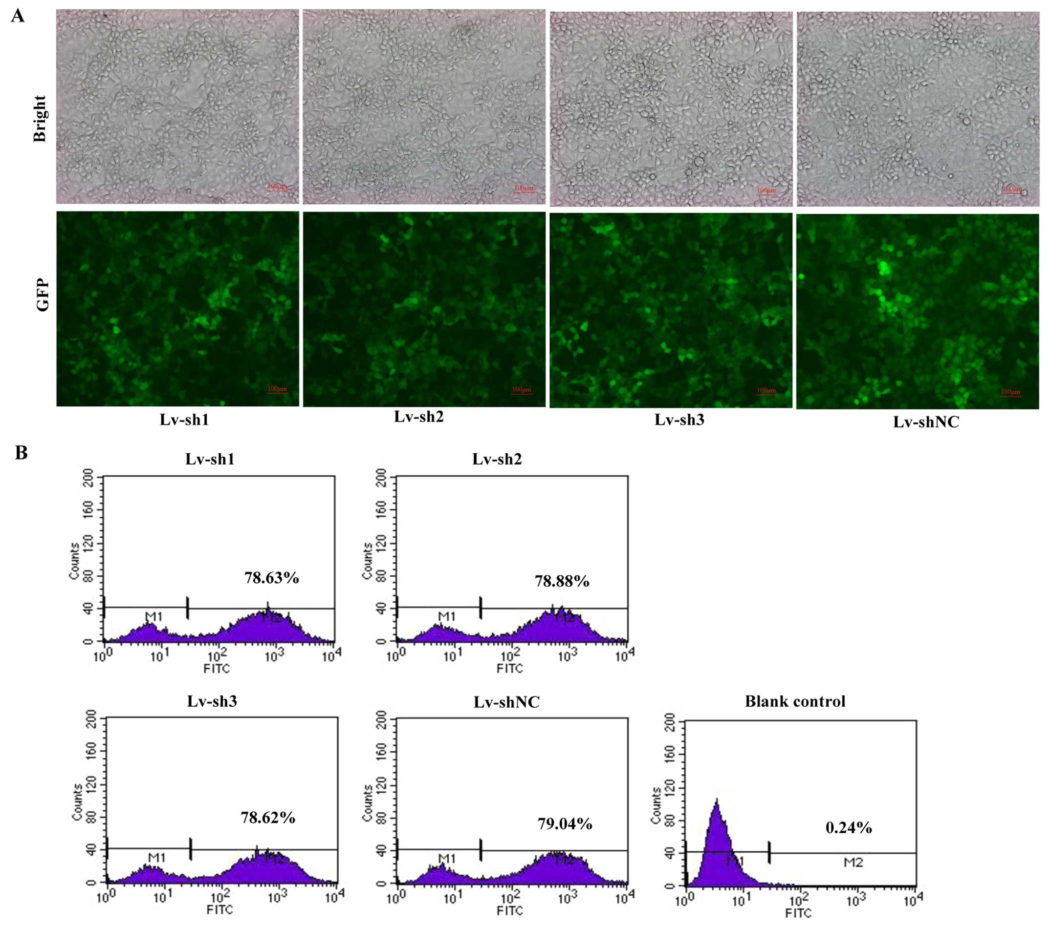
ICAM-1 lentivirus infection of MCF-7
cells and the efficiency
To investigate the function of ICAM-1 in breast
cancer cells, we used lentivirus-mediated RNAi technology, a
powerful method which can knock down the endogenous ICAM-1 gene
expression. Four days after lentivirus infection, MCF-7 cells were
collected to calculate the number of fluorescent-positive cells by
the FACS Calibur Flow Cytometry System. Fuorescence of GFP showed a
high percentage of cells being infected by lentivirus (Fig. 2A) and results of FCM indicated the
infection efficiency was ~80% (Fig.
2B).
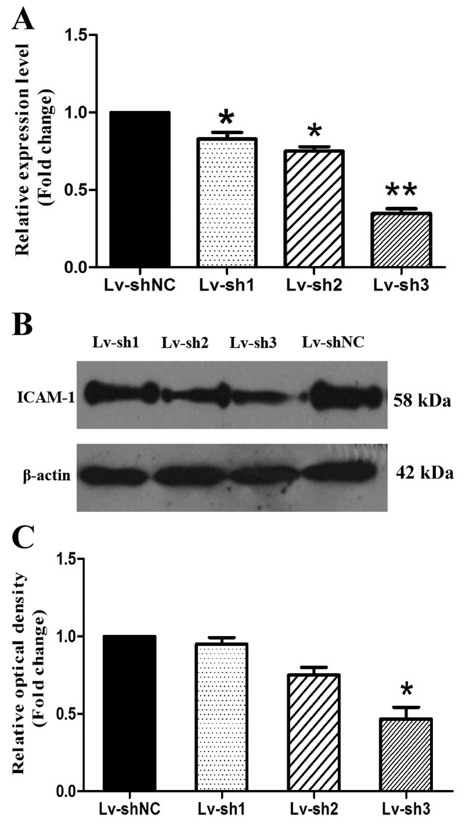
Lentivirus-mediated RNAi knockdown of
ICAM-1 expression in MCF-7 cells
Four days after lentivirus infection, MCF-7 cells
were harvested and the mRNA or protein level were detected by
real-time PCR or western blotting method. Real-time PCR results
indicated that the ICAM-1 mRNA level of MCF-7 cells in Lv-sh3 group
were significantly reduced compared with Lv-shNC group (Fig. 3A, P<0.05 or P<0.01), Western
blotting confirmed the silencing of ICAM-1 in MCF-7 cells (Fig. 3B and C, P<0.05). These results
suggest that the Lv-sh3 constructed efficiently and specifically
knocked down expression of ICAM-1 in MCF-7 cells.
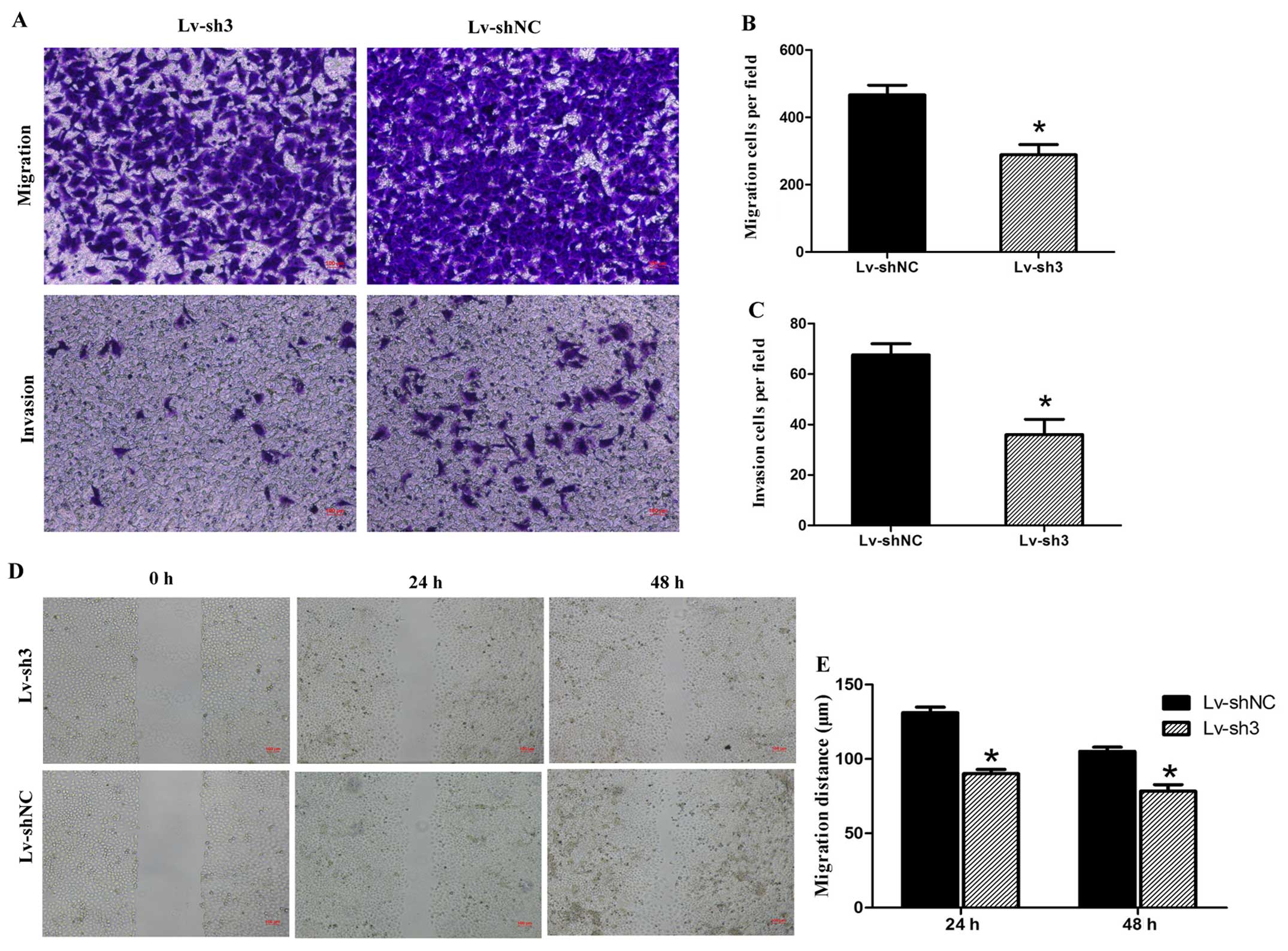
Suppression of ICAM-1 inhibits the
ability of MCF-7 cell metastasis in vitro
To clarify the relationship between ICAM-1 and the
metastatic capability of MCF-7 cells, we stably suppressed ICAM-1
expression using lentivirus mediated RNAi technology. Suppression
of ICAM-1 can inhibits metastasis ability of MCF-7 cells in
vitro. In the migration test, on average 288.67±53.15 cells in
Lv-sh3 group migrated to the lower chamber, whereas 466.67±50.33
cells in Lv-shNC group. In invasion test, there was on average
36±10.53 cells in Lv-sh3 group and 67.67±7.5 in Lv-shNC group. The
cell numbers of Lv-sh3 group that migrated through the membrane in
migration or invasion test were significantly less than that of
Lv-shNC group (Fig. 4A–C,
P<0.05). In wound-healing assay, the average migration distance
in Lv-sh3 group was 90±5 µm (24 h) and 78.33±7.63 µm
(48 h), significantly shorter than Lv-shNC group which migrated
131±6.55 µm (24 h) and 105±5 µm (48 h), respectively
(Fig. 4D and E, P<0.05).
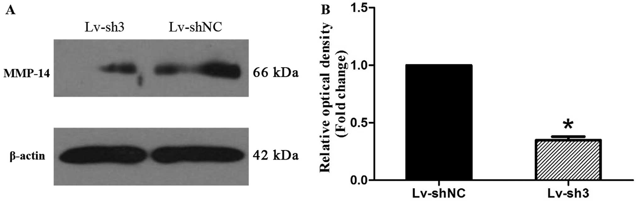
ICAM-1 gene silencing affects MMP-14
protein expression levels in MCF-7 cells
Matrix metalloproteinase (MMPs) are critical factors
involved in tumor metastasis and we detected the influence of
ICAM-1 gene silencing on MMP-14 protein expression levels by
western blot analysis. As revealed in Fig. 5, we found that gene silencing of
ICAM-1 can caused apparent decrease in MMP-14 expression levels,
indicating that ICAM-1 mediated breast cancer cell metastasis might
be linked with matrix metalloproteinase (MMP-14) (Fig. 5A and B, P<0.05).
ICAM-1 silencing reduces the colony
formation ability of MCF-7 cells
Giemsa staining showed that the colony number in
Lv-sh3 group was 58±7.94 and significantly less than that in
Lv-shNC group (87.33±8.74). Moreover, the size of the colony in
Lv-sh3 group was reduced markedly compared with Lv-shNC group,
indicating that ICAM-1 gene silencing could inhibit the colony
formation ability of MCF-7 cells in vitro (Fig. 6A and B, P<0.05).
Discussion
Tumor invasion or metastasis are major factors
causing death of patients and majority of cancer patients succumb
to the complications from distant metastases. Growing evidence has
demonstrated that adhesion molecules expressed on tumor cell
surface can mediate the adhesion between tumor cells and stromal or
vascular endothelial cells and play an important role in the
occurrence and development of tumors (16).
ICAM-1 is a cell surface glycoprotein that belongs
to immunoglobulin superfamily, and expressed in a wide variety of
lymphocytes or tumor cells. The ligand of ICAM-1 is lymphocyte
function associated antigen 1 (LFA-1) and the interaction between
ICAM-1 and its ligand can promote tumor metastasis through
modulating cell-cell adhesion (17). However, the exact role of and
function of ICAM-1 in the development of tumors has not been fully
elucidated. It has been reported that the upregulation of ICAM-1
expression can increase the invasion and metastasis ability of
tumor cells by promoting the adhesion between tumor cells and
vascular endothelial or lymphatic endothelial cells, ultimately
resulting in the distant metastasis of tumor cells (18). ICAM-1 is highly expressed in
different human malignancies and believed to be involved in their
pathogenesis. Previous research has shown that upregulation of
ICAM-1 has been demonstrated in different malignant tumors
including liver cancer, esophageal cancer, renal cell carcinoma,
squamous carcinoma, gastric cancer or pancreatic cancer and
associated with poor prognosis (14,15,19–22).
Under inflammatory environment, TGF (transforming growth factor)
promoted osteosarcoma cell metastasis through the increasement of
ICAM-1 by Akt signal pathway (23).
Downregulation of ICAM-1 by short hairpin RNA (shRNA) transduction
methods mitigated mouse colon cancer cell MC38 invasion ability
(24). Proto oncogene K-ras could
increase ICAM-1 expression in pancreatic acinar cells and the
formation of precancerous lesions (25). ICAM-1 was found to be expressed at
high levels in tumor-associated endothelium or tumor cells and was
associated with epithelial carcino genesis (26). Blocking NF-κB pathway by Bay11-7082
can downregulate ICAM-1 expression and inhibit the disintegration
of the lymph-endothelial barrier triggered by MCF-7 cells (27). However, data from other experiments
regard that high expression of ICAM-1 enhances the anti-tumor
ability through mediating the adhesion between tumor cells and
cytotoxic T cells or NK cells (28), low expression of ICAM-1 allowing
tumors to escape immune recognition (9). Breast cancer tissue or breast cancer
cell lines showed low expression of ICAM-1 than that of normal
breast epithelium or benign breast cells, the overexpression of
ICAM-1 after TNF stimulation was able to help lymphokine-stimulated
killer (LAK) cells to recognize breast cancer cells (29). The expression of ICAM-1 can be
regulated by transcription factor E2F1 in prostate cells through
NF-κB signaling pathway, silencing of E2F1 can increase ICAM-1
mediated leucocyte infiltration and inhibits tumor growth (30).
Other substances such as cannabidiol (CBD) elicits
upregulation of ICAM-1 and inhibits lung cancer cell line A549,
H358 or H460 metastasis, as a intermediate link in the CBD
antimetastatic action on human lung cancer cells. ICAM-1 can
increase cancer cell susceptibility to LAK cell-mediated cytolysis
(8,31). ICAM-1 also reduces ovarian cancer
cell growth in the absence of immune cells (32). Other studies confirmed that
upregulation of endogenous ICAM-1 can also reduce ovarian cancer
cell growth. To date, the exact role of ICAM-1 in breast cancer
cells still remains unclear. In this study, we screened the
expression level of ICAM-1 in breast cancer tissues with
microarrays tissue chip by immunohistochemistry method, the results
indicated that ICAM-1 was highly expressed in breast cancer tissues
compared with normal breast tissues. We also gathered 35 breast
cancer tissues with adjacent non-tumor tissues for further
confirmation and the results were in accordance with that of
microarrays tissue chip. We selected human breast cancer line MCF-7
with high ICAM-1 expression level as our study subjects and knocked
down ICAM-1 expression by lentivirus-mediated RNA interference
(RNAi).
We demonstrated that ICAM-1 silencing could inhibit
MCF-7 cell metastatic ability in vitro compared with cells
infected with Lv-shNC group and MMP-14 expression leval was
significantly decreased. One of the key steps in tumor invasion and
metastasis is the degradation of the extracellular matrix. MMP-14
known as MT1-MMP was the first identified membrane type MMP
(MT-MMPs) that highly expressed in many tumor cell surfaces and
play a role in biological processes such as angiogenesis, invasion
or proliferation (33), MMP-14 has
been recognized as an important MMP involved in dissemination of
tumor cells and cancer progression, blockade of MMP-14 can inhibit
the metastatic ability of tumor cells (12). Studies have shown that there is a
close link between ICAM and MMPs and ICAM-1 can be mediated by
MMP-14, and the ectodomain of ICAM-1 is mediated by MMP-14
(34,35). Furthermore, the number of colonies
in Lv-sh3 group was markedly reduced compared with Lv-shNC group.
Giemsa staining showed that the size of a single colony was
smaller, indicating that silencing ICAM-1 could downregulate the
adhesion ability between tumor cells and inhibited the colony
formation ability of breast cancer cells in vitro.
In conclusion, we showed in the present study that
ICAM-1 is overexpress in human breast cancers, while the knockdown
of ICAM-1 can suppresses the invasion of breast cancer cells
through MMP-14 expression, suggesting that ICAM-1 may serve as a
novel and useful prognostic marker and a potential therapeutic
target for the treatment of breast cancer.
Acknowledgments
This study was supported by grants from the National
Natural Science Foundation of China (81373185), the Natural Science
Foundation of Shandong, China (ZR2009CM019, ZR2014HL058), Shandong
Province Department of Education Foundation of China (no. J10LF62);
Shandong Province Health Department (no. 2013WS0287,
2014WS0462).
References
|
1
|
Shaukat U, Ismail M and Mehmood N:
Epidemiology, major risk factors and genetic predisposition for
breast cancer in the Pakistani population. Asian Pac J Cancer Prev.
14:5625–5629. 2013. View Article : Google Scholar : PubMed/NCBI
|
|
2
|
Ortiz-Martínez F, Sanmartín E,
Pomares-Navarro E, Pérez-Balaguer A, Andrés L, Sánchez-Payá J,
Aranda FI, Lerma E and Peiró G: Osteopontin regulates VEGFA and
ICAM-1 mRNA expression in breast carcinoma. Am J Clin Pathol.
143:812–822. 2015. View Article : Google Scholar : PubMed/NCBI
|
|
3
|
Fan L, Strasser-Weipp K, Li J-J, St Louis
J, Finkelstein DM, Yu K-D, Chen W-Q, Shao Z-M and Goss PE: Breast
cancer in China. Lancet Oncol. 15:pp. e279–e289. 2014, http://www.thelancet.com/journals/lanonc/article/PIIS1470-2045%2813%2970567-9/abstract.
View Article : Google Scholar
|
|
4
|
Kaulfuss S, Herr AM, Büchner A, Hemmerlein
B, Günthert AR and Burfeind P: Leupaxin is expressed in mammary
carcinoma and acts as a transcriptional activator of the estrogen
receptor α. Int J Oncol. 47:106–114. 2015.PubMed/NCBI
|
|
5
|
Müller V, Fuxius S, Steffens CC,
Lerchenmüller C, Luhn B, Vehling-Kaiser U, Hurst U, Hahn LJ,
Soeling U, Wohlfarth T, et al: Quality of life under capecitabine
(Xeloda®) in patients with metastatic breast cancer:
Data from a German non-interventional surveillance study. Oncol Res
Treat. 37:748–755. 2014. View Article : Google Scholar
|
|
6
|
Ramos TN, Bullard DC and Barnum SR:
ICAM-1: Isoforms and phenotypes. J Immunol. 192:4469–4474. 2014.
View Article : Google Scholar : PubMed/NCBI
|
|
7
|
Veitonmäki N, Hansson M, Zhan F, Sundberg
A, Löfstedt T, Ljungars A, Li ZC, Martinsson-Niskanen T, Zeng M,
Yang Y, et al: A human ICAM-1 antibody isolated by a function-first
approach has potent macrophage-dependent antimyeloma activity in
vivo. Cancer Cell. 23:502–515. 2013. View Article : Google Scholar : PubMed/NCBI
|
|
8
|
Haustein M, Ramer R, Linnebacher M, Manda
K and Hinz B: Cannabinoids increase lung cancer cell lysis by
lymphokine-activated killer cells via upregulation of ICAM-1.
Biochem Pharmacol. 92:312–325. 2014. View Article : Google Scholar : PubMed/NCBI
|
|
9
|
Kotteas EA, Boulas P, Gkiozos I, Tsagkouli
S, Tsoukalas G and Syrigos KN: The intercellular cell adhesion
molecule-1 (ICAM-1) in lung cancer: Implications for disease
progression and prognosis. Anticancer Res. 34:4665–4672.
2014.PubMed/NCBI
|
|
10
|
Martin ES: The BCSC-1 locus at chromosome
11q23-q24 is a candidate tumor suppressor gene. Proc Natl Acad Sci
USA. 100:11517–11522. 2003. View Article : Google Scholar : PubMed/NCBI
|
|
11
|
Anghel SI, Correa-Rocha R, Budinska E,
Boligan KF, Abraham S, Colombetti S, Fontao L, Mariotti A, Rimoldi
D, Ghanem GE, et al: Breast cancer suppressor candidate-1 (BCSC-1)
is a melanoma tumor suppressor that down regulates MITF. Pigment
Cell Melanoma Res. 25:482–487. 2012. View Article : Google Scholar : PubMed/NCBI
|
|
12
|
Rossé C, Lodillinsky C, Fuhrmann L,
Nourieh M, Monteiro P, Irondelle M, Lagoutte E, Vacher S, Waharte
F, Paul-Gilloteaux P, et al: Control of MT1-MMP transport by
atypical PKC during breast-cancer progression. Proc Natl Acad Sci
USA. 111:E1872–E1879. 2014. View Article : Google Scholar : PubMed/NCBI
|
|
13
|
Sugiyama N, Gucciardo E, Tatti O,
Varjosalo M, Hyytiäinen M, Gstaiger M and Lehti K: EphA2 cleavage
by MT1-MMP triggers single cancer cell invasion via homotypic cell
repulsion. J Cell Biol. 201:467–484. 2013. View Article : Google Scholar : PubMed/NCBI
|
|
14
|
Yan J, Jiang Y, Ye M, Liu W and Feng L:
The clinical value of lymphatic vessel density, intercellular
adhesion molecule 1 and vascular cell adhesion molecule 1
expression in patients with oral tongue squamous cell carcinoma. J
Cancer Res Ther. 10(Suppl): C125–C130. 2014. View Article : Google Scholar : PubMed/NCBI
|
|
15
|
Alasehirli B, Oğuz E, Oksuzler E, Koruk I,
Oztuzcu S, Ozkara E, Karakok M, Erbagcı AB and Demiryurek AT:
Investigation of intercellular adhesion molecules (ICAMs) gene
expressions in patients with Barrett's esophagus. Tumour Biol.
35:4907–4912. 2014. View Article : Google Scholar : PubMed/NCBI
|
|
16
|
Lu J and Jin ML: Short-hairpin
RNA-mediated MTA2 silencing inhibits human breast cancer cell line
MDA-MB231 proliferation and metastasis. Asian Pac J Cancer Prev.
15:5577–5582. 2014. View Article : Google Scholar : PubMed/NCBI
|
|
17
|
Liu S, Han L, Wang X, Liu Z, Ding S, Lu J,
Bi D, Mei Y and Niu Z: Nephroblastoma overexpressed gene (NOV)
enhances RCC cell motility through upregulation of ICAM-1 and COX-2
expression via Akt pathway. Int J Clin Exp Pathol. 8:1302–1311.
2015.PubMed/NCBI
|
|
18
|
Guo P, Huang J, Wang L, Jia D, Yang J,
Dillon DA, Zurakowski D, Mao H, Moses MA and Auguste DT: ICAM-1 as
a molecular target for triple negative breast cancer. Proc Natl
Acad Sci USA. 111:14710–14715. 2014. View Article : Google Scholar : PubMed/NCBI
|
|
19
|
Jenkinson C, Elliott V, Menon U,
Apostolidou S, Fourkala OE, Gentry-Maharaj A, Pereira SP, Jacobs I,
Cox TF, Greenhalf W, et al: Evaluation in pre-diagnosis samples
discounts ICAM-1 and TIMP-1 as biomarkers for earlier diagnosis of
pancreatic cancer. J Proteomics. 113:400–402. 2015. View Article : Google Scholar
|
|
20
|
Liu S, Li N, Yu X, Xiao X, Cheng K, Hu J,
Wang J, Zhang D, Cheng S and Liu S: Expression of intercellular
adhesion molecule 1 by hepatocellular carcinoma stem cells and
circulating tumor cells. Gastroenterology. 144:1031–1041.e10. 2013.
View Article : Google Scholar : PubMed/NCBI
|
|
21
|
Shi X, Jiang J, Ye X, Liu Y, Wu Q and Wang
L: Prognostic prediction and diagnostic role of intercellular
adhesion molecule-1 (ICAM1) expression in clear cell renal cell
carcinoma. J Mol Histol. 45:427–434. 2014. View Article : Google Scholar : PubMed/NCBI
|
|
22
|
Dong Z, Fu S, Xu X, Yang Y, Du L, Li W,
Kan S, Li Z, Zhang X, Wang L, et al: Leptin-mediated regulation of
ICAM-1 is Rho/ROCK dependent and enhances gastric cancer cell
migration. Br J Cancer. 110:1801–1810. 2014. View Article : Google Scholar : PubMed/NCBI
|
|
23
|
Hou CH, Lin FL, Tong KB, Hou SM and Liu
JF: Transforming growth factor alpha promotes osteosarcoma
metastasis by ICAM-1 and PI3K/Akt signaling pathway. Biochem
Pharmacol. 89:453–463. 2014. View Article : Google Scholar : PubMed/NCBI
|
|
24
|
Howard K, Lo KK, Ao L, Gamboni F, Edil BH,
Schulick R and Barnett CC Jr: Intercellular adhesion molecule-1
mediates murine colon adenocarcinoma invasion. J Surg Res.
187:19–23. 2014. View Article : Google Scholar
|
|
25
|
Liou GY, Döppler H, Necela B, Edenfield B,
Zhang L, Dawson DW and Storz P: Mutant KRAS-induced expression of
ICAM-1 in pancreatic acinar cells causes attraction of macrophages
to expedite the formation of precancerous lesions. Cancer Discov.
5:52–63. 2015. View Article : Google Scholar :
|
|
26
|
Strell C, Lang K, Niggemann B, Zaenker KS
and Entschladen F: Neutrophil granulocytes promote the migratory
activity of MDA-MB-468 human breast carcinoma cells via ICAM-1. Exp
Cell Res. 316:138–148. 2010. View Article : Google Scholar
|
|
27
|
Viola K, Kopf S, Huttary N, Vonach C,
Kretschy N, Teichmann M, Giessrigl B, Raab I, Stary S, Krieger S,
et al: Bay11-7082 inhibits the disintegration of the
lymph-endothelial barrier triggered by MCF-7 breast cancer
spheroids; the role of ICAM-1 and adhesion. Br J Cancer.
108:564–569. 2013. View Article : Google Scholar :
|
|
28
|
Ogawa Y, Hirakawa K, Nakata B, Fujihara T,
Sawada T, Kato Y, Yoshikawa K and Sowa M: Expression of
intercellular adhesion molecule-1 in invasive breast cancer
reflects low growth potential, negative lymph node involvement, and
good prognosis. Clin Cancer Res. 4:31–36. 1998.PubMed/NCBI
|
|
29
|
Budinsky AC, Brodowicz T, Wiltschke C,
Czerwenka K, Michl I, Krainer M and Zielinski CC: Decreased
expression of ICAM-1 and its induction by tumor necrosis factor on
breast-cancer cells in vitro. Int J Cancer. 71:1086–1090. 1997.
View Article : Google Scholar : PubMed/NCBI
|
|
30
|
Ren Z, Kang W, Wang L, Sun B, Ma J, Zheng
C, Sun J, Tian Z, Yang X and Xiao W: E2F1 renders prostate cancer
cell resistant to ICAM-1 mediated antitumor immunity by NF-κB
modulation. Mol Cancer. 13:842014. View Article : Google Scholar
|
|
31
|
Ramer R, Bublitz K, Freimuth N, Merkord J,
Rohde H, Haustein M, Borchert P, Schmuhl E, Linnebacher M and Hinz
B: Cannabidiol inhibits lung cancer cell invasion and metastasis
via intercellular adhesion molecule-1. FASEB J. 26:1535–1548. 2012.
View Article : Google Scholar
|
|
32
|
de Groote ML, Kazemier HG, Huisman C, van
der Gun BTF, Faas MM and Rots MG: Upregulation of endogenous ICAM-1
reduces ovarian cancer cell growth in the absence of immune cells.
Int J Cancer. 134:280–290. 2014. View Article : Google Scholar
|
|
33
|
Lu H, Hu L, Yu L, Wang X, Urvalek AM, Li
T, Shen C, Mukherjee D, Lahiri SK, Wason MS, et al: KLF8 and FAK co
operatively enrich the active MMP14 on the cell surface required
for the metastatic progression of breast cancer. Oncogene.
33:2909–2917. 2014. View Article : Google Scholar :
|
|
34
|
Essick E, Sithu S, Dean W and D'Souza S:
Pervanadate–induced shedding of the intercellular adhesion molecule
(ICAM)-1 ectodomain is mediated by membrane type-1 matrix
metallopro-teinase (MT1-MMP). Mol Cell Biochem. 314:151–159. 2008.
View Article : Google Scholar : PubMed/NCBI
|
|
35
|
Park JK, Park SH, So K, Bae IH, Yoo YD and
Um HD: ICAM-3 enhances the migratory and invasive potential of
human non-small cell lung cancer cells by inducing MMP-2 and MMP-9
via Akt and CREB. Int J Oncol. 36:181–192. 2010.
|