Introduction
Osteosarcoma is one of the most common malignant
bone tumor in childhood (1), but it
is difficult to cure, especially during the later stages.
Chemoresistance is a main cause of unsatisfactory survival
outcomes, although chemotherapy has become more effective in recent
years (2,3). Several factors are implicated in the
development of chemoresistance including genetic alterations, drug
uptake and increased activation of DNA repair mechanisms and
evasion of chemotherapy-induced apoptosis (4,5).
Additionally, our previous results showed that Beclin-1-mediated
autophagy contributed to osteosarcoma chemoresistance (6). However, the molecular regulating
mechanism of autophagy in chemoresistance is largely unclear.
MicroRNA (miRNA), an endogenous noncoding RNA with
18–25 nucleotides, is able to regulate gene expression by binding
to the 3′-untranslated region (3′-UTR) of their target mRNAs,
modulating mRNA and protein expression. A series studies on miRNAs
in osteosarcoma have been performed and several miRNAs contribute
to the development and progression of osteosarcoma (2,7–9).
miR-30a, an intronic transcriptional unit, was widely expressed in
various tissues (10). Recent
studies have suggested that miR-30a played a tumor-suppressive role
in various malignant tumors, including lung cancer (11), breast cancer (12), colon cancer (13), and nasopharyngeal carcinoma
(14). Moreover, a negative
correlation was observed between miR-30a expression and malignant
grade in osteosarcoma. Importantly, overexpression of miR-30a
reduced proliferation, migration, and invasion and further studies
revealed that runt-related transcription factor 2 was a regulative
target gene of miR-30a (7).
However, the role of miR-30a in osteosarcoma chemoresistance
remains elusive.
Autophagy is essential for the maintenance of
cellular biosynthesis, growth and differentiation. Accumulating
evidence supports that activation of autophagy was also a crucial
step in the process of chemoresistance (15). Since autophagy promoted tumor cells
to survive under adverse stress conditions, such as hypoxia,
ischemia and chemotherapy (16).
Recent studies indicated microRNA-143 (17), miR-101 (18), and miR-22 (19) to be involved in chemoresistance
through regulating autophagy. Therefore, a better understanding of
the role of miRNA-mediated autophagy in osteosarcoma is essential
for future treatment in chemoresistance.
In this study, we investigated the expression of
miR-30a in osteosarcoma cell lines. We demonstrated autophagy was
activated by chemotherapy and miR-30a was downregulated following
chemotherapy. Further study showed that overexpression of miR-30a
reduced chemoresistance of osteosarcoma cells through
downregulating autophagy. In addition, Beclin-1 was identified as
the target gene of miR-30a in osteosarcoma. The above results
indicate miR-30a and its downstream target gene Beclin-1 can be
used in treatment of osteosarcoma chemoresistance in the
future.
Materials and methods
Cell culture and doxorubicin-resistant
MG-63 cell line
Human osteosarcoma MG-63 cell line was preserved in
our laboratory as previously described (20). Cells was cultured in high glucose
DMEM medium supplemented with 10% fetal bovine serum (Gibco,
Carlsbad, CA, USA), 100 U/ml penicillin, and 100 µg/ml
streptomycin, and were cultured in a humidified atmosphere
containing 5% Co2 at 37°C. A doxorubicin (Dox)-resistant
cell line (MG-63/Dox) was established from its parental cell line
MG-63 by gradually increasing the concentration of Dox over 6
months. The concentration of Dox added to MG-63 cells was from 10
to 100 nM, after which the cells were maintained in culture medium
containing 100 nM Dox, it displayed 6-fold resistance to Dox
compared with the corresponding parental sensitive cells.
Quantitative real-time RT-PCR
Total RNA containing miRNA was extracted from cells
with TRIzol Reagent (Invitrogen, Carlsbad, CA, USA) according to
the manufacturer's instructions. miR-30a and U6 small nuclear 2
(U6) expression was detected from the cDNA product using TaqMan
miRNA sequence-specific probes (Takara, Japan) and ABI Prism 7500
sequence detection system (Applied Biosystems, Foster City, CA,
USA). The fold-change in gene expression was calculated by
2−ΔΔCT.
Overexpression of miR-30a
To generate the stable cell line, MG-63/Dox
osteosarcoma cells were transfected with lenti-viruses containing
miR-30a gene (Lenti-miR-30a) or a blank lentivirus expression
vectors (Lenti-NC) GeneChem Corp. (Shanghai, China). The infection
efficiency was confirmed by qRT-PCR after transfection 72 h.
miRNA target prediction
Candidate targets of miR-30a and Beclin-1 were
predicted by miRBase (http://www.mirbase.org/) and TargetScan (http://www.Targetscan.org/).
Cell proliferation and apoptosis
assays
Cell proliferation was evaluated using Cell Counting
Kit-8 (CCK-8, Beyotime Institute of Biotechnology, haimen, China).
Cells (2×10³) were seeded in 96-well plates and cultured
for 1, 2, 3, 4 and 5 days after transfection. CCK-8 solution (10
µl) was added to each well and followed by incubation for 2
h. Absorbance was measured at 450 nm with a Microplate Autoreader
(Bio-Rad, Hercules, CA, USA). For apoptosis detection, miR-30a
transfected cells in the presence or absence of chemotherapy agents
were stained with Annexin V/PI double staining kit (BD Biosciences,
Bedford, MA, USA) according to the manufacturer's protocol.
Apoptotic cells were examined by flow cytometry (6).
Western blot analysis
Proteins were separated by 10% SDS pages and then
performed as previously described (6). Antibodies against Beclin-1, p62,
LC3-I, LC3-II, cleaved caspase-3, Total caspase-3, and β-actin were
purchased from Cell Signaling Technology, Inc. (Danvers, MA, USA).
Then the band were incubated by horseradish peroxidase-labeled goat
anti-rabbit IgG (Santa Cruz Biotechnology, Santa Cruz, CA, USA) and
detected by chemiluminescence. β-actin was used as a protein
loading control.
Stably expressing GFP-LC3
The adenovirus vector containing the GFP-LC3
reporter was purchased from Hanbio (Shanghai, China). In the
presence or absence of chemotherapy, the cells were analyzed by a
fluorescence microscope (Olympus, Tokyo, Japan).
Luciferase reporter assay
According to the expression of miR-30a with the
Beclin-1 3′-UTR, 3′-UTR sequence or three copies of the mutated
3′-UTR sequence of Beclin-1 were amplified by PCR from human
genomic DNA and immediately inserted into pGL3-control vector
(Promega Corp., WI, USA). MG-63/Dox cells in 6-well plates were
transfected with 1 µg of the firefly luciferase report
vector, 1 h post-transfection with 25 nM of mimic NC, miR-30a
mimics, anti-NC or anti-miR-30a. At 24 h post-transfection, firefly
luciferase activities were measured consecutively using luciferase
assays (Promega Corp.).
Statistical analysis
The data are expressed as means ± standard error
(Se). A two-tailed Student's t-test was used to determine the
significance of differences between groups. Results with P-values
<0.05 were considered statistically significant and SPSS 13.0
was used for the statistical analysis.
Results
Autophagy is increased in Dox-resistant
osteosarcoma cells and miR-30a is downregulated
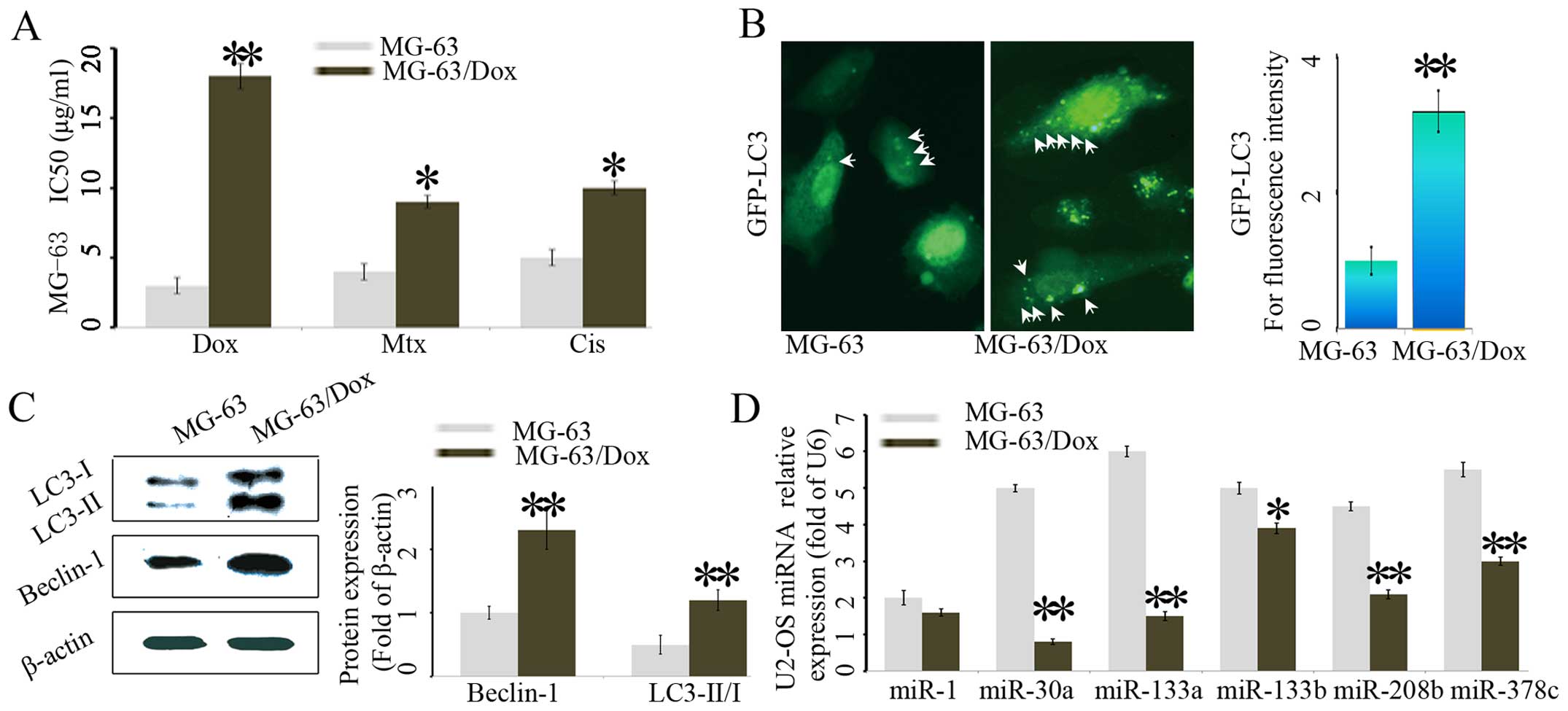
Dox has been widely used in clinical chemotherapy of
osteosarcoma patients, we established the Dox-resistant MG-63
cells. In terms of IC50, the Dox-selected MG-63/Dox
subline showed 6-fold higher resistance to doxorubicin (Dox),
2.2-fold, and 2-fold higher resistance to cisplatin (Cis), and
methotrexate (Mtx), respectively, when compared with parental
MG-63cells. The results indicated that MG-63/Dox subline was
cross-resistant to the conventional chemotherapeutic agents
(Fig. 1A).
Next, we assessed the autophagy activity of the
above two different osteosarcoma cells, by using LC3-GFP-labeled
autophagosomes, which indicated that the level of autophagy was
significantly increased in MG-63/Dox subline cells (Fig. 1B). Western blot analysis confirmed
that the autophagy-related protein Beclin-1 increased in MG-63/Dox
cells compared with the control cells (Fig. 1C). The autophagy-related protein
LC3-I and LC3-II was also higher in the MG-63/Dox cells as compared
with the control cells (Fig.
1C).
Using a TaqMan probe-based qRT-PCR assay, we
determined the expression levels of a panel of miRNAs that have
been reported to be associated with osteosarcoma cellular autophagy
and chemoresistance, particularly miR-1, miR-30a, miR-133a,
miR-133b, miR-208b and miR-378c (21). As shown in Fig. 1D, among the 6 miRNAs tested, a
largest reduction of miR-30a was observed in MG-63/Dox cells
compared with the control cells.
miR-30a overexpression suppresses
Beclin-1 expression and autophagy is induced by chemotherapy
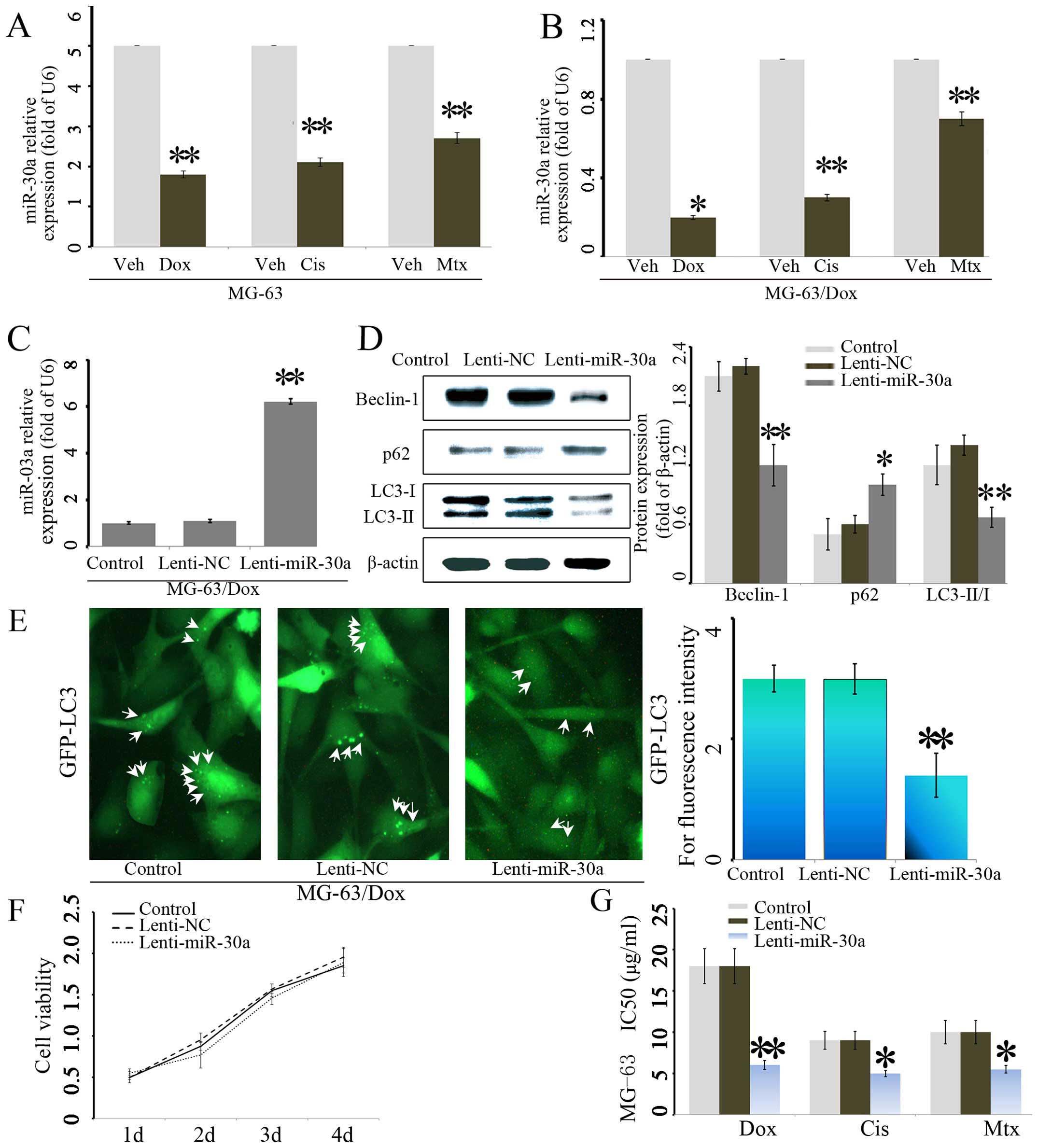
Subsequently, we assayed the effects of the
anticancer agents on the expression of miR-30a. As shown in
Fig. 2A and B, anticancer agents
significantly suppressed the expression of miR-30a in both MG-63
and MG-63/Dox cell lines. These findings show that miR-30a
expression was downregulated during chemotherapy in osteosarcoma
cells.
To further investigate the possible role of miR-30a
in the MG-63/Dox osteosarcoma cells, we applied lentivirus system
to make stable cell lines to overexpress miR-30a on the base of
osteosarcoma cell line MG-63/Dox including a blank group
(untransfected cells), a Lenti-NC group (cells transfected with the
control lentivirus), and a Lenti-miR-30a group (overexpressing of
miR-30a). The miR-30a expression levels in these groups were
evaluated using qRT-PCR. As expected, the expression of miR-30a was
increased in Lenti-miR-30a group cells (P<0.05) (Fig. 2C). Next, we investigated the effect
of miR-30a on autophagy. Results showed that miR-30a overexpression
significantly decreased Beclin-1, LC3-I and LC3-II expression,
while miR-30a increased p62 protein level (Fig. 2D). A significantly decreased
accumulation of LC3-GFP punctae were also confirmed by confocal
assay in MG-63/miR-30a cells. These results revealed that
overexpressed miR-30a inhibited autophagy in MG-63/Dox cells
(Fig. 2E).
To further explore the function of miR-30a in the
chemoresistance of osteosarcoma, cell proliferation assays were
performed to determine cell growth curve and 50% inhibition of
growth (IC50) values following chemotherapy treatment.
Overexpression of miR-140-5p did not alter cell growth (Fig. 2F). Remarkably, overexpression of
miR-30a dramatically reduced the IC50 values for the
three chemotherapeutic agents in MG-63 cells (Fig. 2G).
miR-30a promotes chemotherapy-induced
osteosarcoma cell apoptosis via repressing Beclin-1-mediated
osteosarcoma autophagy
As shown in Fig. 3A and
B, after chemotherapy treatments for 12 h, the ratio of LC3-II
versus LC3-I was increased from 2.2–3.5 in the lenti-NC cells in
relative to 1.3–1.5 range in lenti-miR-30a group (Fig. 1A). Consistently, the level of
Beclin-1 proteins was greatly increased about 1.5-, 1.8- and
2.7-fold in lenti-NC group as compared with 1.1-, 1.23- and 1.28-
fold in lenti-miR-30a group, respectively. The above data suggested
that miR-30a overexpression led to a down regulation of autophagy
responding to chemotherapy in MG-63/Dox osteosarcoma cells.
Importantly, apoptosis in MG-63/miR-30a cells was assessed after
chemotherapy. Chemotherapy treatment for 12 h showed 2.6-fold,
1.8-fold, 1.9-fold increase in apoptosis incidence in the
Lenti-miR-30a group as compared with the counterparts,
respectively.
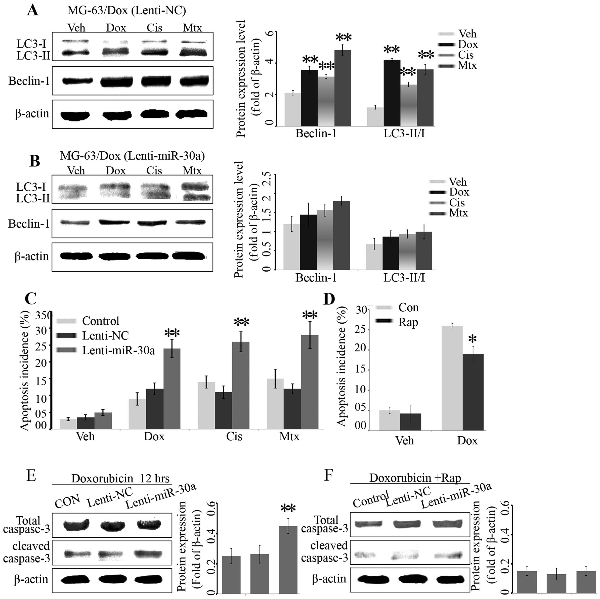 | Figure 3miR-30a promotes chemotherapy-induced
osteosarcoma cell apoptosis via repressing Beclin-1-mediated
osteosarcoma autophagy. Western blot analysis of LC3-I, LC3-II and
Beclin-1. MG63/Dox cells in Lenti-NC group (A) and Lenti- miR-30a
group (B) were treated with vehicle (veh, distilled water), Dox
(0.2 mg/ml), Cis (20 µmol/l), and Mtx (50 mmol/l) for 12 h.
p<0.05; **p<0.01 vs. Veh group. (C) Annexin
V-FITC/PI staining for apoptotic cells. MG63/Dox cells in CON,
Lenti-NC group and Lenti-miR-30a group were treated with veh
(distilled water), Dox (0.2 mg/ml), Cis (20 mmol/l), and Mtx (50
mmol/l) for 12 h. Subsequently, the apoptosis incidence were
quantified by flow cytometry. (D) Annexin V-FITC/PI staining for
apoptosis. Cells in Lenti-miR-30a group were pretreated with
rapamycin (Rap; 100 nmol/l) or without (Con) for 6 h and then
treated with veh (distilled water) or 0.2 mg/ml Dox for 12 h. The
apoptosis was analyzed by Annexin V-FITC/PI staining using flow
cytometry. (E) Western blot analysis of Total caspase-3 and cleaved
caspase-3 in Control, Lenti-NC and Lenti-miR-30a groups after 0.2
mg/ml Dox treatment for 12 h. (F) Western blot for total caspase-3
and cleaved caspase-3. Cells in Control, Lenti-NC and Lenti-miR-30a
groups were pretreated with 100 nmol/l Rap for 6 h and then treated
with 0.2 mg/ml Dox for 12 h, then analyzed by western blotting. The
data are presented as the mean ± SE for three independent
experiments. *p<0.05; **p<0.01 vs.
Control group. |
Furthermore, to determine the role of downregulated
autophagy in MG-63/miR-30a cells, autophagy was activated by
rapamycin, an autophagy promoter. As a result, rapamycin could
partly decrease the Dox-induced apoptosis, suggesting that
activating autophagy reversed the effect of miR-30a. Additionally,
the apoptotic markers were assessed by western blotting. Compared
with the lenti-NC group, the level of cleaved caspase-3 was
increased in Lenti-miR-30a group following Dox treatment,
suggesting that miR-30a promoted Dox-induced apoptosis.
Consistently, rapamycin could partly reduce the expression of
cleaved caspase-3. These results showed that miR-30a overexpression
could promote Dox-induced apoptosis of osteosarcoma cells through
suppressing autophagy.
Beclin-1 is a target of miR-30a in
osteosarcoma
It has been reported that Beclin-1 is the direct
target of miR-30a in the breast cancer cell line MCF-7 (22), and we reasoned that miR-30a may have
a similar regulating effect on osteosarcoma. Binding sites for
miR-30a in the 3′-UTR of Beclin-1 were identified by bioinformatics
analysis (Fig. 4A).
To further assess whether miR-30a was directly
targeting Beclin-1 expression through the target site in the 3′-UTR
of Beclin-1, reporter constructs containing either the wild-type
(WT) Beclin-1 3′-UTR or Beclin-1 3′-UTR with mutation at the
predicted miR-30a target sequence were cotransfected into MG-63
cells together with mimic NC, miR-30a mimics, anti-NC or
anti-miR-30a. Luciferase reporter assays showed that transduction
of miR-30a mimics substantially inhibited the luciferase activity
of the wild-type Beclin-1 3′-UTR by ~42% in MG-63 cells, but had no
effect on mutant MG-63 3′-UTR relative to the control (Fig. 4B), and cotransfection with
anti-miR-30a significantly increased luciferase activity of WT
Beclin-1 3′UTR-compared with anti-NC control (Fig. 4B). Whereas, transfection of the
mutant Beclin-1 3′-UTR with anti-miR-30a had no effect on
luciferase activity. The above results strongly suggested that
Beclin-1 was a direct target of miR-30a in osteosarcoma cells.
To further examine whether miR-30a modulated the
expression of Beclin-1 in osteosarcoma, cells were transfected with
mimic NC, miR-30a mimics, anti-NC or anti-miR-30a and then the mRNA
expressions of miR-30a and Beclin-1 were analyzed by real-time PCR.
Transfection of the miR-30a mimics significantly increased the
miR-30a expression and decreased the mRNA level of Beclin-1.
Consistently, anti-miR-30a had the opposite effects on expression
in miR-30a and Beclin-1 (Fig. 4C and
D).
Discussion
With the advancement of neoadjuvent chemotherapy,
the global survival rate of osteosarcoma improved to ~70% (23). However, like most malignant cancers,
~30% of patients will develop relapse and chemoresistance, which is
the major reason for recurrence of osteosarcoma (24). Previously, we investigated the role
of Beclin-1 in osteosarcoma, and suggested that the knockdown of
Beclin-1 rendered osteosarcoma cells more sensitive to chemotherapy
(6). In this study, we observed
that chemotherapy induced downregulation of miR-30a in osteosarcoma
cells, suggesting that miR-30a might participate in osteosarcoma
chemoresistance. Consistently, downregulation of miR-30a has been
reported in diverse cancers, and to be involved in tumor cell
proliferation and metastasis (11–14).
However, little is known about contribution of miR-30a to
chemoresistance of osteosarcoma. Thus, we further investigated the
expression of miR-30a in Dox-resistant osteosarcoma. Our results
showed there was a significant downregulation of miR-30a and
increased autophagy in Dox-resistant osteosarcoma cell line MG-63,
which suggested that miR-30a might be a suppressor for
autophagy.
Next, to further confirm the biological role of
miR-30a in osteosarcoma, miR-30a was overexpressed in
Dox-resistance osteosarcoma cell line MG-63/Dox. Real-time PCR
confirmed that miR-30a was lowly expressed in MG-63/Dox cells.
Overexpression of miR-30a did not alter osteosarcoma cell growth,
but it significantly suppressed the autophagy activity of
osteosarcoma cells, indicating that miR-30a should be a suppressor
for autophagy. Given that autophagy is implicated in
chemoresistance (25–27), it was likely that miR-30a
downregulation contributed to chemoresistance of osteosarcoma cells
by suppressing autophagy, which might explain the parallel
downregulation of miR-30a and elevated autophagy in Dox-resistance
MG-63 cells.
Autophagy is an essential pathway for cellular
homeostasis and recent studies demonstrated that autophagy is
implicated in chemoresistance (25–27).
However, the regulating mechanism of autophagy in osteosarcoma is
still unclear. Our data showed downregulation of miR-30a in
Dox-resistance osteosarcoma, consistent with the result in mouse
human macrophages (28). Then, it
was necessary to clarify whether miR-30a affects the
chemoresistance of osteosarcoma. Thus, we next examined the effects
of miR-3a on osteosarcoma cell death in the presence of
chemotherapy. The data showed that miR-30a strongly reduced the
IC50 values and significantly promoted
chemotherapy-induced cell death, suggesting that overexpression of
miR-30a rendered osteosarcoma cells more sensitive to
chemotherapy.
Notably, western blot analysis demonstrated that
overexpression of miR-30a enhanced doxorubicin-induced caspase-3
cleavage, whereas autophagy promoter, rapamycin attenuated
doxorubicin-induced caspase-3 cleavage and reversed the effect of
miR-30a. These results strongly suggested that overexpression of
miR-30a decreased chemoresistance by suppressing autophagy, leading
to increased doxorubicin-induced cell death. Several studies have
demonstrated that inhibiting autophagy sensitizes tumor cells to
chemotherapy-induced cell death, suggesting a key role for the
miR-30a/autophagy loop in chemoresistance of osteosarcoma (25,29).
Therefore, it was reasonable that downregulation of miR-30a
contributed to chemoresistance of osteosarcoma cells through
regulating autophagy. Furthermore, to investigate the mechanism of
miR-130a in regulating autophagy, bioinformatics analysis was
performed. The results showed that the 3′-UTR region of Beclin-1
were the binding sites for miR-30a. Consistently, previous studies
demonstrated that Beclin-1 was the directly target of miR-30a
(30).
The study of the miR-30a/autophagy loop is based on
our previous results and this is the first study of miR-30a in the
chemoresistance of osteosarcoma. Our work led to the identification
of a novel functional pathway controlled by miR-30a and its direct
target, Beclin-1, that regulated chemoresistance in osteosarcoma.
Therefore, miR-30a and its target gene pathway may represent new
therapeutic opportunities for chemoresistant patients.
Acknowledgments
This study was supported by the grants from National
Natural Science Foundation of Youth Program (no. 81101394),
Shanghai Special Fund for Outstanding Young Teachers in
Universities (no. JDY10080), Shanghai Renji hospital Fund for
outstanding Youth (no. RJPY10-010), Shanghai Fund for Young
Physician Development (no. 20141051), Medical-Engineering Joint
Fund of Shanghai Jiao Tong University (no. YG2014MS51), Shanghai
Pujiang Program (no. 15PJD026).
References
|
1
|
Jemal A, Bray F, Center MM, Ferlay J, Ward
E and Forman D: Global cancer statistics. CA Cancer J Clin.
61:69–90. 2011. View Article : Google Scholar : PubMed/NCBI
|
|
2
|
Zhang Y, Duan G and Feng S: MicroRNA-301a
modulates doxorubicin resistance in osteosarcoma cells by targeting
AMP-activated protein kinase alpha 1. Biochem Biophys Res Commun.
459:367–373. 2015. View Article : Google Scholar : PubMed/NCBI
|
|
3
|
Xu M, Jin H, Xu CX, Sun B, Mao Z, Bi WZ
and Wang Y: miR-382 inhibits tumor growth and enhance
chemosensitivity in osteosarcoma. Oncotarget. 5:9472–9483. 2014.
View Article : Google Scholar : PubMed/NCBI
|
|
4
|
Gillet JP and Gottesman MM: Overcoming
multidrug resistance in cancer: 35 years after the discovery of
ABCB1. Drug Resist Updat. 15:2–4. 2012. View Article : Google Scholar : PubMed/NCBI
|
|
5
|
He Y, Wang Y, Liu H, Xu X, He S, Tang J,
Huang Y, Miao X, Wu Y, Wang Q, et al: Pyruvate kinase isoform M2
(PKM2) participates in multiple myeloma cell proliferation,
adhesion and chemoresistance. Leuk Res. 39:1428–1436. 2015.
View Article : Google Scholar : PubMed/NCBI
|
|
6
|
Zhang W, Li Q, Song C and Lao L: Knockdown
of autophagy-related protein 6, Beclin-1, decreases cell growth,
invasion, and metastasis and has a positive effect on
chemotherapy-induced cytotoxicity in osteosarcoma cells. Tumour
Biol. 36:2531–2539. 2015. View Article : Google Scholar
|
|
7
|
Zhang R, Yan S, Wang J, Deng F, Guo Y, Li
Y, Fan M, Song Q, Liu H, Weng Y, et al: MiR-30a regulates the
proliferation, migration, and invasion of human osteosarcoma by
targeting Runx2. Tumour Biol. 2015.
|
|
8
|
Jin H and Wang W: MicroRNA-539 suppresses
osteosarcoma cell invasion and migration in vitro and targeting
Matrix metal-lopeptidase-8. Int J Clin Exp Pathol. 8:8075–8082.
2015.
|
|
9
|
Mori F, Sacconi A, Canu V, Ganci F,
Novello M, Anelli V, Covello R, Ferraresi V, Muti P, Biagini R, et
al: miR-181c associates with tumor relapse of high grade
osteosarcoma. Oncotarget. 6:13946–13961. 2015. View Article : Google Scholar : PubMed/NCBI
|
|
10
|
Rodriguez A, Griffiths-Jones S, Ashurst JL
and Bradley A: Identification of mammalian microRNA host genes and
transcription units. Genome Res. 14(10A): 1902–1910. 2004.
View Article : Google Scholar : PubMed/NCBI
|
|
11
|
Wen XP, Ma HL, Zhao LY, Zhang W and Dang
CX: MiR-30a suppresses non-small cell lung cancer progression
through AKT signaling pathway by targeting IGF1R. Cell Mol Biol
(Noisy-le-grand). 61:78–85. 2015.
|
|
12
|
Fu J, Xu X, Kang L, Zhou L, Wang S, Lu J,
Cheng L, Fan Z, Yuan B, Tian P, et al: miR-30a suppresses breast
cancer cell proliferation and migration by targeting Eya2. Biochem
Biophys Res Commun. 445:314–319. 2014. View Article : Google Scholar : PubMed/NCBI
|
|
13
|
Zhang Q, Tang Q, Qin D, Yu L, Huang R, Lv
G, Zou Z, Jiang XC, Zou C, Liu W, et al: Role of microRNA 30a
targeting insulin receptor substrate 2 in colorectal tumorigenesis.
Mol Cell Biol. 35:988–1000. 2015. View Article : Google Scholar : PubMed/NCBI
|
|
14
|
Wang HY, Li YY, Fu S, Wang XP, Huang MY,
Zhang X, Shao Q, Deng L, Zeng MS, Zeng YX, et al: MicroRNA-30a
promotes invasiveness and metastasis in vitro and in vivo through
epithelial-mesenchymal transition and results in poor survival of
nasopharyngeal carcinoma patients. Exp Biol Med (Maywood).
239:891–898. 2014. View Article : Google Scholar
|
|
15
|
Yang HZ, Ma Y, Zhou Y, Xu LM, Chen XJ,
Ding WB and Zou HB: Autophagy contributes to the enrichment and
survival of colorectal cancer stem cells under oxaliplatin
treatment. Cancer Lett. 361:128–136. 2015. View Article : Google Scholar : PubMed/NCBI
|
|
16
|
Wu H, Huang S, Chen Z, Liu W, Zhou X and
Zhang D: Hypoxia-induced autophagy contributes to the invasion of
salivary adenoid cystic carcinoma through the HIF-1α/BNIP3
signaling pathway. Mol Med Rep. 12:6467–6474. 2015.PubMed/NCBI
|
|
17
|
Zhou J, Wu S, Chen Y, Zhao J, Zhang K,
Wang J and Chen S: microRNA-143 is associated with the survival of
ALDH1+CD133+ osteosarcoma cells and the
chemoresistance of osteosarcoma. Exp Biol Med (Maywood).
240:867–875. 2015. View Article : Google Scholar
|
|
18
|
Chang Z, Huo L, Li K, Wu Y and Hu Z:
Blocked autophagy by miR-101 enhances osteosarcoma cell
chemosensitivity in vitro. Sci World J. 2014:7947562014. View Article : Google Scholar
|
|
19
|
Guo S, Bai R, Liu W, Zhao A, Zhao Z, Wang
Y, Wang Y, Zhao W and Wang W: miR-22 inhibits osteosarcoma cell
proliferation and migration by targeting HMGB1 and inhibiting
HMGB1-mediated autophagy. Tumour Biol. 35:7025–7034. 2014.
View Article : Google Scholar : PubMed/NCBI
|
|
20
|
Zhou X, Yuan B, Yuan W, Wang C, Gao R and
Wang J: The expression and clinical significance of high mobility
group nucleosome binding domain 5 in human osteosarcoma. Tumour
Biol. 35:6539–6547. 2014. View Article : Google Scholar : PubMed/NCBI
|
|
21
|
Zhao H, Li M, Li L, Yang X, Lan G and
Zhang Y: MiR-133b is down-regulated in human osteosarcoma and
inhibits osteosarcoma cells proliferation, migration and invasion,
and promotes apoptosis. PLoS One. 8:e835712013. View Article : Google Scholar
|
|
22
|
Zou Z, Wu L, Ding H, Wang Y, Zhang Y, Chen
X, Chen X, Zhang CY, Zhang Q and Zen K: MicroRNA-30a sensitizes
tumor cells to cis-platinum via suppressing beclin 1-mediated
autophagy. J Biol Chem. 287:4148–4156. 2012. View Article : Google Scholar :
|
|
23
|
Luetke A, Meyers PA, Lewis I and Juergens
H: Osteosarcoma treatment - where do we stand? A state of the art
review. Cancer Treat Rev. 40:523–532. 2014. View Article : Google Scholar
|
|
24
|
Huang J, Ni J, Liu K, Yu Y, Xie M, Kang R,
Vernon P, Cao L and Tang D: HMGB1 promotes drug resistance in
osteosarcoma. Cancer Res. 72:230–238. 2012. View Article : Google Scholar
|
|
25
|
Pan B, Feng B, Chen Y, Huang G, Wang R,
Chen L and Song H: MiR-200b regulates autophagy associated with
chemoresistance in human lung adenocarcinoma. Oncotarget.
6:32805–32820. 2015.PubMed/NCBI
|
|
26
|
Ran X, Yang J, Liu C, Zhou P, Xiao L and
Zhang K: MiR-218 inhibits HMGB1-mediated autophagy in endometrial
carcinoma cells during chemotherapy. Int J Clin Exp Pathol.
8:6617–6626. 2015.PubMed/NCBI
|
|
27
|
Shteingauz A, Boyango I, Naroditsky I,
Hammond E, Gruber M, Doweck I, Ilan N and Vlodavsky I: Heparanase
enhances tumor growth and chemoresistance by promoting autophagy.
Cancer Res. 75:3946–3957. 2015. View Article : Google Scholar : PubMed/NCBI
|
|
28
|
Chen Z, Wang T, Liu Z, Zhang G, Wang J,
Feng S and Liang J: Inhibition of autophagy by miR-30A induced by
mycobacteria tuberculosis as a possible mechanism of immune escape
in human macrophages. Jpn J Infect Dis. 68:420–424. 2015.
View Article : Google Scholar : PubMed/NCBI
|
|
29
|
Wang L, Zhang H, Sun M, Yin Z and Qian J:
high mobility group box 1-mediated autophagy promotes neuroblastoma
cell chemoresistance. Oncol Rep. 34:2969–2976. 2015.PubMed/NCBI
|
|
30
|
Zhu H, Wu H, Liu X, Li B, Chen Y, Ren X,
Liu CG and Yang JM: Regulation of autophagy by a beclin 1-targeted
microRNA, miR-30a, in cancer cells. Autophagy. 5:816–823. 2009.
View Article : Google Scholar : PubMed/NCBI
|