Identification of PCAG1 as a novel prostate cancer-associated gene

  • Authors:
    • Yongqing Lai
    • Zuhu Yu
    • Yadong Wang
    • Jiongxian Ye
  • View Affiliations

  • Published online on: December 24, 2012     https://doi.org/10.3892/mmr.2012.1249
  • Pages: 755-760
Metrics: Total Views: 0 (Spandidos Publications: | PMC Statistics: )
Total PDF Downloads: 0 (Spandidos Publications: | PMC Statistics: )


Abstract

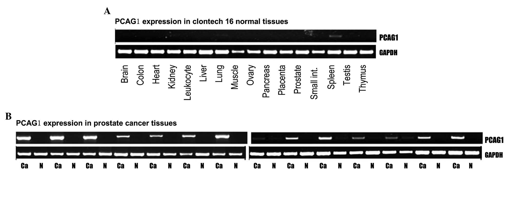
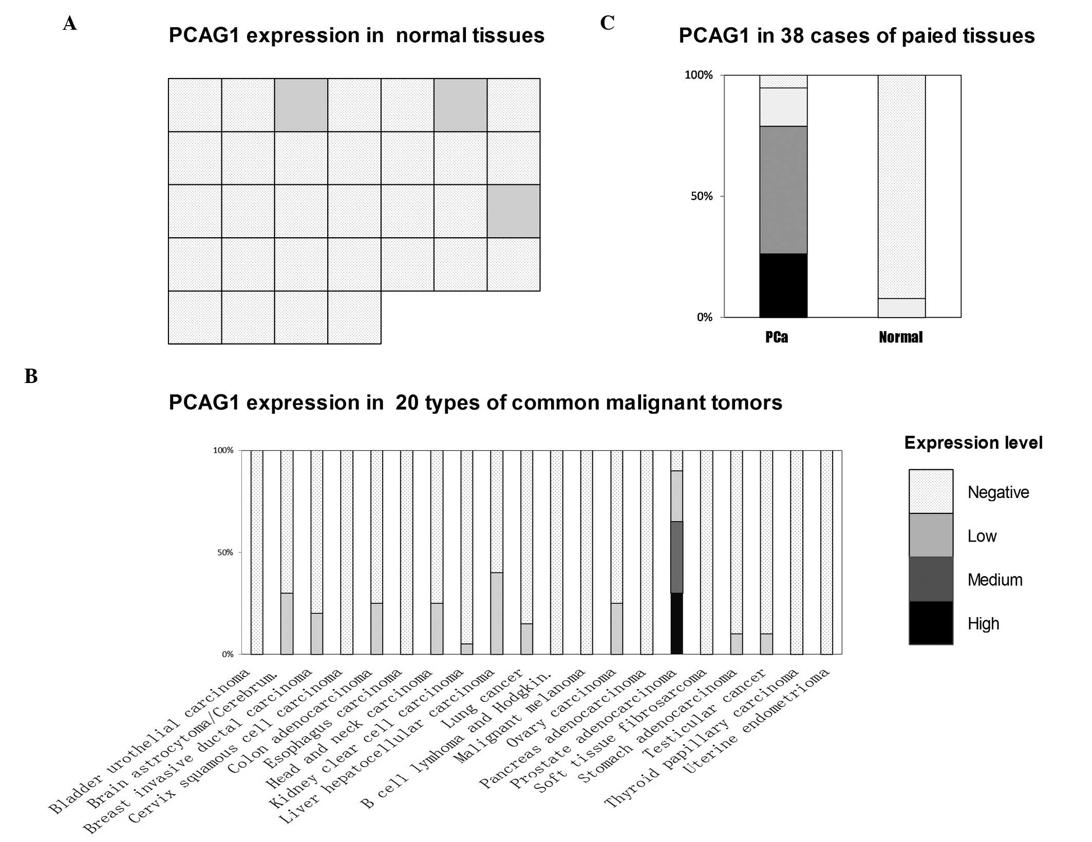
The aim of the present study was to identify a new prostate cancer‑associated gene and analyze its expression pattern. Comprehensive expression analysis of expressed sequence tags (ESTs) and microarray data and serial analysis of gene expression (SAGE) were conducted to screen in silico for candidate prostate cancer‑associated genes. Reverse transcription (RT)-PCR was performed to validate prostate cancer specificity. Prostate cancer‑associated gene 1 (PCAG1) was identified. The expression of PCAG1 mRNA and protein was evaluated in common human normal tissues, common malignant tumors, prostate adenocarcinoma and paired adjacent normal prostate tissues. An immunofluorescence assay was conducted to determine the subcellular location of PCAG1. PCAG1 mRNA was absent in the 15 pooled normal tissues (including normal prostate tissue) but registered at low levels in the spleen tissue (+). By contrast, PCAG1 mRNA was significantly higher than in the adjacent normal tissues in each of the 14 cases of prostate cancer, with ~50% scoring a high degree of expression (+++). Of the 32 types of normal tissues, 29 (including normal prostate tissue) demonstrated negative PCAG1 protein staining while the remaining tissues of the adrenal gland, parathyroid gland and liver expressed low levels. While 18/20 cases of prostate adenocarcinoma showed positive expression results, PCAG1 protein expression in the remaining types of cancer was scarce when present at all; only 41/380 other cancer cases demonstrated positive results at a low level. The most substantial PCAG1-positive expression results were identified by cytoplasmic staining in 36/38 prostate adenocarcinoma cases, with 10 cases showing high expression levels, 20 showing medium levels and 6 showing low levels. In the paired adjacent normal prostate tissues, only 3/38 cases showed low level positive staining, while 35/38 cases were negative. Immunofluorescent staining of the human prostate cancer PC3 cell line showed positive PCAG1 expression results in the mitochondria. The present study demonstrated that while PCAG1 mRNA was highly expressed in prostate cancer tissues, it was almost absent in all common normal tissues and paired adjacent normal prostate tissues. Furthermore, PCAG1 protein was also highly expressed in prostate cancer tissues, while few common normal tissues, other common malignant tumors and paired adjacent normal prostate tissues had even low levels of expression. Clarification of the function and transcriptional mechanism of PCAG1 may aid the elucidation of the mechanisms of carcinogenesis and progression of prostate cancer. The unique expression pattern of PCAG1 suggests its potential in certain clinical applications.
View Figures
View References

Related Articles

Journal Cover

March 2013
Volume 7 Issue 3

Print ISSN: 1791-2997
Online ISSN:1791-3004

Sign up for eToc alerts

Recommend to Library

Copy and paste a formatted citation
x
Spandidos Publications style
Lai Y, Yu Z, Wang Y and Ye J: Identification of PCAG1 as a novel prostate cancer-associated gene. Mol Med Rep 7: 755-760, 2013
APA
Lai, Y., Yu, Z., Wang, Y., & Ye, J. (2013). Identification of PCAG1 as a novel prostate cancer-associated gene. Molecular Medicine Reports, 7, 755-760. https://doi.org/10.3892/mmr.2012.1249
MLA
Lai, Y., Yu, Z., Wang, Y., Ye, J."Identification of PCAG1 as a novel prostate cancer-associated gene". Molecular Medicine Reports 7.3 (2013): 755-760.
Chicago
Lai, Y., Yu, Z., Wang, Y., Ye, J."Identification of PCAG1 as a novel prostate cancer-associated gene". Molecular Medicine Reports 7, no. 3 (2013): 755-760. https://doi.org/10.3892/mmr.2012.1249