|
1
|
Sherin JE and Nemeroff CB: Post-traumatic
stress disorder: the neurobiological impact of psychological
trauma. Dialogues Clin Neurosci. 13:263–278. 2011.PubMed/NCBI
|
|
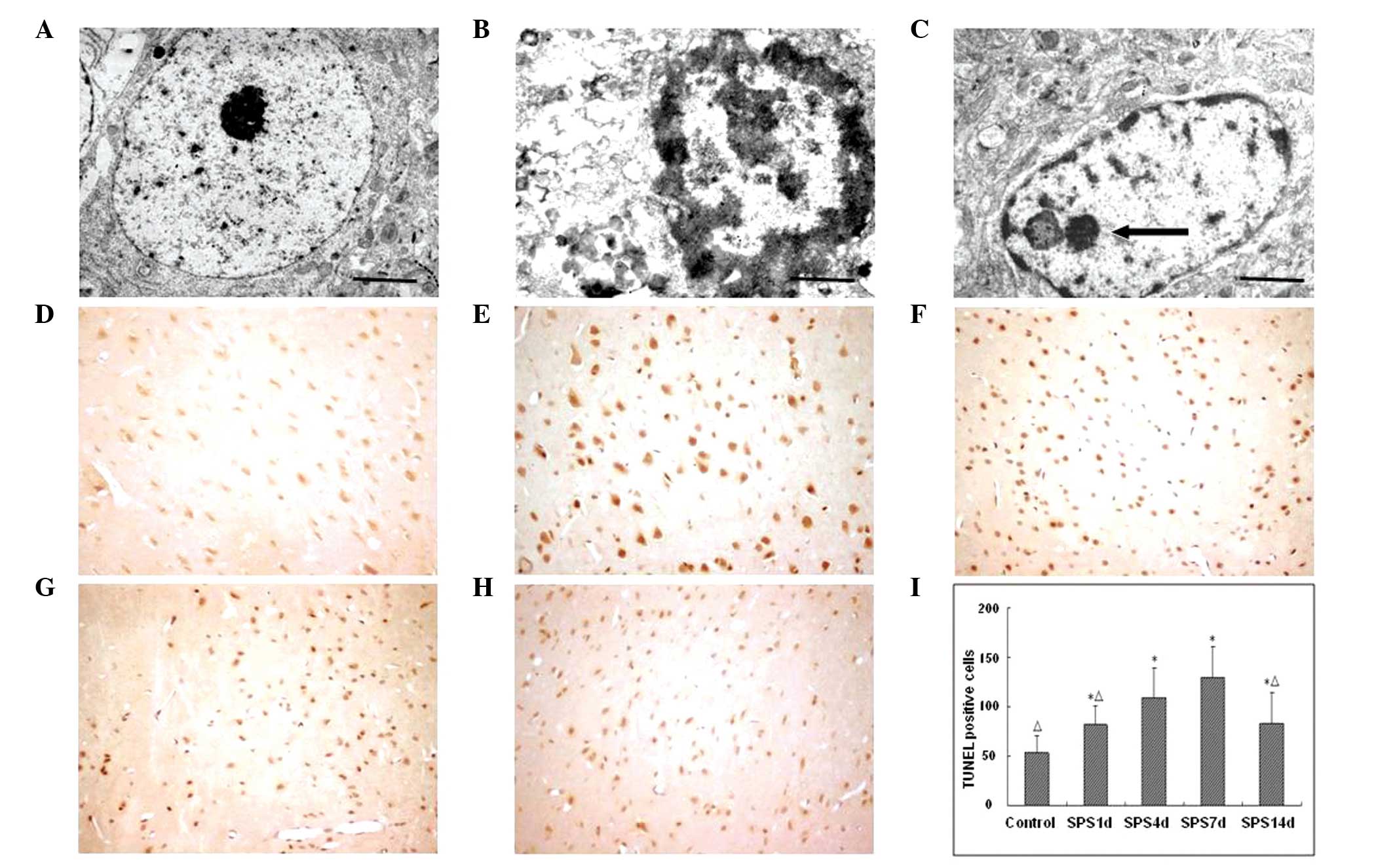
2
|
Vermetten E: Stress, trauma, and
post-traumatic stress disorder. Tijdschr Psychiatr. 51:595–602.
2009.(In Dutch).
|
|
3
|
Liberzon I, Krstov M and Young EA:
Stress-restress: effects on ACTH and fast feedback.
Psychoneuroendocrinology. 22:443–453. 1997. View Article : Google Scholar : PubMed/NCBI
|
|
4
|
Roozendaal B, Griffith QK, Buranday J, De
Quervain DJ and McGaugh JL: The hippocampus mediates
glucocorticoid-induced impairment of spatial memory retrieval:
dependence on the basolateral amygdala. Proc Natl Acad Sci USA.
100:1328–1333. 2003. View Article : Google Scholar : PubMed/NCBI
|
|
5
|
Wen Y, Li B, Han F, Wang E and Shi Y:
Dysfunction of calcium/calmodulin/CaM kinase IIα cascades in the
medial prefrontal cortex in post-traumatic stress disorder. Mol Med
Rep. 6:1140–1144. 2012.PubMed/NCBI
|
|
6
|
Bremner JD: Neuroimaging studies in
post-traumatic stress disorder. Curr Psychiatry Rep. 4:254–263.
2002. View Article : Google Scholar : PubMed/NCBI
|
|
7
|
Rauch SL, Shin LM and Phelps EA:
Neurocircuitry models of posttraumafic stress disorder and
extinction: human neuroimaging research - past, present, and
future. Biol Psychiatry. 60:376–382. 2006. View Article : Google Scholar : PubMed/NCBI
|
|
8
|
Xiao B, Yu B, Wang HT, Han F and Shi YX:
Single-prolonged stress induces apoptosis by activating cytochrome
C/caspase-9 pathway in a rat model of post-traumatic stress
disorder. Cell Mol Neurobiol. 31:37–43. 2011. View Article : Google Scholar
|
|
9
|
Fink G: Stress controversies:
post-traumatic stress disorder, hippocampal volume, gastroduodenal
ulceration. J Neuroendocrinol. 23:107–117. 2011. View Article : Google Scholar
|
|
10
|
Wang HT, Han F and Shi YX: Activity of the
5-HT1A receptor is involved in the alteration of glucocorticoid
receptor in hippocampus and corticotropin-releasing factor in
hypothalamus in SPS rats. Int J Mol Med. 24:227–231.
2009.PubMed/NCBI
|
|
11
|
Becchetti A, Pillozzi S, Morini R, Nesti E
and Arcangeli A: New insights into the regulation of ion channels
by integrins. Int Rev Cell Mol Biol. 279:135–190. 2010. View Article : Google Scholar : PubMed/NCBI
|
|
12
|
Sheppard D: Roles of alphav integrins in
vascular biology and pulmonary pathology. Curr Opin Cell Biol.
16:552–557. 2004. View Article : Google Scholar : PubMed/NCBI
|
|
13
|
Chatzizacharias NA, Kouraklis GP and
Theocharis SE: The role of focal adhesion kinase in early
development. Histol Histopathol. 25:1039–1055. 2010.PubMed/NCBI
|
|
14
|
Khan S and Liberzon I: Topiramate
attenuates exaggerated acoustic startle in an animal model of PTSD.
Psychopharmacology (Berl). 172:225–229. 2004. View Article : Google Scholar
|
|
15
|
Takahashi T, Morinobu S, Iwamoto Y and
Yamawaki S: Effect of paroxetine on enhanced contextual fear
induced by single prolonged stress in rats. Psychopharmacology
(Berl). 189:165–173. 2006. View Article : Google Scholar
|
|
16
|
Zhu CZ, Situ MJ, Zhang Y, Fang H, Jing LS,
Wang D, Yan J and Huang Y: Influence factors of post-traumatic
stress disorder (PTSD) and depression symptoms in children and
adolescents after Wenchuan earthquake in China. Zhonghua Yu Fang Yi
Xue Za Zhi. 45:531–536. 2011.(In Chinese). PubMed/NCBI
|
|
17
|
Milner R and Campbell IL: The integrin
family of cell adhesion molecules has multiple functions within the
CNS. J Neurosci Res. 69:286–291. 2002. View Article : Google Scholar : PubMed/NCBI
|
|
18
|
Gold AL, Shin LM, Orr SP, Carson MA, Rauch
SL, Macklin ML, Lasko NB, Metzger LJ, Dougherty DD, Alpert NM,
Fischman AJ and Pitman RK: Decreased regional cerebral blood flow
in medial prefrontal cortex during trauma-unrelated stressful
imagery in Vietnam veterans with post-traumatic stress disorder.
Psychol Med. 13:1–10. 2011.
|
|
19
|
Su TP, Zhang L, Chung MY, Chen YS, Bi YM,
Chou YH, Barker JL, Barrett JE, Maric D, Li XX, Li H, Webster MJ,
Benedek D, Carlton JR and Ursano R: Levels of the potential
biomarker p11 in peripheral blood cells distinguish patients with
PTSD from those with other major psychiatric disorders. J Psychiatr
Res. 43:1078–1085. 2009. View Article : Google Scholar : PubMed/NCBI
|
|
20
|
Mierke CT: The role of vinculin in the
regulation of the mechanical properties of cells. Cell Biochem
Biophys. 53:115–126. 2009. View Article : Google Scholar : PubMed/NCBI
|
|
21
|
Brohawn KH, Offringa R, Pfaff DL, Hughes
KC and Shin LM: The neural correlates of emotional memory in
posttraumatic stress disorder. Biol Psychiatry. 68:1023–1030. 2010.
View Article : Google Scholar : PubMed/NCBI
|
|
22
|
Jordà EG, Verdaguer E, Jimenez A, Arriba
SG, Allgaier C, Pallàs M and Camins A: Evaluation of the neuronal
apoptotic pathways involved in cytoskeletal disruption-induced
apoptosis. Biochem Pharmacol. 70:470–480. 2005. View Article : Google Scholar : PubMed/NCBI
|
|
23
|
Gary DS and Mattson MP: Integrin signaling
via the PI3-kinase-Akt pathway increases neuronal resistance to
glutamate-induced apoptosis. J Neurochem. 76:1485–1496. 2001.
View Article : Google Scholar : PubMed/NCBI
|
|
24
|
Demir O, Singh S, Klimaschewski L and
Kurnaz IA: From birth till death: neurogenesis, cell cycle, and
neurodegeneration. Anat Rec (Hoboken). 292:1953–1961. 2009.
View Article : Google Scholar
|
|
25
|
Leerberg JM and Yap AS: Vinculin, cadherin
mechanotransduction and homeostasis of cell-cell junctions.
Protoplasma. 250:817–829. 2013. View Article : Google Scholar : PubMed/NCBI
|
|
26
|
Klee P and Meda P: Connexin signaling: a
new mechanism for protection of insulin-producing cells against
apoptosis. Med Sci (Paris). 28:41–44. 2012.(In French). View Article : Google Scholar
|
|
27
|
Nandrot EF, Silva KE, Scelfo C and
Finnemann SC: Retinal pigment epithelial cells use a
MerTK-dependent mechanism to limit the phagocytic particle binding
activity of αvβ5 integrin. Biol Cell. 104:326–341. 2012. View Article : Google Scholar : PubMed/NCBI
|
|
28
|
Kawaguchi SY and Hirano T: Integrin
alpha3beta1 suppresses long-term potentiation at inhibitory
synapses on the cerebellar Purkinje neuron. Mol Cell Neurosci.
31:416–426. 2006. View Article : Google Scholar
|
|
29
|
Farwell AP, Tranter MP and Leonard JL:
Thyroxine-dependent regulation of integrin-laminin interactions in
astrocytes. Endocrinology. 136:3909–3915. 1995.PubMed/NCBI
|
|
30
|
Mobley AK and McCarty JH: Use of Cre-lox
technology to analyze integrin functions in astrocytes. Methods Mol
Biol. 814:555–570. 2012. View Article : Google Scholar
|
|
31
|
Carisey A and Ballestrem C: Vinculin, an
adapter protein in control of cell adhesion signalling. Eur J Cell
Biol. 90:157–163. 2010. View Article : Google Scholar : PubMed/NCBI
|
|
32
|
Li WE and Nagy JI: Connexin43
phosphorylation state and intercellular communication in cultured
astrocytes following hypoxia and protein phosphatase inhibition.
Eur J Neurosci. 12:2644–2650. 2000. View Article : Google Scholar : PubMed/NCBI
|
|
33
|
Solan JL and Lampe PD: Connexin43
phosphorylation: structural changes and biological effects. Biochem
J. 419:261–272. 2009. View Article : Google Scholar : PubMed/NCBI
|
|
34
|
Irie A: Integrin family. Nihon Rinsho.
68:163–166. 2010.(In Japanese).
|
|
35
|
Lv X, Su L, Yin D, Sun C, Zhao J, Zhang S
and Miao J: Knockdown of integrin beta4 in primary cultured mouse
neurons blocks survival and induces apoptosis by elevating NADPH
oxidase activity and reactive oxygen species level. Int J Biochem
Cell Biol. 40:689–699. 2008. View Article : Google Scholar
|