Introduction
Ventricular fibrillation (VF) is one of the most
common causes of sudden cardiac death (1). In recent years, along with the
development of electrophysiological mapping techniques and in-depth
research on cardiac fibrillation, the understanding of the
mechanism of VF has greatly progressed (2,3).
Re-entry is still considered to be the most important mechanism
responsible for tachyarrhythmias (4,5).
Conditions for re-entry include: i) The objective existence of an
anatomical re-entry ring; ii) a unidirectional conduction block;
iii) inconsistent conduction velocity and refractory period. For
these three basic conditions, the refractory period is determined
by the electrical activity of single cells. The spread of
electrical activity from a single cell to its neighboring cells,
thereby causing the synchronization of cardiac tissue excitability,
is determined by the electrical coupling via gap junctions (GJ)
between cells (6–8). GJs are important for the passive
electrical activity of myocardial tissues.
There are two connection types between GJs:
End-to-end and side-to-side. The structural basis for myocardial
anisotropy depends on GJ connections. Normal myocardium is fusiform
in arrangement and most GJs are end-to-end type at the long axis of
the cardiomyocytes, with the lower junction resistance facilitating
easy conduction of the ionic current and rapid electrical
excitation. However, in the direction of the short axis, where GJs
are of the side-to-side type, conduction of electrical excitation
is slow and spreads in a 'zig-zag' style shape, causing increased
probability of re-entry (9,10).
In theory, the occurrence and maintenance of arrhythmias increase
with the proportion of side-to-side GJs.
Connexins (Cx) are transmembrane proteins that
assemble to form intercellular GJs. Reduced expression and abnormal
distribution of Cx can slow down the overall cardiac conduction
velocity and change conduction anisotropy, thereby forming the
anatomical basis of a re-entrant loop that induces arrhythmia. In
adult ventricles, GJs contain only Cx43 and are distributed mainly
in the intercalated disk (11).
Previous studies by our group have demonstrated that GJ remodeling
caused by decreased expression and abnormal distribution of
myocardial Cx43 is involved in the occurrence and maintenance of VF
(12,13). Furthermore, a previous study by our
group showed that the gap junction enhancer ZP123 can reduce or
reverse the Cx43 degradation and derangement, thereby reducing
defibrillation energy and improving cardioversion in VF (14).
In cardiomyocytes, only phosphorylated Cx43 (p-Cx43)
constitutes a functional GJ and dephosphorylated Cx43 does not have
this function (15). Therefore,
the aim of the present study was to investigate changes in Cx43
phosphorylation and the effects of Z123 in cardioversion in an
animal model of VF.
Materials and methods
Animal preparation
All animal care and experimental protocols received
approval from the Animal Care and Use Committee of Qilu Hospital,
Shandong University, (Jinan, China). Mongrel dogs (n=56, male or
female, weighing 11–17 kg) were obtained from the Center for
Experimental Animals of Qilu Hospital of Shandong University.
Animals were randomly divided into seven groups (n=8/group): Sham
control, 8-min VF + rotigaptide (ZP123; Shanghai Fuhe Chemistry
Technology Co., Ltd., Shanghai, China), 8-min VF + normal saline
(NS), 12-min VF + ZP123, 12-min VF + NS, 30-min VF + ZP123 and
30-min VF + NS. The three ZP123 groups were given ZP123 1
μg/kg bolus + 10 μg/kg/h by micropump (Perfusor
Basic; B. Braun Melsungen AG, Melsungen, Germany) intravenous (iv)
infusion for 30 min; the NS groups were given saline instead of
ZP123. The animals were anesthetized with pentobarbital-Na (30
mg/kg iv, repetition when necessary; Merck Millipore, Darmstadt,
Germany). After anesthesia, dogs were placed in the supine position
and restrained at the four extremities. Animals were intubated with
a 5.0 cuffed tracheal attached to a ventilator (Newport E-100M,
Newport Medical Instruments, Costa Mesa, CA, USA). Ventilation
began at a tidal volume of 10–15 ml/kg, with a ventilator rate of
16–20 breaths/min, and an inspiration to expiration ratio of
1:1.5–2.0. Three surface electrodes were placed separately under
three limbs to correspond to standard lead II electrocardiogram
(ECG). The right femoral artery and bilateral femoral veins were
cannulated. 6-F catheters (Cordis Corp, Miami, FL, USA) were
positioned in the intrathoracic ascending aorta and the right
atrium under fluoroscopic guidance (CGO-3000; Beijing Wandong
Medical Equipment,Beijing, China). The remaining cannulated femoral
vein was used for drug infusion. Heparin (Shenzhen Hepalink
Pharmaceutical Co., Ltd., Shenzhen, China) was administered at 100
U/kg for anti-coagulation, and added when necessary.
Creation of the VF model
VF was induced by delivering a 5-sec alternating
current at 80 V across the thorax through two needles after the
completion of drug delivery. Successful VF was defined as a
decrease in aortic blood pressure below 25 mmHg and the presence of
a VF waveform on the ECG. Dogs in the sham control group were
anesthetized and intubated tracheally without the induction of VF.
Untreated VF lasted for 8, 12 or 30 min in other groups. All
measurements were performed by an investigator blinded to the
experimental group assignment. At the end of the experimental
protocol, the surviving animals were sacrificed by KCl (Tianjin
XuanChi International Trade Co., Ltd., Tianjin, China)
infusion.
Transthoracic echocardiography (TTE)
study
Prior to VF and after return of spontaneous
circulation (ROSC), all animals were examined by transeophageal
echocardiography with a 2.5–3.5 MHz transducer (Philips Sonos 7500,
Philips Corp, Eindhoven, The Netherlands) to measure the left
atrium dimension (LAD), left ventricular diastolic dimensions
(LVDd) and left ventricular ejection fraction (LVEF), in a
parasternal long axis and M-mode view.
Hemodynamic measurements
Catheter positions were confirmed by X-ray
fluoroscopy. Catheters in the intrathoracic ascending aorta and the
right atrium were connected to the pressure transducers attached to
a PRO EP recording system (PowerLab/16sp; AD Instruments, Sydney,
Australia) by which aortic systolic pressure (AOSP), diastolic
aortic pressure (AODP) and right atrial diastolic pressure (RADP)
were recorded throughout the study. The coronary perfusion pressure
(CPP) was calculated as AODP minus RADP and the mean aortic
pressure (MAP) was calculated as 1/3 AOSP plus 2/3 AODP.
Cardiac arrest and defibrillation
protocols
Cardiopulmonary resuscitation (CPR) was begun at the
start of the VF period. All animals were given 2 min of CPR (chest
compression rate, 100/min; chest compression-to-ventilation ratio,
30:2) prior to transthoracic countershock. If ROSC was not
achieved, an immediate defibrillation using a 70-J biphasic shock
was delivered by an external defibrillator (M4735; Philips). If
ROSC was not achieved, another 2 min of CPR was performed
immediately and the defibrillation energy was increased to a 100-J
biphasic shock. If ROSC was still not achieved, this process was
repeated with increased defibrillation energy (up to 150-J biphasic
shock). The third CPR was continued until ROSC was achieved. If VF
persisted, additional epinephrine (0.02 mg/kg) was administered and
CPR was continued for 2 min with equal defibrillation energy (150-J
biphasic shock)until the animals achieved ROSC or the entire rescue
process reached 30 min. ROSC was defined as AOSP ≥80 mmHg, lasting
for at least 1 min (16). The
average defibrillation energy, defibrillation success rate, the
success rate of the previous three defibrillations, ROSC and
survival (1 h) rate were calculated.
Tissue collection
Samples of the left ventricular myocardium were
collected within 15 sec of death and immediately snap-frozen in
liquid nitrogen (stored at −80°C).
Western blot analysis
Myocardial membrane proteins were extracted by
differential centrifugation (4°C; 1,000 × g for 10 min then 10,000
× g for 1 h). Equal amounts of protein from each sample were
subjected to SDS-PAGE (10% polyacrylamide; EMD Millipore, Bedford,
MA, USA) and transferred onto polyvinylidene difluoride (PVDF)
membranes (EMD Millipore). The membranes were blocked for 2 h with
1% bovine serum albumin (cat. no. ST023; Huayan Biological
Technology Co., Ltd., Shanghai, China) and then incubated overnight
at 4°C with either rabbit polyclonal anti-Cx43 (cat. no. AB1727) or
anti-p-Cx43 (cat. no. P3859Rb-p) diluted 1:400 (USCN Life, Wuhan,
China). After being washed, membranes were incubated with
horseradish peroxidase-conjugated goat anti-rabbit IgG secondary
antibody diluted 1:2,000 (cat. no. sc-2004; Santa Cruz
Biotechnology, Inc., Dallas, TX, USA) for 2 h. Immunoreactive bands
were visualized with the SuperSignal West Pico enhanced
chemiluminescence kit (Pierce, Rockford, IL, USA) according to the
manufacturer's instructions. A monoclonal mouse antibody against
β-actin (cat. no. A1978; Sigma-Aldrich, St. Louis, MO, USA) was
used in every experiment as an internal control. Band intensities
were quantified with Quantity One (Bio-Rad Laboratories, Inc.
Hercules, CA, USA).
Immunofluorescence and confocal laser
scanning microscopy of Cx43 and phosphorylated (p-)Cx43
Left ventricular preparations were fixed in 4%
paraformaldehyde for 24 h. Tissue samples were embedded in paraffin
(Santa Cruz Biotechnology, Inc., Santa Cruz, CA, USA), cut into
4-μm sections and mounted on gelatin-coated slides, prior to
dewaxing, and rehydration with graded alcohols (Santa Cruz
Biotechnology, Inc.). The slides were microwaved (700 watts for 15
min at 96°C) in boiling 0.01 M sodium citrate buffer (Santa Cruz
Biotechnology, Inc.) for 15 min to enhance specific immunostaining.
For blocking, a goat serum/phosphate-buffered saline (PBS; 1:20;
Santa Cruz Biotechnology, Inc.) solution was added to the slides
for 20 min at room temperature. The polyclonal rabbit anti-dog
primary antibodies for Cx43 (AB1727; 1:100 dilution in PBS; EMD
Millipore) and p-Cx43 (P3859Rb-p; 1:100 dilution in PBS; USCN Life
Science, Inc., Wuhan, China) detection were added and slides were
incubated overnight at 4°C. Finally, the slides were incubated in
fluorescein isothiocyanate (FITC)-conjugated goat anti-rabbit
secondary IgG antibody (sc-2012; 1:500 dilution; Santa Cruz
Biotechnology, Inc.) for 30 min at 37°C. Samples were examined by
laser scanning confocal microscopy (TCS SP2; Leica, Wetzlar,
Germany). Primary antibodies were replaced with PBS in
immunostaining controls. FITC excitation was performed at 488 nm
and fluorescence was emitted at 519 nm. High-intensity signals were
measured and analyzed with Image-Pro plus 6.0 software (Media
Cybernetics, Inc., Rockville, MD, USA).
Statistical analysis
Values are expressed as the mean ± standard error of
the mean. All variables were normally distributed and were analyzed
by analysis of variance, correlation between variables was
performed by linear correlation analysis, and non-normal
distribution data were subjected to the rank correlation analysis,
with P-values <0.05 considered to indicate statistically
significant differences between values. Statistical analysis was
performed with SPSS 13.0 (SPSS, Inc., Chicago, IL, USA).
Results
Baseline characteristics
There were no statistically significant differences
among the animals in terms of body weight, heart rate, LAD, LVDd,
LVEF and hemodynamic variables at baseline (Table I).
 | Table IBaseline characteristics in
experimental groups. |
Table I
Baseline characteristics in
experimental groups.
| Characteristic | 8-min VF
| 12-min VF
| 30-min VF
|
|---|
| ZP123 | NS | ZP123 | NS | ZP123 | NS |
|---|
| Subjects (n) | 8 | 8 | 8 | 8 | 8 | 8 |
| Weight (kg) | 13.15±1.43 | 13.29±1.61 | 13.36±1.26 | 13.19±1.72 | 13.42±1.32 | 13.31±1.52 |
| Heart (bpm) | 158±12 | 153±10 | 154±11 | 156±13 | 157±9 | 152±12 |
| Echocardiography |
| LAD (mm) | 15.12±1.24 | 15.41±1.53 | 16.65±1.73 | 17.52±1.46 | 17.16±1.58 | 16.27±1.35 |
| LVDd (mm) | 23.16±1.38 | 24.64±1.58 | 23.46±1.51 | 25.33±1.71 | 24.82±1.62 | 23.67±1.38 |
| LVEF (%) | 0.68±0.08 | 0.67±0.06 | 0.67±0.05 | 0.69±0.07 | 0.69±0.04 | 0.68±0.09 |
| Hemodynamics |
| AOSP (mmHg) | 134.52±15.27 | 132.75±16.3 | 132.48±16.51 | 137.25±20.16 | 135.37±18.26 | 133.62±19.43 |
| AODP (mmHg) | 109.32±25.67 | 112.62±28.36 | 108.45±23.26 | 114.63±21.53 | 110.51±22.35 | 107.83±24.63 |
| MAP (mmHg) | 117.72±20.63 | 119.33±24.07 | 116.46±23.66 | 122.17±19.40 | 118.8±21.16 | 116.43±22.32 |
| RADP (mmHg) | 6.62±2.71 | 7.98±2.54 | 6.77±2.25 | 7.85±2.07 | 7.56±1.93 | 8.15±1.86 |
| CPP (mmHg) | 102.7±26.35 | 104.64±33.96 | 101.68±22.5 | 106.78±22.5 | 102.95±23.53 | 99.68±28.16 |
Comparison of TTE and hemodynamic index
of animals achieving ROSC in the groups
Compared with the pre-VF values, LAD and LVDd in the
8-min and 12-min VF groups were significantly increased, while LVEF
was significantly decreased (P<0.05). LAD and LVEF in the 12-min
VF ZP123 and NS groups were different compared with those in the
8-min VF groups (P<0.05). The difference in LVDd among groups
did not reach statistical significance. With the exception of RADP,
all other hemodynamic indexes (AOSP, AODP, MAP and CPP) in the four
groups were significantly lower than those prior to VF. Too few
animals survived to determine any statistically significant
differences between the 30-min VF groups (Table II).
| Table IICharacteristics of animals which
achieved return of spontaneous circulation in experimental
groups. |
Table II
Characteristics of animals which
achieved return of spontaneous circulation in experimental
groups.
| Content | 8-min VF
| 12-min VF
| 30-min VF
|
|---|
| ZP123 | NS | ZP123 | NS | ZP123 | NS |
|---|
| Subjects (n) | 7 | 6 | 5 | 4 | 0 | 1 |
| Echocardiography |
| LAD (mm) | 17.73±1.52a | 17.62±1.98a | 21.82±2.04a,b | 22.15±1.82a,b | – | 19.57 |
| LVDd (mm) | 27.76±2.03a | 28.75±1.72a | 30.67±1.93a | 31.32±2.46a | – | 26.28 |
| LVEF (%) | 0.51±0.07a | 0.53±0.09a | 0.37±0.07a,b | 0.33±0.06a,b | – | 0.20 |
| Hemodynamics |
| AOSP (mmHg) | 86.92±18.65a | 89.31±17.32a | 80.26±15.69a | 81.27±13.52a | – | 80.43 |
| AODP (mmHg) | 63.33±21.34a | 66.23±19.3a | 55.95±20.56a | 57.81±17.94a | – | 53.47 |
| MAP (mmHg) | 71.19±17.25a | 73.92±13.78a | 63.72±18.22a | 65.63±16.82a | – | 62.46 |
| RADP (mmHg) | 7.97±1.56 | 8.02±1.33 | 8.27±1.54 | 8.99±2.06 | – | 9.02 |
| CPP (mmHg) | 55.36±17.15a | 58.21±16.2a | 47.68±14.36a | 48.82±18.73a | – | 44.45 |
Comparison of defibrillation success
rate, average defibrillation energy and survival rate among
groups
Successful defibrillation was achieved in all eight
animals in the 8-min VF + ZP123 group, including full ROSC and
survival achieved during the first three defibrillations in seven
of these animals. Successful defibrillation was achieved in seven
animals in the 8-min VF + NS group, including four in the first
three defibrillations and six in total achieving ROSC and survival
for >1 h. Successful defibrillation was achieved in seven
animals in the 12-min VF + ZP123 group, with five achieving ROSC
and four surviving for >1 h. However, in the 12-min NS group,
successful defibrillation was achieved in only five animals, with
four achieving ROSC and three surviving for >1 h. Successful
defibrillation was achieved in only two animals in the 30-min ZP123
group, although none but none survived, while successful
defibrillation and survival was achieved in one animal in the
30-min NS group.
Statistical analysis showed that the success rates
of the first three defibrillations in the 8-min and 12-min ZP123
groups were significantly higher than those in the NS control group
(P<0.05), and the average defibrillation energy was
significantly lower than that in the NS group (P<0.05). Although
the difference in the ROSC and survival rates between the ZP123 and
control groups did not reach statistical significance (P>0.05),
the rates were higher in the three ZP123 treatment groups than
those in the NS groups (Table
III).
| Table IIIComparison of indexes at the end of
the experiment. |
Table III
Comparison of indexes at the end of
the experiment.
| Index | 8-min VF
| 12-min VF
| 30-min VF
|
|---|
| ZP123 | NS | ZP123 | NS | ZP123 | NS |
|---|
| Defibrillation
success rate | 8/8 | 7/8 | 7/8 | 5/8 | 2/8 | 1/8 |
| First 3
defibrillations success rate | 7/8a | 4/8 | 5/8a | 2/8 | 0 | 0 |
| Average
defibrillation energy (J) | 239±141a | 406±170 | 577±207a | 745±221 | – | – |
| ROSC rate | 7/8 | 6/8 | 5/8 | 4/8 | 0 | 1/8 |
| Survival rate | 7/8 | 6/8 | 4/8 | 3/8 | 0 | 1/8 |
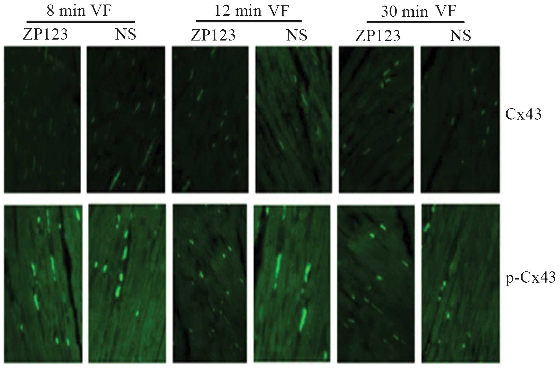
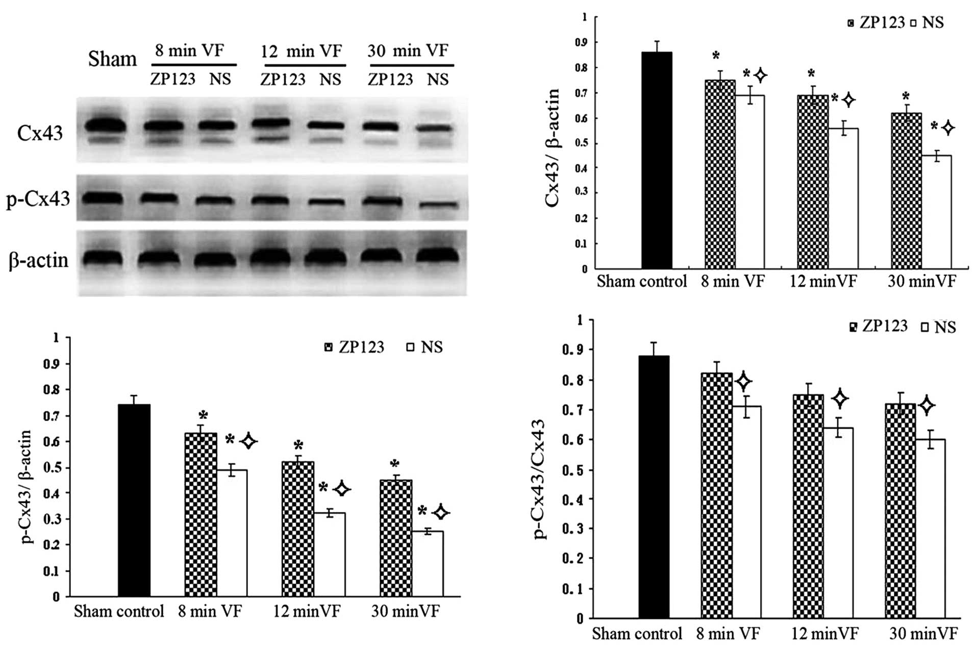
Immunofluorescence and western blot
analysis results of Cx43 and p-Cx43
High-intensity specific immunoreactive signals for
the detection of Cx43 and p-Cx43 were clearly identified with
regular distribution in the ZP123 groups compared to those in the
control groups. With the duration of VF, the Cx43 and p-Cx43
signals became progressively weaker with more irregular
distribution (Fig. 1).
Western blot analysis showed that Cx43 expression in
the VF groups was significantly lower than that in the sham control
group (P<0.05). Cx43 expression was higher in the 12-min and
30-min ZP123 groups compared with that in the control groups
(P<0.05), while no difference was detected in the 8-min groups.
With the duration of VF, p-Cx43 expression decreased, while p-Cx43
in the ZP123 groups was significantly higher than that in the
control groups (P<0.05). Furthermore, the ratio of p-Cx43/Cx43
decreased, while the p-Cx43/Cx43 ratio in the ZP123 groups was
significantly higher than that in the control groups (P<0.05)
(Fig. 2A–D).
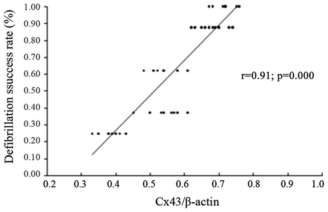
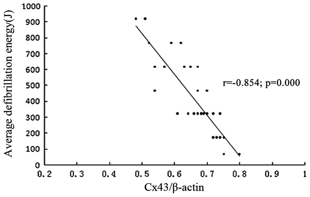
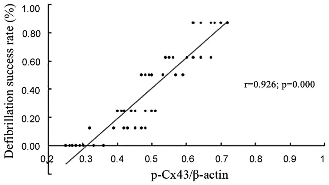
Correlation analysis
Cx43 expression was positively correlated with the
defibrillation success rate (r=0.91; P<0.01) (Fig. 3), and negatively with the average
defibrillation energy (r=−0.854; P<0.01) (Fig. 4), while p-Cx43 expression
correlated positively with the success rate of the first three
defibrillations (r=0.926; P<0.01) (Fig. 5).
Discussion
The main finding of the present study was that
pre-treatment with ZP123 reduced Cx43 remodeling through
upregulation of Cx43 expression, downregulation of Cx43
dephosphorylation and by unifying the arrangement and distribution
of Cx43 expression. These effects improved the electrical coupling
between gap junctions, thereby reducing the defibrillation energy
required for successful cardioversion (12). ~80% of cardiac arrests are caused
by VF and although numerous types of anti-arrhythmic drugs have
been evaluated, the therapeutic effects on ventricular tachycardia
and VF arrhythmia are poor. Most traditional anti-arrhythmic drugs
are myocardial cell membrane ion channel blockers, while some also
have potential arrhythmogenic effects, and even increase mortality
(17,18). Therefore, the identification of
safer and more effective anti-arrhythmic drugs has become a focus
of research in recent years.
ZP123 is a novel anti-arrhythmic peptide, which acts
specifically on gap junctions (19,20).
Although there are numerous studies on ZP123 and ventricular
arrhythmias, the main focus has been on the prevention of acute
myocardial infarction with re-entrant ventricular tachycardia and
ischemic reperfusion arrhythmia (21–23).
Studies on the effects of ZP123 on VF, particularly prolonged VF,
are rare. A previous study by our group conducted in an 8-min VF
model in pigs showed that ZP123 treatment upregulated Cx43
expression with a uniform arrangement and distribution (14). Another study by our group using a
4-min VF rabbit model confirmed the effects of ZP123 in reducing
defibrillation energy without significant effects on hemodynamic
and electrophysiological indicators, including systolic blood
pressure, right atrial pressure, heart rate, QRS width, QT interval
and one-way action potential (24).
In the present study, 8-min, 12-min and 30-min
models of shock-induced VF were established in dogs. It was found
that, compared with the NS control group subjected to the same VF
process, ZP123 increased Cx43 expression in myocardial membranes,
particularly the phosphorylated form (p-Cx43), while the disruption
of Cx43 distribution was alleviated. In the ZP123 groups, Cx43
expression in the myocardial cell membrane increased with VF
compared with that in the NS control group; however, despite the
8-min VF, the increase did not reach the level of statistical
significance, possibly due to the small sample size. In addition,
ZP123 lowered the defibrillation energy and improved the
defibrillation success rate, particularly in the first three
defibrillations, compared with that in the NS control group. A
correlation was identified between Cx43 expression in myocardial
cell membranes and the defibrillation energy and success rate
(r=−0.854, 0.91; P<0.001). Specifically, increased Cx43
expression levels significantly reduced the defibrillation energy
and improved defibrillation success rates. A significant
correlation was also identified between the success rate of the
first three cardiac defibrillations and p-Cx43 expression levels in
the myocardial cell membrane (r=0.926; P<0.001), indicating that
the cardiac defibrillation success rate is dependent on Cx43
phosphorylation.
The specific molecular and electrophysiological
mechanism underlying the effects of ZP123 on GJs remain elusive.
However, according to a previous study, ZP123 enhances electrical
coupling between GJs in ventricular myocytes without affecting ion
channels and accelerates conduction between cells while reducing
the heterogeneity of myocardial cell repolarization, thus
eliminating the effects generated by the arrhythmia (25).
In conclusion, the present study indicated that
prolonged VF leading to myocardial ischemia, hypoxia and acidosis,
can induce Cx43 remodeling and the loss of GJ coupling. These
factors lead to slowing of the electrical conduction velocity and
increased re-entry, which facilitate the maintenance of VF. ZP123
treatment upregulated the expression of Cx43, particularly the
phosphorylated form, in cardiac cell membranes, and reduced the
heterogeneity in Cx43 arrangement and distribution, thus
alleviating the effects of myocardial GJ remodeling and enhancing
GJ electrical coupling. Thereby, these effects reduced re-entry and
improved cardiac electrical conduction velocity, thus facilitating
cardioversion in VF. It can be speculated that the molecular
mechanism underlying the ZP123-mediated reduction in defibrillation
energy and improved defibrillation success rate are mediated
through regulation of the expression of Cx43 and phosphorylated
Cx43 in particular.
Acknowledgments
This study was sponsored by the Natural Science
Foundation of China (no. 81270238) and the Scientific Research
Foundation for the Doctoral Degree, State Education Ministry of
China (no. 20100131110059), and supported by the Scientific
Development Plan of Shandong Province of China (no.
2012G0021850).
References
|
1
|
Hovdenes J, Laake JH, Aaberge L, Haugaa H
and Bugge JF: Therapeutic hypothermia after out-of-hospital cardiac
arrest: experiences with patients treated with percutaneous
coronary intervention and cardiogenic shock. Acta Anaesthesiol
Scand. 51:137–142. 2007. View Article : Google Scholar
|
|
2
|
Osadchii OE: Mechanisms of
hypokalemia-induced ventricular arrhythmogenicity. Fundam Clin
Pharmacol. 24:547–559. 2010. View Article : Google Scholar : PubMed/NCBI
|
|
3
|
Bae JH, Park CW, Cho JH, Kim YS, Lee HY
and Won MH: The potential mechanism of the detrimental effect of
defibrillation prior to cardiopulmonary resuscitation in prolonged
cardiac arrest model. Lab Anim Res. 30:79–83. 2014. View Article : Google Scholar : PubMed/NCBI
|
|
4
|
Langley P, MacGowan GA and Murray A:
Spatial and temporal organization of the dominant frequencies in
the fibrillating heart: body surface potential mapping in a rare
case of sustained human ventricular fibrillation. Europace.
11:324–327. 2009. View Article : Google Scholar
|
|
5
|
Tribulová N, Knezl V, Okruhlicová L and
Slezák J: Myocardial gap junctions: targets for novel approaches in
the prevention of life-threatening cardiac arrhythmias. Physiol
Res. 57(Suppl 2): S1–S13. 2008.
|
|
6
|
Chaldoupi SM, Loh P, Hauer RN, de Bakker
JM and van Rijen HV: The role of connexin40 in atrial fibrillation.
Cardiovasc Res. 84:15–23. 2009. View Article : Google Scholar : PubMed/NCBI
|
|
7
|
Axelsen LN, Haugan K, Stahlhut M, et al:
Increasing gap junctional coupling: a tool for dissecting the role
of gap junctions. J Membr Biol. 216:23–35. 2007. View Article : Google Scholar : PubMed/NCBI
|
|
8
|
Fialová M, Dlugosová K, Okruhlicová L,
Kristek F, Manoach M and Tribulová N: Adaptation of the heart to
hypertension is associated with maladaptive gap junction
connexin-43 remodeling. Physiol Res. 57:7–11. 2008.
|
|
9
|
Lee PJ and Pogwizd SM: Micropatterns of
propagation. Adv Cardiol. 42:86–106. 2006. View Article : Google Scholar : PubMed/NCBI
|
|
10
|
van Rijen HV, de Bakker JM and van Veen
TA: Hypoxia, electrical uncoupling, and conduction slowing: Role of
conduction reserve. Cardiovasc Res. 66:9–11. 2005. View Article : Google Scholar : PubMed/NCBI
|
|
11
|
Moffitt JA, Henry MK, Welliver KC, Jepson
AJ and Garnett ER: Hindlimb unloading results in increased
predisposition to cardiac arrhythmias and alters left ventricular
connexin 43 expression. Am J Physiol Regul Integr Comp Physiol.
304:R362–R373. 2013. View Article : Google Scholar : PubMed/NCBI
|
|
12
|
Yi SL, Zhong JQ, Zhang J, Su GY, Li JS,
Liu HZ and Zhang Y: ZP123 reduces energy required for
defibrillation by preventing connexin43 remodeling during prolonged
ventricular fibrillation in swine. Tex Heart Inst J. 39:784–791.
2012.
|
|
13
|
Li JS, Zhong JQ, Zeng QX, Liu HZ, Su GY
and Zhang Y: Effect of ZP123, a gap junction modifier, on prolonged
ventricular fibrillation in swine. Cardiology. 118:147–152. 2011.
View Article : Google Scholar : PubMed/NCBI
|
|
14
|
Liu HZ, Zhong JQ, Li JS, Su GY, Wang J and
Zhang Y: Changes in myocardial connexin 43 during ventricular
fibrillation. Zhongguo Wei Zhong Bing Ji Jiu Yi Xue. 22:595–598.
2010.In Chinese. PubMed/NCBI
|
|
15
|
Salameh A and Dhein S: Adrenergic control
of cardiac gap junction function and expression. Naunyn
Schmiedebergs Arch Pharmacol. 383:331–346. 2011. View Article : Google Scholar : PubMed/NCBI
|
|
16
|
Sattur S and Kern KB: Increasing CPR
duration prior to first defibrillation does not improve return of
spontaneous circulation or survival in a swine model of prolonged
ventricular fibrillation. Resuscitation. 80:382author reply
382–383. 2009. View Article : Google Scholar
|
|
17
|
Echt DS, Liebson PR, Mitchell LB, et al:
Mortality and morbidity in patients receiving encainide,
flecainide, or placebo. The Cardiac Arrhythmia Suppression Trial. N
Engl J Med. 324:781–788. 1991. View Article : Google Scholar : PubMed/NCBI
|
|
18
|
Waldo AL, Camm AJ, deRuyter H, et al:
Effect of d-sotalol on mortality in patients with left ventricular
dysfunction after recent and remote myocardial infarction. The
SWORD Investigators. Survival With Oral d-Sotalol. Lancet.
348:7–12. 1996. View Article : Google Scholar : PubMed/NCBI
|
|
19
|
Dhein S, Larsen BD, Petersen JS and Mohr
FW: Effects of the new antiarrhythmic peptide ZP123 on epicardial
activation and repolarization pattern. Cell Commun Adhes.
10:371–378. 2003. View Article : Google Scholar : PubMed/NCBI
|
|
20
|
Xing D, Kjølbye AL, Petersen JS and
Martins JB: Pharmacological stimulation of cardiac gap junction
coupling does not affect ischemia-induced focal ventricular
tachycardia or triggered activity in dogs. Am J Physiol Heart Circ
Physiol. 288:H511–H516. 2005. View Article : Google Scholar : PubMed/NCBI
|
|
21
|
Xing D, Kjølbye AL, Nielsen MS, et al:
ZP123 increases gap junctional conductance and prevents reentrant
ventricular tachycardia during myocardial ischemia in open chest
dogs. J Cardiovasc Electrophysiol. 14:510–520. 2003. View Article : Google Scholar : PubMed/NCBI
|
|
22
|
Haugan K, Marcussen N, Kjølbye AL, Nielsen
MS, Hennan JK and Petersen JS: Treatment with the gap junction
modifier rotigaptide (ZP123) reduces infarct size in rats with
chronic myocardial infarction. J Cardiovasc Pharmacol. 47:236–242.
2006. View Article : Google Scholar : PubMed/NCBI
|
|
23
|
Hennan JK, Swillo RE, Morgan GA, et al:
Rotigaptide (ZP123) prevents spontaneous ventricular arrhythmias
and reduces infarct size during myocardial ischemia/reperfusion
injury in open-chest dogs. J Pharmacol Exp Ther. 317:236–243. 2006.
View Article : Google Scholar
|
|
24
|
Zhong JQ, Laurent G, So PP, Hu X, Hennan
JK and Dorian P: Effects of rotigaptide, a gap junction modifier,
on defibrillation energy and resuscitation from cardiac arrest in
rabbits. J Cardiovasc Pharmacol Ther. 12:69–77. 2007. View Article : Google Scholar : PubMed/NCBI
|
|
25
|
Eloff BC, Gilat E, Wan X and Rosenbaum DS:
Pharmacological modulation of cardiac gap junctions to enhance
cardiac conduction: evidence supporting a novel target for
antiarrhythmic therapy. Circulation. 108:3157–3163. 2003.
View Article : Google Scholar : PubMed/NCBI
|