Introduction
Colorectal cancer constitutes a notable global
public health challenge. It ranks as the 3rd most prevalent
malignancy globally, following only lung and breast cancer, and
concurrently represents the 4th leading cause of malignant
neoplasm-related mortality. Thus, the adoption of comprehensive
measures encompassing prevention, early detection, therapeutic
intervention and prognosis is imperative to effectively address
this substantial threat (1–3).
In recent years, next-generation sequencing (seq)
technology has promoted cancer research progress at the genomic,
transcriptomic and epigenetic levels (4). Consequently, a wealth of sequencing
and clinical data for different types of cancer is now available
(5). By using public databases such
as The Cancer Genome Atlas (TCGA) and Gene Expression Omnibus
(GEO), bioinformatics can be used to perform pan-cancer analysis at
the DNA, RNA, protein and epigenetic level to reveal the roles of
genes in different cancers (6–9).
Nck-associated protein 5-like (NCKAP5L), also known
as Cep169, is a centrosomal protein with a molecular weight of 169
kDa. Research has demonstrated the pivotal role served by NCKAP5L
as a microtubule plus-end tracking protein, involved in the
regulation of microtubule dynamics and stability (10–12).
Previous studies have reported that NCKAP5L is involved in certain
cellular processes associated with tumorigenesis and development
including proliferation, invasion, and so on. (13–16).
However, despite the abundance of available gene and clinical data,
the value of NCKAP5L as a biomarker for the diagnosis and prognosis
of human cancers, especially colorectal cancer, is not well
understood. In the present study, the role of NCKAP5L in human
cancers was assessed by databases including TCGA, GEO and The
University of California Santa Cruz Genome Browser, Tumor Immune
Estimation Resource (version 2) and Human Protein Atlas from
multiple perspectives, including gene expression and alteration,
immune infiltration, protein phosphorylation, and survival
prognosis and gene enrichment analyses. The findings of the present
study may be valuable for understanding the role of NCKAP5L in
colorectal and other human cancers.
Materials and methods
Analysis of gene expression and
functions
The specific location of the NCKAP5L gene on the
chromosome was identified, as well as its expression in 54 tissues
using RNA-seq data from 17,382 samples and 948 donors
[Genotype-Tissue Expression (GTEx) project; version 8, using the
University of California Santa Cruz (UCSC) Genome Browser Human
Dec. 2013 (GRCh38/hg38) Assembly (http://genome.ucsc.edu/) (17).
To assess the differences in NCKAP5L expression
between distinct tumors of TCGA cohorts and their adjacent normal
tissues, the ‘Exploration’ module of the Tumor Immune Estimation
Resource version 2.0 (TIMER2.0) webserver (http://timer.cistrome.org/) (18) was used. The distribution of gene
expression levels was displayed using a boxplot.
The online Human Protein Atlas (HPA) portal (version
21.0, http://www.proteinatlas.org/) was
also used, and NCKAP5L was inputted in the ‘Tissue’ module to
assess the normalized expression (NX) levels of 55 normal tissue
types. The row data was TPM normalized, and the transcript
expression values were represented as NX. ‘Low specificity’
indicated that NX was ≥1 in ≥1 cell/region/tissue type, but not
significantly elevated in any cell/region/tissue.
The Gene Expression Profiling Interactive Analysis
version 2 (GEPIA2) webserver (http://gepia2.cancer-pku.cn/#index) (19) was used to assess the difference in
NCKAP5L expression between normal and tumor tissues for specific
tumor types where a normal control group was not available in TCGA
database, including cholangiocarcinoma (CHOL), lymphoid neoplasm
diffuse large B-cell lymphoma (DLBC), glioblastoma multiforme
(GBM), lung adenocarcinoma (LUAD), pancreatic adenocarcinoma
(PAAD), stomach adenocarcinoma (STAD), thymoma (THYM), clear cell
RCC, liver hepatocellular carcinoma (LIHC), testicular germ cell
tumors (TGCT). Boxplots that compared the NCKAP5L expression levels
between normal and tumor tissues were generated by configuring the
primary parameters, such as P-value cutoff=0.01 and
log2fold change cutoff=1 and selecting the option ‘Match
TCGA normal and GTEx data’ in the ‘Box Plots’ module of GEPIA2.
Additionally, the ‘Pathological Stage Plot’ module of GEPIA2 was
used to generate violin plots that showed the NCKAP5L expression
levels across different pathological stages of all TCGA tumors. The
expression data for the boxplots and violin plots were
log2-transformed transcripts per million (TPM) values
with a ‘+1’ offset.
Finally, the ‘National Cancer Institute's Clinical
Proteomic Tumor Analysis Consortium (CPTAC) analysis’ module of the
University of Alabama at Birmingham Cancer Data Analysis (UALCAN)
portal (http://ualcan.path.uab.edu/analysis-prot.html), an
interactive web portal for the analysis of TCGA gene expression,
was used and the ‘CPTAC analysis’ module was applied to assess the
difference in the total NCKAP5L protein expression level between
tumors and normal tissues (20).
Survival prognosis analysis
To assess the potential role of NCKAP5L in cancer
prognosis, multiple online tools were used. First, the ‘Survival
Map’ module of GEPIA2 was used and cut-off high (50%) and cut-off
low (50%) values were set to generate survival maps for NCKAP5L
expression in all types of TCGA tumors, including overall survival
(OS) and disease-free survival (DFS) significance map data. This
allowed the identification of high- and low-expression cohorts of
NCKAP5L in certain types of cancer (19).
Subsequently, the Kaplan-Meier Plotter (https://kmplot.com/analysis/) was used to assess the
prognosis of NCKAP5L in four types of cancer: Breast, ovarian, lung
and gastric cancer, at the level of gene chip or RNA-seq. ‘NCKAP5L’
was also entered into UALCAN in ‘TCGA’ module, and the ‘Survival’
section was used to obtain information on the effects of NCKAP5L
expression levels on the survival of patients with cancer based on
relevant cancer data in TCGA database.
Clinical tissue samples
In the present study, clinical samples of tumor
tissue (n=3) and non-tumor tissue (n=3) were collected from
patients with colorectal cancer who underwent surgical operations
at The Second Affiliated Hospital of Guilin Medical College
(Guilin, China). The clinical samples were collected from September
to November 2022. The present study was approved (approval no.
NO.ZLXM-2022001) by the Ethics Committee of The Second Affiliated
Hospital of Guilin Medical College (Guilin, China) and all the
patients have signed an informed consent form.. The clinical tissue
samples used for reverse transcription (RT)-quantitative (q)PCR
were stored at −80°C after adding the RNAlater™
Stabilization Solution (Invitrogen; Thermo Fisher Scientific,
Inc.).
RT-qPCR
Total RNA was extracted from tumor and non-tumor
tissues obtained from patients with colorectal cancer using
TRIzol® reagent (Invitrogen; Thermo Fisher Scientific,
Inc.). The extracted RNA was reverse transcribed into cDNA using
RevertAid Master Mix, with DNase I (Invitrogen; Thermo Fisher
Scientific, Inc.) and RT-qPCR was performed using the ViiA 7
real-time PCR system (Applied Biosystems; Thermo Fisher Scientific,
Inc.). The reaction mixture consisted of 5 µl 2X SYBR®
Green Real-time qPCR Master Mix (Arraystar Inc), 2 µl cDNA, 2 µl
nuclease-free water and 0.5 µl primer, in a total volume of 10 µl.
The RT-qPCR conditions included 40 cycles of amplification with the
following parameters per cycle: 95°C for 10 min, 95°C for 10 sec
and 60°C for 60 sec. The primer sequences used were as follows:
NCKAP5L, forward (F): 5′-AGATGCTGAGTGCCCTGTTTC-3′ and reverse (R):
5′-GTGGCTGGAGTGGAGTGAGTG-3′; β-actin, F: 5′-GTGGCCGAGGACTTTGATTG-3′
and R: 5′-CCTGTAACAACGCATCTCATATT-3′. NCKAP5L expression was
quantified using the 2−ΔΔCq method (21), and statistical analysis was
performed using GraphPad Prism (version 9; Dotmatics) and a paired
Student's t-test.
Genetic alteration analysis
cBioportal (http://www.cbioportal.org) is a multidimensional
cancer genomics dataset that provides interactive exploration,
visualization and analysis of multivariate genetic alterations
(22–24). ‘TCGA Pan-Cancer Atlas Studies’
section of the cBioPortal was used to obtain data on the type of
NCKAP5L mutation, frequency of alteration and copy number
alteration in different TCGA-based tumors.
Immune infiltration analysis
To assess the association between NCKAP5L expression
and immune infiltration, the ‘Immune-Gene’ module of TIMER2.0 and
specifically selected ‘cancer-associated fibroblasts’ were used.
EPIC (18,25,26),
MCPCOUNTER (18,25,27),
XCELL (18,25,28)
and TIDE (18,25,29)
algorithms were then applied with the ‘Purity Adjustment’ option to
assess the immune infiltration of NCKAP5L in several TCGA cancer
types (30).
NCKAP5L-related gene enrichment
analysis
The Search Tool for the Retrieval of Interacting
Genes/Proteins (STRING, version: 11.5) web server (https://string-db.org/) was used to identify proteins
that interact with NCKAP5L by inputting ‘NCKAP5L’ as the single
protein name for ‘Homo sapiens’ organism and setting the following
parameters: ‘Low confidence (0.150)’ for minimum required
interaction score; ‘evidence’ for the meaning of network edges; ‘no
more than 50 interactors’ for max number of interactors to show;
and ‘experiments’ for active interaction sources. The ‘Similar Gene
Detection’ module of GEPIA2 was then used to obtain the top 100
NCKAP5L-correlated genes across all tumor and normal tissues of
TCGA, and a correlation analysis was performed using the
‘correlation analysis’ module of GEPIA2 to visualize the results in
a scatter plot with log2TPM values. Additionally, the
‘Gene Corr’ function of TIMER2.0 was used to obtain and visualize
the association between NCKAP5L and the screened genes in a
heatmap.
To further narrow down the candidate genes, the
interactive Venn diagram viewer Jvenn (http://bioinformatics.psb.ugent.be/webtools/Venn/) was
used to intersect the top 100 genes correlated with NCKAP5L, and
the 10 genes that interacted with NCKAP5L-binding proteins,
resulting in the identification of one gene (LZTS2).
To perform functional enrichment analysis of
NCKAP5L, the Metascape web server (https://metascape.org/gp/index.html) and the Gene
Ontology (GO) database were used. GO is a community-based
bioinformatics resource that provides information on the function
of gene products across different species (31,32).
Statistical analysis
The significance of differences in NCKAP5L
expression was assessed using the Wilcoxon rank-sum test, comparing
tumor and normal tissues from different TCGA cohorts. Additionally,
NCKAP5L expression was analyzed in specific tumor tissues without
normal controls using one-way ANOVA and Tukey's Honest Significant
Difference post-hoc test, using combined data from TCGA and GTEx
databases through GEPIA2. In UALCAN, the overall protein expression
of NCKAP5L between phosphorylation sites in normal and tumor
tissues was assessed, represented by Z-scores tailored to specific
cancer types. Log2 spectral count ratio values from the
CPTAC dataset were normalized within and across samples, and
Wilcoxon rank-sum test was used to assess the statistical
significance of these differences. Furthermore, NCKAP5L expression
analysis was conducted using GraphPad Prism with the paired t-test.
Moreover, the Mantel-Cox test in GEPIA2 analysis was used to
evaluate the prognostic association between NCKAP5L expression and
both OS and DFS across certain tumor types. Kaplan-Meier analysis
and log-rank tests were used to assess the prognostic significance
of NCKAP5L expression in pan-cancer studies. Finally, Spearman's
correlation coefficient was used to assess the correlation between
NCKAP5L expression and immune cell infiltration levels. P<0.05
was considered to indicate a statistically significant
difference.
Results
Analysis of gene expression and
functions
Based on the UCSC Genome Browser on Human Dec. 2013
(GRCh38/hg38) Assembly, NCKAP5L was found to be located on
chromosome 12 at position chr12:49791146-49828750 (Fig. 1A). Analysis of the NCKAP5L gene
expression data from 54 normal tissues of GTEx RNA-seq from 17,382
samples and 948 donors, revealed that NCKAP5L expression was
notably higher in tissues such as ‘Nerve-Tibial’,
‘Cervix-Endocervix’, ‘Adrenal Gland’ and ‘Testis’ compared with
other normal tissues including liver and pancreas (Fig. 1B).
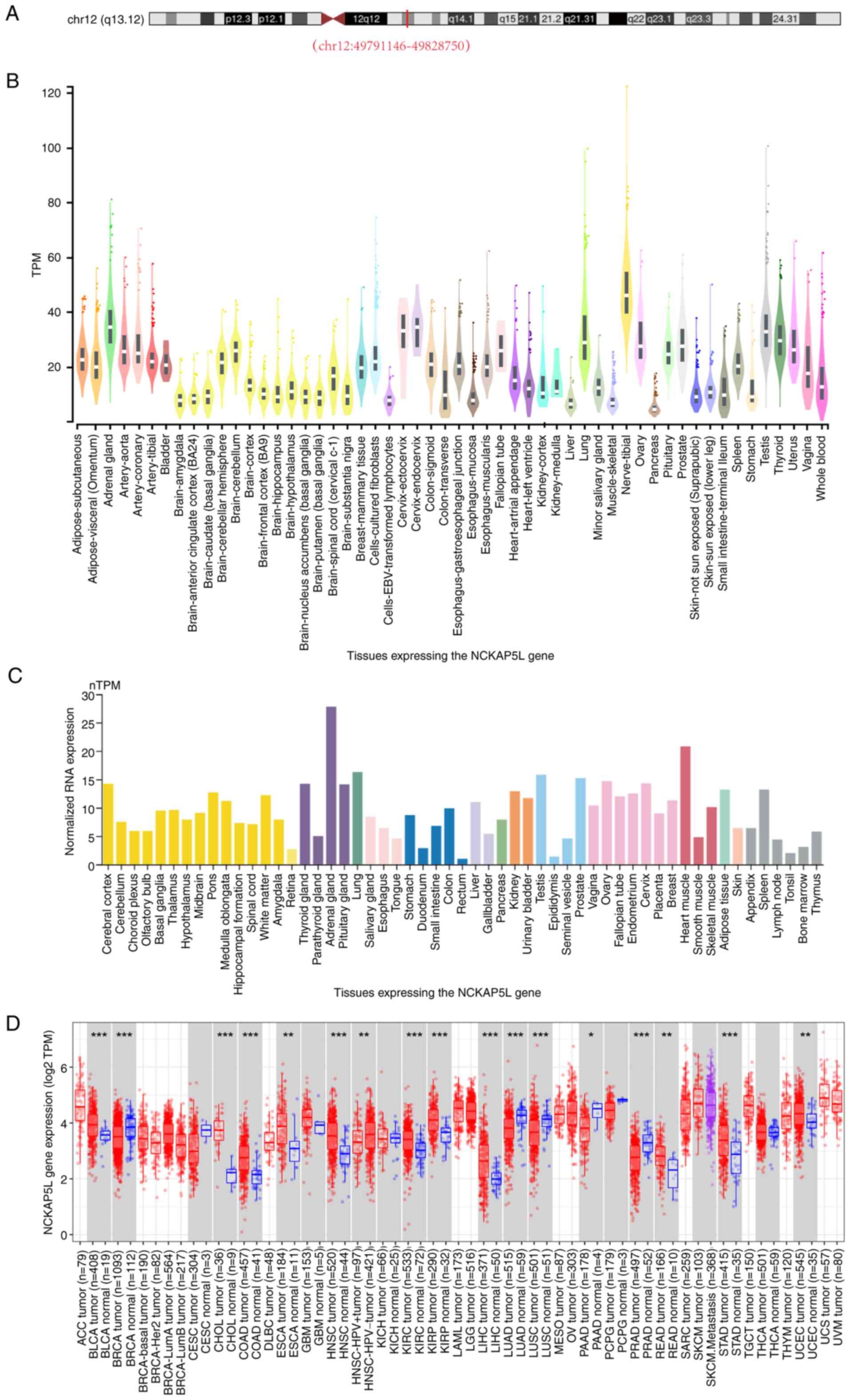 | Figure 1.Analysis of NCKAP5L gene expression
and genetic location in human tissues and tumors. (A) Genetic
location of human NCKAP5L using data from The University of
California Santa Cruz Genome Browser Genome Browser on Human Dec.
2013 (GRCh38/hg38) Assembly. (B) Expression of NCKAP5L gene using
GTEx RNA-sequencing data from 54 tissues from 17,382 samples and
948 donors. (C) Consensus NCKAP5L dataset consists of normalized
expression levels in 55 tissue types, created by combining the
Human Protein Atlas and GTEx transcriptomics datasets; (D)
Differences in NCKAP5L expression between distinct tumors in The
Cancer Genome Atlas cohorts and their adjacent normal tissues using
the Tumor Immune Estimation Resource, version 2. *P<0.05;
**P<0.01; ***P<0.001. NCKAP5L, Nck-associated protein 5-like;
GTEx, Genotype-Tissue Expression project; TPM, transcripts per
million; nTPM, normalized TPM, ACC, adrenocortical carcinoma; BLCA,
Bladder Urothelial Carcinoma; BRCA, Breast invasive carcinoma;
CESC, Cervical squamous cell carcinoma and endocervical
adenocarcinoma; CHOL, Cholangiocarcinoma; COAD, Colon
adenocarcinoma; DLBC, Lymphoid Neoplasm Diffuse Large B-cell
Lymphoma; ESCA, Esophageal carcinoma; GBM, Glioblastoma multiforme;
HNSC, Head and Neck squamous cell carcinoma; KICH, Kidney
Chromophobe; KIRC, Kidney renal clear cell carcinoma; KIRP, Kidney
renal papillary cell carcinoma; LAML, Acute Myeloid Leukemia; LGG,
Brain Lower Grade Glioma; LIHC, Liver hepatocellular carcinoma;
LUAD, Lung adenocarcinoma; LUSC, Lung squamous cell carcinoma;
MESO, mesothelioma; OV, Ovarian serous cystadenocarcinoma; PAAD,
Pancreatic adenocarcinoma; PCPG, Pheochromocytoma and
Paraganglioma; PRAD, Prostate adenocarcinoma; READ, Rectum
adenocarcinoma; SARC, Sarcoma; SKCM, Skin Cutaneous Melanoma; STAD,
Stomach adenocarcinoma; TGCT, Testicular Germ Cell Tumors; THCA,
Thyroid carcinoma; THYM, Thymoma; UCEC, Uterine Corpus Endometrial
Carcinoma; UCS, Uterine Carcinosarcoma; UVM, Uveal Melanoma. |
A consensus dataset on NCKAP5L was obtained from the
online HPA, which included expression levels of 55 tissue types.
This consensus dataset, which combined HPA and GTEx transcriptomics
datasets, employed an internal normalization pipeline and
color-coded tissue groups based on similar functional features. As
a consensus dataset, NCKAP5L expression level was relatively high
in the adrenal gland, lung, testis, prostate and myocardium, and
was detected to varying degrees in the other 50 tissue types,
indicating that NCKAP5L mRNA has low tissue specificity (Fig. 1C).
A boxplot was generated using TIMER2.0 to visualize
the differences in NCKAP5L expression between different tumors and
their corresponding adjacent normal tissues in TCGA (Fig. 1D). In colon adenocarcinoma (COAD),
CHOL and bladder urothelial carcinoma tissue, the expression level
of NCKAP5L was significantly increased compared with that in
adjacent normal tissues. Additionally, head and neck squamous cell
carcinoma (HNSC), GBM and esophageal carcinoma (ESCA) tissue also
had notably increased levels of NCKAP5L expression, compared with
their corresponding adjacent normal tissues. Moreover, ovarian
serous cystadenocarcinoma (OV), kidney renal papillary cell
carcinoma (KIRP), kidney renal clear cell carcinoma (KIRC), rectum
adenocarcinoma (READ), thyroid carcinoma (THCA), uterine corpus
endometrial carcinoma (UCEC), STAD and LIHC tissue demonstrated
notably increased NCKAP5L expression compared with their adjacent
normal tissue. However, the expression level of NCKAP5L in breast
invasive carcinoma (BRCA), kidney chromophobe), lung squamous cell
carcinoma (LUSC), prostate adenocarcinoma (PRAD), cervical squamous
cell carcinoma and endocervical adenocarcinoma (CESC), LUAD, PAAD
and pheochromocytoma and paraganglioma (PCPG) tissue had a notably
lower expression level of NCKAP5L than the corresponding adjacent
tissues (Fig. 1D).
Boxplots were generated using the ‘Box Plots’ module
of GEPIA2 that compared the NCKAP5L expression levels in tumor and
normal tissues for several cancer types. Fig. 2A demonstrates that the NCKAP5L gene
was significantly upregulated in the tumor tissues of CHOL, DLBC,
GBM, LUAD, PAAD, STAD and THYM, compared with that in normal
tissues. The heightened expression of NCKAP5L in these tissues may
be associated with the development and progression of these
cancers, thereby suggesting the potential candidacy of NCKAP5L as a
putative biomarker for these cancer types.
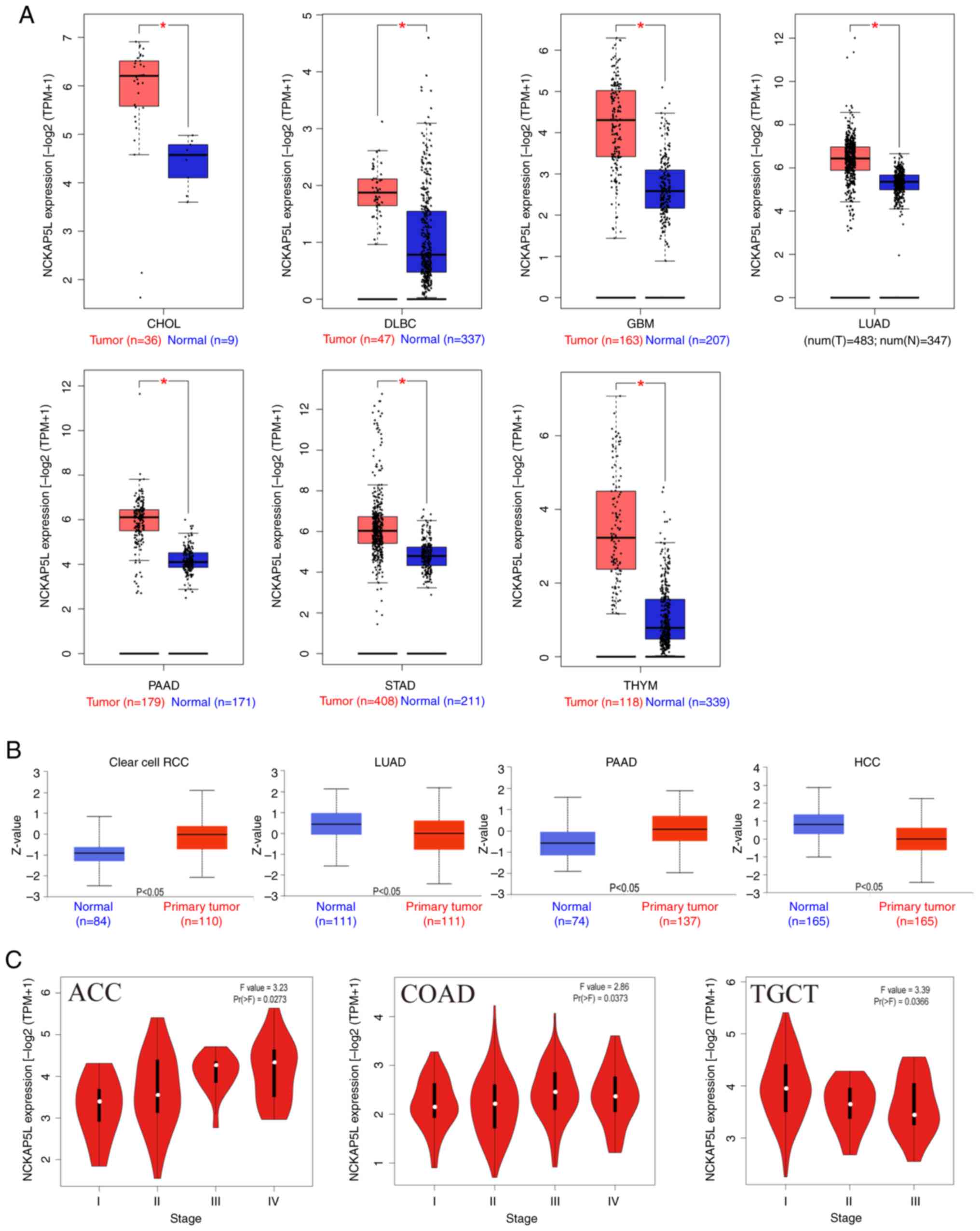 | Figure 2.NCKAP5L gene expression patterns in
tumor tissues and pathological stages across different cancer
types. (A) Difference in NCKAP5L expression in certain tumor
tissues (which have no normal control group in TCGA database) and
normal tissue, obtained through the combined analysis of data from
TCGA and GTEx database using the GEPIA2. (B) Protein expression
levels of NCKAP5L in primary tumors and normal tissues in clear
cell RCC, PAAD, HCCand LUAD using data from the University of
Alabama at Birmingham Cancer Data Analysis portal. (C) Violin plots
demonstrating NCKAP5L expression in different pathological stages
of ACC, COAD and TGCT, assessed using the GEPIA2. *P<0.05.
NCKAP5L, Nck-associated protein 5-like; TCGA, The Cancer Genome
Atlas; GTEx, Genotype-Tissue Expression project; GEPIA2, Gene
Expression Profiling Interactive Analysis, version 2; CHOL,
cholangiocarcinoma; DLBC, lymphoid neoplasm diffuse large B-cell
lymphoma; GBM, glioblastoma multiforme; HCC, hepatocellular
carcinoma; PAAD, pancreatic adenocarcinoma; STAD, stomach
adenocarcinoma; THYM, thymoma; RCC, renal carcinoma; ACC,
adrenocortical carcinoma; COAD, colon adenocarcinoma; TGCT,
testicular germ cell tumors. |
The difference in the total NCKAP5L protein
expression level between tumors and normal tissues from the UALCAN
portal was assessed. As shown in Fig.
2B, clear cell renal cell carcinoma and PAAD had significantly
higher protein expressions of NCKAP5L in primary tumor tissues,
compared with that in normal tissues, whereas hepatocellular
carcinoma (HCC) and LUAD had significantly lower protein expression
of NCKAP5L in primary tumor tissues, compared with that in normal
tissues. The results indicate that the variation in NCKAP5L protein
expression among primary tissues in different cancer types may
serve a significant role in cancer progression. These findings of
differential expression sets the stage for further investigations
into the specific functional roles of NCKAP5L within numerous
cancer subtypes and its potential use as a biomarker.
Furthermore, the ‘Pathological Stage Plot’ module of
GEPIA2 was used to generate violin plots of the NCKAP5L expression
in different pathological stages of all TCGA tumors, helping detect
the clinical stage of the patient. As shown in Fig. 2C, NCKAP5L expression levels were
demonstrated to significantly increase with increases in clinical
stage in patients with adrenocortical carcinoma (ACC), but
significantly decrease with increases in the clinical stage in
patients with TGCT. Additionally, NCKAP5L expression levels were
significantly higher in stage III of COAD, compared with that in
stages I, II and IV.
Survival prognosis analysis
To further assess the association between the level
of NCKAP5L gene expression and prognosis for patients with cancer,
GEPIA2 was used to generate survival maps for NCKAP5L expression in
all types of TCGA tumors. Subsequently, the association between
high and low expression of NCKAP5L and OS and DFS was assessed.
Fig. 3A demonstrates that high
NCKAP5L expression was significantly associated with lower OS in
several cancer types, including ACC, KIRC, OV, COAD, LIHC and skin
cutaneous melanoma (SKCM). Similarly, high NCKAP5L expression was
significantly associated with lower DFS in ACC, KIRC, SKCM, COAD,
LIHC and STAD (Fig. 3B). Therefore,
the results of the present study indicate a significant association
between high expression of the NCKAP5L gene and unfavorable
survival rates across multiple cancer types. Specifically, for
cancer types such as ACC, KIRC, OV, COAD and SKCM, patients in the
high-expression group consistently exhibited a markedly higher risk
of mortality at any given time point compared with the
low-expression group. Furthermore, in the case of patients with
LIHC, even when the analyzed period of time was <100 months, the
high-expression group also demonstrated an notably elevated risk of
mortality. Additionally, the DFS analysis indicates that high
expression of the NCKAP5L gene was associated with higher mortality
rates in numerous cancer types, including ACC, SKCM, COAD, LIHC and
STAD. Regardless of the time points of observation, patients in the
high-expression group consistently displayed a significantly higher
risk of mortality compared with those in the low-expression group.
Moreover, for patients with KIRC, a notably elevated risk of
mortality was also observed when the analyzed period of time ≤120
months. These findings suggest that the expression levels of
NCKAP5L may represent a critical influencing factor in the survival
rates of patients across multiple cancer types.
Furthermore, the association between OS,
relapse-free survival (RFS), first progression (FP),
post-progression survival (PPS), disease-specific survival and
progress-free survival, and the level of expression of NCKAP5L in
patients with breast, ovarian, lung and gastric cancer was assessed
using Kaplan-Meier Plotter based on gene chip (Fig. 4A) or RNA-seq data (Fig. 4B). The findings obtained from gene
chip data demonstrated significant associations between NCKAP5L
gene expression and patient prognosis across several cancer types.
It was demonstrated that for ovarian cancer, the high NCKAP5L
expression group exhibited a significantly lower RFS, OS and PPS
than that of the low-expression group. Conversely, for breast
cancer, gene chip data demonstrated that low NCKAP5L expression was
significantly associated with lower RFS, compared with that of the
high-expression group. Gene chip data, analyzed within a period of
≤150 months, also demonstrated a significant association between
high NCKAP5L expression and a worse prognosis in lung cancer,
compared with that of the low-expression group. Furthermore, gene
chip data from patients with gastric cancer demonstrated a
signification association between high NCKAP5L expression and an
adverse prognosis, compared with that in the low-expression group,
a trend reflected in OS, FP and PPS outcomes. Lastly, OS results
obtained from breast cancer RNA-seq data also indicated that low
NCKAP5L expression was significantly associated with an unfavorable
prognosis, corroborating the findings from the chip data.
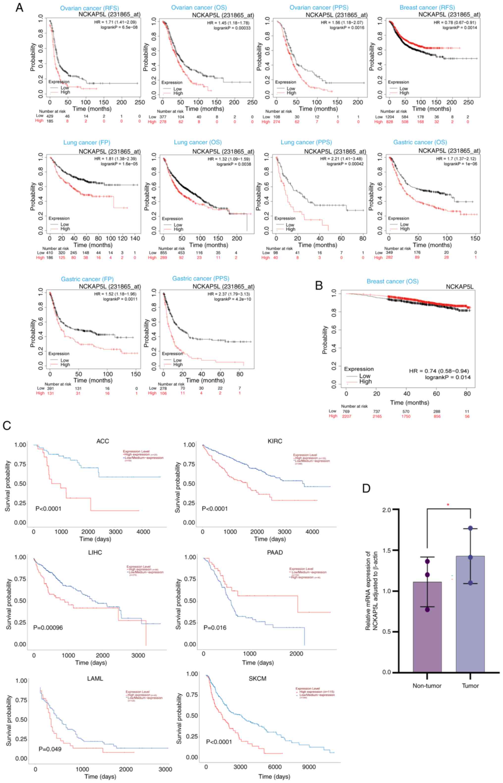 | Figure 4.NCKAP5L expression and prognostic
significance in pan-cancer studies from gene chip, RNA-seq and TCGA
data analyses. Pan-cancer survival prognosis analysis of NCKAP5L
expression from (A) gene chip and (B) RNA-seq data, assessed using
the Kaplan-Meier Plotter. (C) Effect of NCKAP5L expression levels
on survival of patients with cancer, based on relevant cancer data
in the TCGA database. (D) NCKAP5L expression is significantly
higher in colorectal cancer tumor tissues compared with non-tumor
tissues from the same patients, assessed using reverse
transcription-quantitative PCR. *P<0.05. NCKAP5L, Nck-associated
protein 5-like; seq, sequencing; TCGA, The Cancer Genome Atlas;
RFS, relapse-Free Survival; OS, Overall Survival; PPS,
Post-Progression Survival FP, First Progression; ACC,
Adrenocortical carcinoma; KIRC, Kidney renal clear cell carcinoma;
LIHC, Liver hepatocellular carcinoma; PAAD, pancreatic
adenocarcinoma; LAML, acute myeloid leukemia; SKCM, Skin Cutaneous
Melanoma. |
UALCAN was then used to assess the effects of
NCKAP5L expression levels on survival of patients with cancer based
on relevant cancer data in TCGA database. As shown in Fig. 4C, high expression levels of NCKAP5L
in ACC, KIRC, acute myeloid leukemia, PAAD and SKCM were
significantly associated with a lower survival probability,
compared with that in the low/medium expression group.
Additionally, within a 3,000-day analyzed period, elevated NCKAP5L
expression in patients with LIHC was significantly associated with
reduced survival probabilities, compared with those that had
low/medium expression levels. These results collectively indicate
that the expression levels of the NCKAP5L gene may serve as a
pivotal determinant of patient survival across multiple cancer
types, underscoring the potential significance of NCKAP5L as a
prospective biomarker for prognostic predictions in diverse cancer
types.
Furthermore, the relative expression of NCKA5L in
clinical tissue samples of patients with colorectal cancer was
quantified using RT-qPCR, obtaining differential expression of
NCKAP5L in tumor and non-tumor tissues. As shown in Fig. 4D, the level of NCKAP5L mRNA
expression in tumor tissues was significantly higher than in
non-tumor tissue (P=0.0301), which was in agreement with
bioinformatics analyses.
Genetic alteration analysis
As gene changes were demonstrated to be associated
with tumorigenesis, ‘TCGA Pan Cancer Atlas Studies’ module of
cBioPortal was used to analyze the genetic changes of NCKAP5L in
different TCGA-based tumors. As shown in Fig. 5A, SKCM had the highest NCKAP5L gene
alteration frequency (>7%), mainly comprising of mutations.
Across TCGA-based tumors, the types of NCKAP5L gene alterations
were predominantly mutations, which could be found in nearly all
TCGA tumors. COAD, LIHC, HNSC, uveal melanoma, mesothelioma, THYM
and KIRC had only one type of gene alteration, mutations. In
addition, amplification was the second most frequently observed
genetic alteration, with the highest alteration frequency in DLBC
and ACC. TGCT and PCPG only had one type of gene alteration,
amplification. Moreover, Fig. 5B
demonstrates the type, site and number of cases of NCKAP5L
mutations. Missense mutations were the most common, and a
translocation mutation due to an I725Yfs*12/Dfs*85 alteration in
one case of UCEC, LUAD, STAD and BRCA was found.
Immune infiltration analysis
The progression of cancer is influenced by behavior
of cancer cells and their interaction with the tumor
microenvironment (33). Serving a
significant role in the microenvironment, tumor-infiltrating immune
cells also affect the development and metastasis of cancer
(34). To assess the potential
correlation between NCKAP5L expression and immune infiltration of
cancer-associated fibroblasts across diverse TCGA cancer types, we
utilized TIMER2.0. A total of four algorithms (EPIC, MCPCOUNTER,
XCELL and TIDE) were used to generate a heatmap (Fig. 6A) and partial scatterplots (Fig. 6B). The heatmap revealed a
statistically significant positive correlation between immune
infiltration of cancer-associated fibroblasts and diverse TCGA
cancer types, including BRCA, CESC, COAD, DLBC, ESCA, HNSC, KIRP,
LUAD, LUSC, OV, PAAD, PRAD, READ, STAD, THCA, THYM and UCEC. The
positive correlation found for COAD and CESC were also visualized
using scatterplots (Fig 6B).
 | Figure 6.Correlation between NCKAP5L
expression and immune infiltration, based on data from four
algorithms: EPIC, MCPCOUNTER, XCELL and TIDE. (A) Immune
infiltration of cancer-associated fibroblasts have a significant
positive correlation with diverse TCGA cancer types. (B) Positive
correlation between immune infiltration of cancer-associated
fibroblasts and COAD and CESC, visualized by scatter plots.
Analysis using four algorithms, EPIC, MCPCOUNTER, XCELL and TIDE,
found that the NCKAP5L gene expression levels of COAD and CESC
tumors were positively correlated with the level of cancer
associated fibroblast infiltration. ACC, Adrenocortical carcinoma;
BLCA, Bladder Urothelial Carcinoma; BRCA, Breast invasive
carcinoma; CESC, Cervical squamous cell carcinoma and endocervical
adenocarcinoma; CHOL, Cholangiocarcinoma; COAD, Colon
adenocarcinoma; DLBC, Lymphoid Neoplasm Diffuse Large B-cell
Lymphoma; ESCA, Esophageal carcinoma; GBM, Glioblastoma multiforme;
HNSC, Head and Neck squamous cell carcinoma; KICH, Kidney
Chromophobe; KIRC, Kidney renal clear cell carcinoma; KIRP, Kidney
renal papillary cell carcinoma; LGG, Brain Lower Grade Glioma;
LIHC, Liver hepatocellular carcinoma; LUAD, Lung adenocarcinoma;
LUSC, Lung squamous cell carcinoma; MESO, Mesothelioma; OV, Ovarian
serous cystadenocarcinoma; PAAD, Pancreatic adenocarcinoma; PCPG,
Pheochromocytoma and Paraganglioma; PRAD, Prostate adenocarcinoma;
READ, Rectum adenocarcinoma; SARC, Sarcoma; SKCM, Skin Cutaneous
Melanoma; STAD, Stomach adenocarcinoma; TGCT, testicular Germ Cell
Tumors; THCA, Thyroid carcinoma; THYM, Thymoma; UCEC, Uterine
Corpus Endometrial Carcinoma; UCS, Uterine Carcinosarcoma; UVM,
Uveal Melanoma, NCKAP5L, Nck-associated protein 5-like; TCGA, The
Cancer Genome Atlas; COAD, colon adenocarcinoma; CESC, cervical
squamous cell carcinoma and endocervical adenocarcinoma; cor,
correlation; TPM, transcripts per million. |
Protein phosphorylation analysis
In the present study, an in-depth analysis of
variations in NCKAP5L phosphorylation levels across a spectrum of
tumor types within the CPTAC dataset was performed. First, a
schematic representation of the NCKAP5L protein domain was
generated (Fig. 7A). Subsequently,
alterations in phosphorylation sites and the corresponding protein
phosphorylation levels of NCKAP5L in different tumor types was
assessed (Fig. 7B). In comparison
with normal tissue counterparts, there were distinctive patterns of
NCKAP5L phosphorylation observed in the tumors from the base CPTAC
dataset: The S477 site for PAAD, LIHC, HCC and HNSC demonstrated
significantly higher phosphorylation levels; the S630 site had a
significantly higher phosphorylation level for LIHC, GBM, HCC and
HNSC; the T1235 site had a significantly higher phosphorylation
level for PPAD; the T733 site had a significantly higher
phosphorylation level for HNSC; the S735 site had a significantly
higher phosphorylation level for GBM; the T1175 site had a
significantly higher phosphorylation level for HNSC; the S440 had a
significantly higher phosphorylation level for PAAD, but a
significantly lower phosphorylation level for HNSC; the S767 site
had a significantly lower phosphorylation level for PAAD; and the
T1109 site had a significantly lower phosphorylation level for
HNSC. These findings provide a deeper understanding of the
intricate and multifaceted landscape of NCKAP5L phosphorylation in
a diverse array of tumor types. The phosphorylation status of
NCKAP5L, as demonstrated by the findings of the present study,
appears to be a finely tuned and dynamic process, showing distinct
patterns and variations across different cancer types. This not
only emphasizes the complexity of the involvement of NCKAP5L in
numerous malignancies, but also underscores its potential
significance in the context of cancer biology.
NCKAP5L-related gene enrichment
analysis
The potential molecular mechanisms underlying the
involvement of NCKAP5L in tumor occurrence and development was
evaluated by performing gene function analysis, screening out the
NCKAP5L-binding proteins and related genes for gene function
analysis (31,32).
Using the STRING online database, a network of 10
NCKAP5L-binding protein interactions with 11 nodes and 13 edges was
generated (Fig. 8A). Nodes
represented genes, and edges represented associations between bound
genes. Subsequently, the top 100 NCKAP5L-correlated targeting genes
were obtained, based on data of tumor and normal tissues from TCGA
with the help of GEPIA2. Fig. 8B
demonstrates the intersection of NCKAP5L-correlated and
co-expressed genes, showing one common member named LZTS2. In most
TCGA tumors, NCKAP5L was significantly positively correlated with
eight genes (RBM5, RBM6, DVL2, PHLDB1, CLCN6, AKAP8L, IFFO1 and
FAM65A), as shown in Fig. 8C. In
addition, Fig. 8D demonstrates the
significant positive correlation between NCKAP5L and the screened
related genes, underscoring their potential interplay in cellular
processes.
 | Figure 8.NCKAP5L-related gene enrichment
analysis. (A) NCKAP5L-binding proteins obtained using the Search
Tool for the Retrieval of Interacting Genes/Proteins web server
tool. (B) Venn diagram demonstrating the intersection analysis of
NCKAP5L-binded and -correlated genes. (C) Heatmap of detailed
cancer types, based on data from the Cancer Genome Atlas. The
partial correlation and P-value were determined using a
purity-adjusted Spearman's rank correlation test. (D) Top 100
NCKAP5L-related genes acquired using the Gene Expression Profiling
Interactive Analysis, version 2. The correlation between the
expression of NCKAP5L and RBM5, RBM6, DVL2, PHLDB1, CLCN6, AKAP8L,
IFFO1 and FAM65A is displayed. (E) Bar chart of the molecular
functions of NCKAP5L, determined using GO analysis based on
Metascape. Darker color signifies a more pronounced enrichment,
suggesting a higher degree of significance for the association
between the gene set and the specific biological process, cellular
component, or molecular function represented by the Goterm.
NCKAP5L, Nck-associated protein 5-like; Cor, correlation; GO, Gene
Ontology; TPM, transcripts per million; ACC, Adrenocortical
carcinoma; BLCA, Bladder Urothelial Carcinoma; BRCA, Breast
invasive carcinoma; CESC, Cervical squamous cell carcinoma and
endocervical adenocarcinoma; CHOL, Cholangiocarcinoma; COAD, colon
adenocarcinoma; DLBC, Lymphoid Neoplasm Diffuse Large B-cell
Lymphoma; ESCA, Esophageal carcinoma; GBM, Glioblastoma multiforme;
HNSC, Head and Neck squamous cell carcinoma; KICH, Kidney
Chromophobe; KIRC, Kidney renal clear cell carcinoma; KIRP, Kidney
renal papillary cell carcinoma; LAML, Acute Myeloid Leukemia; LGG,
Brain Lower Grade Glioma; LIHC, Liver hepatocellular carcinoma;
LUAD, Lung adenocarcinoma; LUSC, Lung squamous cell carcinoma;
MESO, Mesothelioma; OV, Ovarian serous cystadenocarcinoma; PAAD,
Pancreatic adenocarcinoma; PCPG, Pheochromocytoma and
Paraganglioma; PRAD, Prostate adenocarcinoma; READ, Rectum
adenocarcinoma; SARC, Sarcoma; SKCM, Skin Cutaneous Melanoma; STAD,
Stomach adenocarcinoma; TGCT, Testicular Germ Cell Tumors; THCA,
Thyroid carcinoma; THYM, Thymoma; UCEC, Uterine Corpus Endometrial
Carcinoma; UCS, Uterine Carcinosarcoma; UVM, uveal Melanoma.HCC,
hepatocellular carcinoma, RBM5: RNA Binding Motif Protein 5; DVL2,
Dishevelled Segment Polarity Protein 2; PHLDB1, Pleckstrin Homology
Like Domain Family B Member 1; CLCN6, Chloride Voltage-Gated
Channel 6, AKAP8L: A-Kinase Anchoring Protein 8-Like, IFFO1,
Interferon-Induced Transmembrane Protein Family Member 1; FAM65A,
Family with Sequence Similarity 65 Member A. |
For the functional enrichment analysis of NCKAP5L,
the GO framework was used by accessing the Metascape web server. As
shown in Fig. 8E, the results of
the GO enrichment analysis revealed that the collective genes
assessed were predominantly enriched in categories related to
‘metabolic process’ and ‘cellular process’. This enrichment pattern
suggests that NCKAP5L may serve a pivotal role in tumorigenesis and
development through its involvement in these essential biological
pathways. The enrichment of genes in these specific categories also
implies that NCKAP5L may be intricately connected to cellular
metabolism and general cellular activities, further highlighting
its potential significance in cancer biology and disease
progression.
Discussion
Pan-cancer analyses serve key roles in studying
different types of tumors through a multi-dimensional approach and
have notable implications for cancer biomarker discovery, treatment
and prognosis. With advancement of human genome research, abnormal
gene expression, which is caused by factors such as gene mutations
and copy number alterations, has been found to be closely related
to the development and progression of cancers (35,36).
Therefore, the present study focused on a gene that was abnormally
expressed in the tissues of different cancers.
NCKAP5L is a gene of which its roles in most types
of human cancers remains unclear and therefore, a pan-cancer
analysis of this gene from a comprehensive perspective is
necessary. Furthermore, considering colorectal cancer is the 3rd
most common cancer worldwide, the present study performed
pan-cancer analyses and focused on colorectal cancer (1,2).
Using bioinformatics tools, analyses of numerous
cancers, including colorectal cancer, were performed from multiple
perspectives, including gene expression and mutation, immune
invasion, and survival and prognosis analyses.
Data from the present study indicated that the both
the gene and protein expression of NCKAP5L were significantly
upregulated in the tissues of different cancers, in comparison with
that in normal tissues, and the expression level of NCKAP5L was
associated with the stage of colorectal cancer. RT-qPCR analysis of
clinical tissue samples further demonstrated that the expression of
NCKAP5L in colorectal cancer tissues was significantly higher than
that in non-tumor tissues, which was consistent with the results of
the bioinformatics analysis. Therefore, this indicates that the
abnormal expression of NCKAP5L may serve as a promising biomarker
for the diagnosis and prognosis of human cancers, especially in
colorectal cancer.
Tubulin, encoded by NCKAP5L, is an important
component of microtentacles, which are characteristic adhesion
sites providing signaling nodes in epithelial-mesenchymal
transition (EMT) (37). During EMT,
rearrangements of cell adhesion may affect the proliferation and
invasive capacity of cancer cells. Therefore, EMT may serve an
essential role in cancer progression and metastasis by promoting
tumor cell invasion and tissue infiltration (38–42).
It has been reported that EMT can promote the invasion and
metastasis of cancers such as lung, colorectal, liver and ovarian
cancer (43–48). Therefore, NCKAP5L may serve a role
in tumor promotion during EMT.
In addition, the bioinformatics analysis in the
present study identified NCKAP5L mutations in certain cancers.
Moreover, the high expression and mutations of NCKAP5L may closely
associate with EMT and promote the development of cancer, invasion
and metastasis. The present study found that patients with
colorectal cancer with high expression of NCKAP5L had worse OS and
DFS than those with low expression, and the expression level of
NCKAP5L in patients with cancer was associated with the stage of
cancer. Thus, this indicates that NCKAP5L may be a potential
biomarker for the occurrence, development, invasion and metastasis
of colorectal cancer from multi-dimensional analysis.
As an important component of the tumor
microenvironment, fibroblasts are important mesenchymal stem cells
that constitute tissues and organs of organisms and may serve roles
in biological processes such as tissue fibrosis and collagen
secretion (49–51). Cancer-related fibroblasts have been
extensively studied and are associated with the occurrence and
development of cancers (52–55).
There is also an association between NCKAP5L and cancer-related
fibroblast immune infiltration (52–55).
However, the mechanism of the interaction between fibroblasts and
cancer tissue is still unclear. It has been reported that the C-C
chemokine ligand 5 (CCR5) secreted by tumor tissue recruits a large
number of fibroblasts through the CCR5 solute carrier family 25
member 24 (SLC25A24) pathway, and further promotes the invasion and
metastasis of colorectal cancer by using the function of
fibroblasts to promote angiogenesis and collagen synthesis
(2). The present study demonstrated
that the expression of NCKAP5 was positively associated with the
immune infiltration of cancer-related fibroblasts in colorectal
cancer. Therefore, this indicates that NCKAP5L may promote the
secretion of CCR5 in tumor tissue and then recruit a large number
of cancer-related fibroblasts via the SLC25A24 pathway.
Subsequently, cancer-related fibroblasts lead to invasion and
metastasis of colorectal cancer by promoting tumor tissue to
generate blood vessels and collagen (56). In the present study, GO enrichment
analysis was performed on NCKAP5L-binding proteins and
expression-related genes and it was demonstrated that most of them
were enriched in ‘metabolic process’- and ‘cellular
process’-related functions, which further indicated that NCKAP5L
may be associated with the invasion and proliferation of related
cancers. Therefore, the findings indicate that NCKAP5L may be a
valuable biomarker for the prognosis of human cancers.
However, the present study has limitations. Firstly,
databases such as TCGA, CPTAC and GTEx, have a limited number of
cases for each cancer type. Secondly, the present study only
assessed the expression of NCKAP5L in the tissues of patients with
colorectal cancer and did not evaluate other types of tumors.
Bioinformatics tools are limited by restricted customization and
debugging options. These tools typically rely on external
databases, which are constantly evolving. Consequently, this
dynamic nature of data can lead to variations in analysis results
at different time points. Furthermore, online tools often provide
limited analysis choices, constraining users in terms of
personalized customization and debugging capabilities. For example,
when conducting survival analysis for NCKAP5L using online analysis
tools, the inability to set the observation time range prevents
analyzing survival curves within specific time intervals. This, in
turn, limits our ability to conduct in-depth analysis of time
intervals where survival curve crossovers do not occur.
In conclusion, the present study combined
bioinformatics with RT-qPCR to provide a comprehensive
characterization of NCKAP5L in pan-cancer tumor types, which is
beneficial to further understand the role of NCKAP5L in
tumorigenesis, progression and prognosis. The results indicate that
NCKAP5L may be a potential prognostic biomarker in pan-cancer, with
particular relevance to colorectal cancer.
Acknowledgements
Not applicable.
Funding
The present study was supported by the Science and Technology
Plan of Guilin (20220139-8-4) and Guangxi Health Commission Key
Laboratory of Glucose and Lipid Metabolism Disorders (grant no.
19-xkjs-05).
Availability of data and materials
The datasets used and/or analyzed during the current
study are available from the corresponding author on reasonable
request.
Authors' contributions
MO and YS were involved in the conception and design
of the study. MO provided administrative support. YS, WL, CM and XH
contributed to the provision of study materials or patients. YS, WL
and CM were responsible for the collection and assembly of data.
YS, MO, WL and XH performed the data analysis and interpretation.
MO and YS participated in the writing of the manuscript. YS and MO
confirm the authenticity of all the raw data. All authors have read
and approved the final manuscript.
Ethics approval and consent to
participate
Approval was granted by the Ethics Committee of the
Second Affiliated Hospital of Guilin Medical University (Guilin;
China; September 24, 2022; approval no. ZLXM-2022001) and all the
patients have signed an informed consent form.
Patient consent for publication
Not applicable.
Competing interests
The authors declare that they have no competing
interests.
References
|
1
|
Desai A and Gyawali B: Financial toxicity
of cancer treatment: Moving the discussion from acknowledgement of
the problem to identifying solutions. Clin Med. 20:1002692020.
|
|
2
|
Nafisi S, Randel KR, Støer NC, Veierød MB,
Hoff G, Holme Ø, Schult AL and Botteri E: Association between use
of low-dose aspirin and detection of colorectal polyps and cancer
in a screening setting. Dig Liver Dis. 55:1126–1132. 2023.
View Article : Google Scholar : PubMed/NCBI
|
|
3
|
Torre LA, Bray F, Siegel RL, Ferlay J,
Lortet-Tieulent J and Jemal A: Global cancer statistics, 2012. CA
Cancer J Clin. 65:87–108. 2015. View Article : Google Scholar : PubMed/NCBI
|
|
4
|
Jovčevska I: Next generation sequencing
and machine learning technologies are painting the epigenetic
portrait of glioblastoma. Front Oncol. 10:7982020. View Article : Google Scholar : PubMed/NCBI
|
|
5
|
Bao R, Huang L, Andrade J, Tan W, Kibbe
WA, Jiang H and Feng G: Review of current methods, applications,
and data management for the bioinformatics analysis of whole exome
sequencing. Cancer Inform. 13:67–82. 2014.PubMed/NCBI
|
|
6
|
Zhang K and Wang H: Cancer genome atlas
pan-cancer analysis project. Zhongguo Fei Ai Za Zhi. 18:219–223.
2015.(In Chinese). PubMed/NCBI
|
|
7
|
Tang N, Dou X, You X, Shi Q, Ke M and Liu
G: Pan-cancer analysis of the oncogenic role of discs large homolog
associated protein 5 (DLGAP5) in human tumors. Cancer Cell Inter.
21:4572021. View Article : Google Scholar : PubMed/NCBI
|
|
8
|
Liang W, Chen W, Wei J, Yao H, Shi J, Hou
X, Deng Y and Ou M: Zinc finger C3H1-type containing serves as a
novel prognostic biomarker in human pan-cancer. Gene.
820:1462512022. View Article : Google Scholar : PubMed/NCBI
|
|
9
|
Liang W, Mo C, Wei J, Chen W, Gong W, Shi
J, Hou X, Li C, Deng Y and Ou M: FAM65A as a novel prognostic
biomarker in human tumors reveal by a pan-cancer analysis. Discov
Oncol. 12:602021. View Article : Google Scholar : PubMed/NCBI
|
|
10
|
Mori Y, Taniyama Y, Tanaka S, Fukuchi H
and Terada Y: Microtubule-bundling activity of the centrosomal
protein, Cep169, and its binding to microtubules. Biochem Biophys
Res Commun. 467:754–759. 2015. View Article : Google Scholar : PubMed/NCBI
|
|
11
|
Mori Y, Inoue Y, Tanaka S, Doda S,
Yamanaka S, Fukuchi H and Terada Y: Cep169, a novel microtubule
plus-end-tracking centrosomal protein, binds to CDK5RAP2 and
regulates microtubule stability. PLoS One. 10:e01409682015.
View Article : Google Scholar : PubMed/NCBI
|
|
12
|
Shintomi M, Shiratori M, Negishi L and
Terada Y: Identification of Cep169-interacting proteins and the in
vivo modification sites of Cep169 via proteomic analysis. Biochem
Biophys Res Commun. 495:2275–2281. 2018. View Article : Google Scholar : PubMed/NCBI
|
|
13
|
Chen H, Lin Z, Arnst KE, Miller DD and Li
W: Tubulin inhibitor-based antibody-drug conjugates for cancer
therapy. Molecules. 22:12812017. View Article : Google Scholar : PubMed/NCBI
|
|
14
|
Hammond JW, Cai D and Verhey KJ: Tubulin
modifications and their cellular functions. Curr Opin Cell Biol.
20:71–76. 2008. View Article : Google Scholar : PubMed/NCBI
|
|
15
|
Kanakkanthara A and Miller JH:
βIII-tubulin overexpression in cancer: Causes, consequences, and
potential therapies. Biochim Biophys Acta Rev Cancer.
1876:1886072021. View Article : Google Scholar : PubMed/NCBI
|
|
16
|
Zhu ML, Horbinski CM, Garzotto M, Qian DZ,
Beer TM and Kyprianou N: Tubulin-targeting chemotherapy impairs
androgen receptor activity in prostate cancer. Cancer Res.
70:7992–8002. 2010. View Article : Google Scholar : PubMed/NCBI
|
|
17
|
Kent WJ, Sugnet CW, Furey TS, Roskin KM,
Pringle TH, Zahler AM and Haussler D: The human genome browser at
UCSC. Genome Res. 12:996–1006. 2002. View Article : Google Scholar : PubMed/NCBI
|
|
18
|
Li T, Fu J, Zeng Z, Cohen D, Li J, Chen Q,
Li B and Liu XS: TIMER2.0 for analysis of tumor-infiltrating immune
cells. Nucleic Acids Res. 48:W509–W514. 2020. View Article : Google Scholar : PubMed/NCBI
|
|
19
|
Tang Z, Kang B, Li C, Chen T and Zhang Z:
GEPIA2: An enhanced web server for large-scale expression profiling
and interactive analysis. Nucleic Acids Res. 47:W556–W560. 2019.
View Article : Google Scholar : PubMed/NCBI
|
|
20
|
Chen F, Chandrashekar DS, Varambally S and
Creighton CJ: Pan-cancer molecular subtypes revealed by
mass-spectrometry-based proteomic characterization of more than 500
human cancers. Nat Commun. 10:56792019. View Article : Google Scholar : PubMed/NCBI
|
|
21
|
Livak KJ and Schmittgen TD: Analysis of
relative gene expression data using real-time quantitative PCR and
the 2(−Delta Delta C(T)). Methods. 25:402–408. 2001. View Article : Google Scholar : PubMed/NCBI
|
|
22
|
Cerami E, Gao J, Dogrusoz U, Gross BE,
Sumer SO, Aksoy BA, Jacobsen A, Byrne CJ, Heuer ML, Larsson E, et
al: The cBio cancer genomics portal: An open platform for exploring
multidimensional cancer genomics data. Cancer Discov. 2:401–404.
2012. View Article : Google Scholar : PubMed/NCBI
|
|
23
|
Gao J, Aksoy BA, Dogrusoz U, Dresdner G,
Gross B, Sumer SO, Sun Y, Jacobsen A, Sinha R, Larsson E, et al:
Integrative analysis of complex cancer genomics and clinical
profiles using the cBioPortal. Sci Signal. 6:pl12013. View Article : Google Scholar : PubMed/NCBI
|
|
24
|
Wu P, Heins ZJ, Muller JT, Katsnelson L,
de Bruijn I, Abeshouse AA, Schultz N, Fenyö D and Gao J:
Integration and analysis of cptac proteomics data in the context of
cancer genomics in the cBioPortal. Mol Cell Proteomics.
18:1893–1898. 2019. View Article : Google Scholar : PubMed/NCBI
|
|
25
|
Li B, Severson E, Pignon JC, Zhao H, Li T,
Novak J, Jiang P, Shen H, Aster JC and Rodig S: Comprehensive
analyses of tumor immunity: Implications for cancer immunotherapy.
Genome Biol. 17:1742016. View Article : Google Scholar : PubMed/NCBI
|
|
26
|
Racle J, de Jonge K, Baumgaertner P,
Speiser DE and Gfeller D: Simultaneous enumeration of cancer and
immune cell types from bulk tumor gene expression data. Elife.
6:e264762017. View Article : Google Scholar : PubMed/NCBI
|
|
27
|
Becht E, Giraldo NA, Lacroix L, Buttard B,
Elarouci N, Petitprez F, Selves J, Laurent-Puig P, Sautès-Fridman
C, Fridman WH and de Reyniès A: Estimating the population abundance
of tissue-infiltrating immune and stromal cell populations using
gene expression. Genome Biol. 17:1–20. 2016. View Article : Google Scholar
|
|
28
|
Aran D, Hu Z and Butte AJJGb: xCell:
Digitally portraying the tissue cellular heterogeneity landscape.
Genome Biol. 18:2202017. View Article : Google Scholar : PubMed/NCBI
|
|
29
|
De Meulenaere A, Vermassen T, Aspeslagh S,
Huvenne W, Van Dorpe J, Ferdinande L and Rottey S: Turning the
tide: Clinical utility of PD-L1 expression in squamous cell
carcinoma of the head and neck. Oral Oncol. 70:34–42. 2017.
View Article : Google Scholar : PubMed/NCBI
|
|
30
|
Lv M, Luo L and Chen X: The landscape of
prognostic and immunological role of myosin light chain 9 (MYL9) in
human tumors. Immun Inflamm Dis. 10:241–254. 2022. View Article : Google Scholar : PubMed/NCBI
|
|
31
|
Gene Ontology Consortium, . Going forward.
Nucleic Acids Res. 43:D1049–D1056. 2015. View Article : Google Scholar : PubMed/NCBI
|
|
32
|
Chen L, Zhang YH, Wang S, Zhang Y, Huang T
and Cai YD: Prediction and analysis of essential genes using the
enrichments of gene ontology and KEGG pathways. PLoS One.
12:e01841292017. View Article : Google Scholar : PubMed/NCBI
|
|
33
|
Kashiwagi S, Asano Y, Goto W, Takada K,
Takahashi K, Noda S, Takashima T, Onoda N, Tomita S, Ohsawa M, et
al: Use of Tumor-infiltrating lymphocytes (TILs) to predict the
treatment response to eribulin chemotherapy in breast cancer. PLoS
One. 12:e01706342017. View Article : Google Scholar : PubMed/NCBI
|
|
34
|
Fridman WH, Galon J, Dieu-Nosjean MC,
Cremer I, Fisson S, Damotte D, Pagès F, Tartour E and
Sautès-Fridman C: Immune infiltration in human cancer: Prognostic
significance and disease control. Curr Top Microbiol Immunol.
344:1–24. 2011.PubMed/NCBI
|
|
35
|
Zack TI, Schumacher SE, Carter SL,
Cherniack AD, Saksena G, Tabak B, Lawrence MS, Zhsng CZ, Wala J,
Mermel CH, et al: Pan-cancer patterns of somatic copy number
alteration. Nat Genet. 45:1134–1140. 2013. View Article : Google Scholar : PubMed/NCBI
|
|
36
|
Zhang Y, Xu H and Frishman D: Genomic
determinants of somatic copy number alterations across human
cancers. Hum Mol Genet. 25:1019–1030. 2016. View Article : Google Scholar : PubMed/NCBI
|
|
37
|
Greaves D and Calle Y: Epithelial
mesenchymal transition (EMT) and associated invasive adhesions in
solid and haematological tumours. Cells. 11:6492022. View Article : Google Scholar : PubMed/NCBI
|
|
38
|
Luo C, Zhou M, Chen C, Li S, Li Q, Huang Y
and Zhou Z: A liposome-based combination strategy using doxorubicin
and a PI3K inhibitor efficiently inhibits pre-metastatic initiation
by acting on both tumor cells and tumor-associated macrophages.
Nanoscale. 14:4573–4587. 2022. View Article : Google Scholar : PubMed/NCBI
|
|
39
|
Sato M, Shames DS and Hasegawa Y: Emerging
evidence of epithelial-to-mesenchymal transition in lung
carcinogenesis. Respirology. 17:1048–1059. 2012. View Article : Google Scholar : PubMed/NCBI
|
|
40
|
Scheel C and Weinberg RA: Phenotypic
plasticity and epithelial-mesenchymal transitions in cancer and
normal stem cells? Int J Cancer. 129:2310–2314. 2011. View Article : Google Scholar : PubMed/NCBI
|
|
41
|
Thiery JP: EMT: An update. Methods Mol
Biol. 2179:35–39. 2021. View Article : Google Scholar : PubMed/NCBI
|
|
42
|
Zhu Y, Tao Z, Chen Y, Lin S, Zhu M, Ji W,
Liu X, Li T and Hu X: Exosomal MMP-1 transfers metastasis potential
in triple-negative breast cancer through PAR1-mediated EMT. Breast
Cancer Res Treat. 193:65–81. 2022. View Article : Google Scholar : PubMed/NCBI
|
|
43
|
Alrashed MM, Alharbi H, Alshehry AS, Ahmad
M and Aloahd MS: MiR-624-5p enhances NLRP3 augmented gemcitabine
resistance via EMT/IL-1β/Wnt/β-catenin signaling pathway in ovarian
cancer. J Reprod Immunol. 150:1034882022. View Article : Google Scholar : PubMed/NCBI
|
|
44
|
Chang Y, Zhang J, Huo X, Qu X, Xia C,
Huang K, Xie F, Wang N, Wei X and Jia Q: Substrate rigidity
dictates colorectal tumorigenic cell stemness and metastasis via
CRAD-dependent mechanotransduction. Cell Rep. 38:1103902022.
View Article : Google Scholar : PubMed/NCBI
|
|
45
|
Li A, Wu N and Sun J: E2F1-induced
microRNA-224-5p expression is associated with hepatocellular
carcinoma cell migration, invasion and epithelial-mesenchymal
transition via MREG. Oncol Lett. 23:822022. View Article : Google Scholar : PubMed/NCBI
|
|
46
|
Maleki N, Yavari N, Ebrahimi M, Faiz AF,
Ravesh RK, Sharbati A, Panji M, Lorian K, Gravand A, Abbasi M, et
al: Silibinin exerts anti-cancer activity on human ovarian cancer
cells by increasing apoptosis and inhibiting epithelial-mesenchymal
transition (EMT). Gene. 823:1462752022. View Article : Google Scholar : PubMed/NCBI
|
|
47
|
Martinou E, Moller-Levet C, Karamanis D,
Bagwan I and Angelidi AM: HOXB9 overexpression promotes colorectal
cancer progression and is associated with worse survival in liver
resection patients for colorectal liver metastases. Int J Mol Sci.
23:22812022. View Article : Google Scholar : PubMed/NCBI
|
|
48
|
Owczarek C, Ortiz-Zapater E, Kim J,
Papaevangelou E, Santis G and Parsons M: CAR co-operates with
integrins to promote lung cancer cell adhesion and invasion. Front
Oncol. 12:8293132022. View Article : Google Scholar : PubMed/NCBI
|
|
49
|
Koliaraki V, Prados A, Armaka M and
Kollias G: The mesenchymal context in inflammation, immunity and
cancer. Nat Immunol. 21:974–982. 2020. View Article : Google Scholar : PubMed/NCBI
|
|
50
|
Zeng F, Gao M, Liao S, Zhou Z, Luo G and
Zhou Y: Role and mechanism of CD90+ fibroblasts in inflammatory
diseases and malignant tumors. Mol Med. 29:202023. View Article : Google Scholar : PubMed/NCBI
|
|
51
|
Zhou J, Wei T and He Z: ADSCs enhance
VEGFR3-mediated lymphangiogenesis via METTL3-mediated VEGF-C m6A
modification to improve wound healing of diabetic foot ulcers. Mol
Med. 27:1462021. View Article : Google Scholar : PubMed/NCBI
|
|
52
|
Chen WJ, Ho CC, Chang YL, Chen HY, Lin CA,
Ling TY, Yu SL, Yuan SS, Chen YJ, Lin CY, et al: Cancer-associated
fibroblasts regulate the plasticity of lung cancer stemness via
paracrine signalling. Nat Commun. 5:34722014. View Article : Google Scholar : PubMed/NCBI
|
|
53
|
Heichler C, Scheibe K, Schmied A, Geppert
CI, Schmid B, Wirtz S, Thoma OM, Kramer V, Waldner MJ, Büttner C,
et al: STAT3 activation through IL-6/IL-11 in cancer-associated
fibroblasts promotes colorectal tumour development and correlates
with poor prognosis. Gut. 69:1269–1282. 2020. View Article : Google Scholar : PubMed/NCBI
|
|
54
|
Hu JL, Wang W, Lan XL, Zeng ZC, Liang YS,
Yan YR, Song FY, Wang FF, Zhu XH, Liao WJ, et al: CAFs secreted
exosomes promote metastasis and chemotherapy resistance by
enhancing cell stemness and epithelial-mesenchymal transition in
colorectal cancer. Mol Cancer. 18:912019. View Article : Google Scholar : PubMed/NCBI
|
|
55
|
Murata T, Mizushima H, Chinen I, Moribe H,
Yagi S, Hoffman RM, Kimura T, Yoshino K, Ueda Y, Enomoto T and
Mekada E: HB-EGF and PDGF mediate reciprocal interactions of
carcinoma cells with cancer-associated fibroblasts to support
progression of uterine cervical cancers. Cancer Res. 71:6633–6642.
2011. View Article : Google Scholar : PubMed/NCBI
|
|
56
|
Paauwe M, Schoonderwoerd MJ, Helderman RF,
Harryvan TJ, Groenewoud A, van Pelt GW, Bor R, Hemmer DM, Versteeg
HH, Snaar-Jagalska BE, et al: Endoglin expression on
cancer-associated fibroblasts regulates invasion and stimulates
colorectal cancer metastasis. Clin Cancer Res. 24:6331–6344. 2018.
View Article : Google Scholar : PubMed/NCBI
|


