Introduction
Esophageal squamous cell carcinoma (ESCC) is a
lethal malignancy with a 5-year survival rate of 26.2% due to late
diagnosis, rapid growth and metastasis (1). Thus, it is necessary to identify new
effective therapeutic strategies for ESCC, especially molecularly
targeted therapies, based on a better understanding of the
biological events of ESCC cells. Cancer stem cells (CSCs) are a
limited number of cancer cells with a self-renewal potential and
extensive proliferation capacity and play a dominant role in tumor
initiation, metastasis and recurrence (2–4). In
pancreatic cancer, a subpopulation of migrating
CD133+CXCR4+ CSCs was reported to be
essential for tumor metastasis (5).
In glioma, CD133+ CSCs were associated with
radioresistance and contributed to tumor recurrence after
radiotherapy due to preferential activating DNA damage checkpoint
response. In malignant melanoma, the drug transporter and
chemoresistance mediator ABCB5 was identified as a novel molecular
marker for a distinct subset of chemoresistant, stem-cell
phenotype-expressing tumor cells, indicating that ABCB5 may be a
specific target to enhance cytotoxic efficacy (6). Zhang et al also provided
evidence that CSC-like cells may play a role in the progression and
drug resistance of bladder cancer (7). The aforementioned studies showed that
CSCs can be a good therapeutic target for various types of cancer.
Although CSCs are common many types of tumors (8–11), the
data for CSCs from human ESCC are conflicting.
CD271, a member of the tumor necrosis factor
receptor superfamily (12), plays a
role in cell proliferation, survival, and apoptosis (13). It is also known as a nerve growth
factor receptor and interacts with neurotrophins (14). CD271+ cells have been
reported to be genuine CSCs in human melanoma (15). CD271+ cells showed higher
tumorigenecity and metastatic ability in melanoma (16). The high level of CD271 expression
was correlated with a poor prognosis for patients with
hypopharyngeal cancer (17).
Okumura et al suggested CD271 as the human esophageal
keratinocyte stem cell marker, which may be valuable for
prospectively investigating stem cell regulation in association
with different biological processes including the neoplastic
transformation of regenerative epithelia (18). Authors of that study also identified
CD271 as being expressed in 49.2% ESCC and necessary for survival
and maintenance of ESCC tumors (19). Huang et al demonstrated that
CD271+ cells possess some characteristics of CSCs
(20). However, there is no report
with regard to the epigenetic regulation on CD271 expression.
In the present study, we confirmed that
CD271+ ESCC cells exhibited higher self-renewal ability
and chemoresistance. CD271 expression was associated with TNM stage
and metastatic capacity in human ESCC and we found that CD271
expression was regulated by DNA methylation. Our results showed
that CD271+ ESCC cells possess stem-like properties and
their expression is epigenetically regulated.
Materials and methods
Cell line and tissue specimens
The KYSE70 ESCC cell line was preserved in our
laboratory and maintained in RMPI-1640 supplemented with 10% fetal
bovine serum (both from Hyclone, Logan, UT, USA), 100 U/ml of
penicillin, and 100 μg/ml of streptomycin at 37°C, 5%
CO2. Sixty-three paired ESCC tissues and adjacent
non-cancerous tissues were previously collected and stored
(2008–2010). Tissues were provided by the Department of Pathology,
The First Affiliated Hospital of Zhengzhou University, with
confirmed histopathological results. Information pertaining to
clinicopathological parameters were also available.
Flow cytometric analysis and cell
sorting
Adherent KYSE70 cells were trypsinized and
dissociated into single cells suspended in PBS with 3% fetal bovine
serum. The cells were stained with PE-conjugated mouse anti-human
CD271 monoclonal antibody (BD Biosciences, San Jose, CA, USA). The
corresponding isotype immunoglobulins were used as controls. Dead
cells were identified using 7-AAD (Biolegend, San Diego, CA, USA).
Samples were analyzed using BD FACS Canto II cytometer (BD
Biosciences) and sorted with a MoFlo XDP cytometer (Beckman, Brea,
CA, USA).
RNA extraction and cDNA synthesis
Total RNA was extracted from KYSE70 cells and tissue
specimens by TRIzol reagent (Invitrogen Life Technologies,
Carlsbad, CA, USA) according to the manufacturer’s instructions.
The first-strand cDNA was synthesized from 1 μg of total RNA using
PrimeScript RT reagent kit with gDNA Eraser (Takara, Shiga,
Japan).
Quantitative real-time PCR
The cDNA was used as a template to detect the
expression of CD271 in KYSE70 cells and tissue specimens. qPCR was
performed using SYBR Premix Ex Taq II (Takara) and assessed
by Agilent Mx3005P. GAPDH was used as an internal control. The data
were analyzed by 2−ΔΔCt. Primer sequences for qPCR are
shown in Table I.
 | Table IPrimer sequences for qPCR for all the
genes tested. |
Table I
Primer sequences for qPCR for all the
genes tested.
| Gene name | Sequence | Product size
(bp) |
|---|
| qPCR |
| GAPDH-F |
GCACCGTCAAGGCTGAGAAC | 138 |
| GAPDH-R |
TGGTGAAGACGCCAGTGGA | |
| CD271-F |
AACAAGACCTCATAGCCAGCA | 119 |
| CD271-R |
CAGGATGGAGCAATAGACAGG | |
| NANOG-F |
CAAAGGCAAACAACCCACTT | 158 |
| NANOG-R |
TCTGCTGGAGGCTGAGGTAT | |
| FIBRO-F |
CAGTGGGAGACCTCGAGAAGA | 169 |
| FIBRO-R |
GTCCCTCGGAACATCAGAAAC | |
| VIM-F |
GAGAACTTTGCCGTTGAAGC | 163 |
| VIM-R |
GCTTCCTGTAGGTGGCAATC | |
| BAK-F |
CATCAACCGACGCTATGACTC | 192 |
| BAK-R |
GTCAGGCCATGCTGGTAGAC | |
| CASP3-F |
AGAACTGGACTGTGGCATTGAG | 191 |
| CASP3-R |
GCTTGTCGGCATACTGTTTCAG | |
| CASP9-F |
CTCAGACCAGAGATTCGCAAAC | 116 |
| CASP9-R |
GCATTTCCCCTCAAACTCTCAA | |
| BSP |
| CD271-BF |
GAGAGAGAGAGGGTTGAAATTAG | 505 |
| CD271-BR |
AAACATCCTAAATTCCAACAAA | |
Sphere formation assay
The sorted CD271+ and CD271−
cells were resuspended in DMEM/F12 medium (Invitrogen Life
Technologies) supplemented with 4 μg/ml heparin (Sigma, St. Louis,
MO, USA), B27 (1:50, Gibco, Life Technologies, Carlsbad, CA, USA),
20 ng/ml EGF, 20 ng/ml basic FGF (both from PeproTech, Rocky Hill,
NJ, USA), penicillin 100 IU/ml and streptomycin 100 μg/ml and then
seeded in 24-well ultra-low cluster plates (Corning Costar,
Corning, NY, USA). After culturing for 7 days, the number of
spheres was counted under a microscope (Leica, Wetzlar,
Germany).
Anchorage-independent cell growth
The sorted CD271+ and CD271−
cells (2×10³ cells/well) were suspended in complete
medium containing 0.6% low-melting-point agar (Sigma) and then
applied to the top of a 1.2% agar/complete medium layer in six-well
plates. After 14 days, surviving colonies were stained with 0.4%
crystal violet (Sigma) and counted under a microscope (Leica, USA).
The experiments were carried out in triplicate wells.
Representative images were taken.
Drug sensitivity assay and chemotherapy
resistance assay
5-Fluorouracil (5-FU) and Cisplatin (DDP), both from
Sigma, were dissolved according to the manufacturer’s instructions.
The sorted CD271+ and CD271− cells were
seeded at 3×10³ cells/well in 96-well plates and treated
with chemotherapeutic reagents in quadruplicate. Cell viability was
evaluated using CCK-8 assay (Biyuntian, Jiangsu, China) following
treatment with chemotherapeutic reagents for 48 h, and the
absorbance was measured at 450 nm using Multiskan Mk3 (Thermo
Fisher Scientific, San Jose, CA, USA). The percentage of survival
in treated cells was normalized with untreated controls.
KYSE70 cells were continuously treated with DDP (0.5
μM) and 5-FU (0.5 μg/ml) until significant DDP and 5-FU resistance
in KYSE70 cells was observed via cell viability assessment. Flow
cytometry was then used to detect the CD271 expression.
Bisulfite modification and methylation
analysis
Genomic DNA extracted from the KYSE70 cell line
using the Takara MiniBEST Universal Genomic DNA Extraction kit
(Takara), was modified by sodium bisulfite using the EpiTect
Bisulfite kit (Qiagen, Germany) according to the manufacturer’s
instructions. Methylation status was analyzed by bisulfite genomic
sequencing (BSP) of the CpG islands. The region was amplified using
the primers shown in Table I.
Amplified products were cloned into pMD-18T simple vector (Takara),
transformed into DH5α competent cells (Takara), and plated under
ampicilin selection. Five independent clones were sequenced.
Statistical analysis
Data were expressed as mean ± SD and analyzed using
the Student’s t-test. Paired t-test was used for paired samples.
Non-parametric test was performed for samples of non-normal
distribution. Statistical analyses were conducted with SPSS 17.0
software. P<0.05 was considered to indicate a statistically
significant difference.
Results
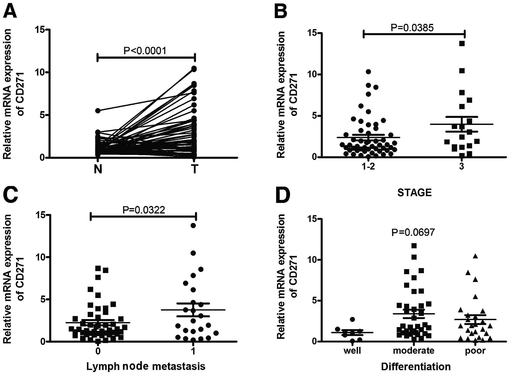
CD271 expression is associated with stage
and lymph node metastasis in human ESCC specimens
CD271 is considered crucial to maintain
tumorigenecity and stem-like properties of cancer cells including
melanoma, hypopharyngeal cancer and esophageal cancer. We compared
the expression of CD271 in paired human ESCC specimens and adjacent
non-cancerous tissues. As shown in Fig.
1A, qPCR analysis revealed frequent upregulation of CD271 mRNA
expression in carcinoma tissues compared with adjacent
non-cancerous tissues, indicating that CD271 may act as an oncogene
in human ESCC. Moreover, the expression of CD271 was significantly
correlated with TNM stage and lymph node metastasis but not other
variables such as age, gender and differentiation (Fig. 1B–D).
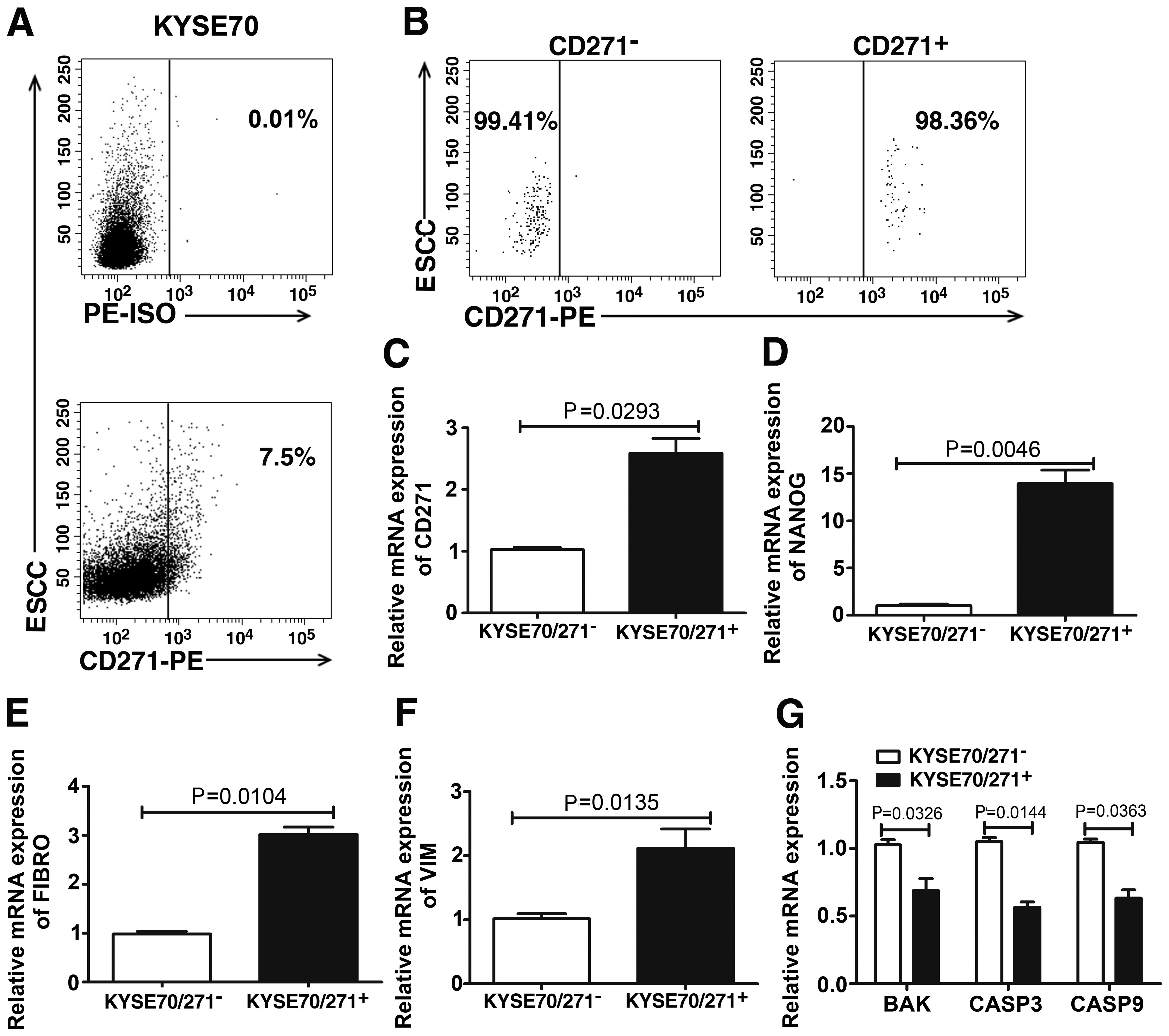
CD271+ cells overexpress
stem-related gene NANOG and EMT markers
To study the stem cell-like properties of isolated
CD271+ cells from the KYSE70 ESCC cell line, we first
detected the CD271 expression by flow cytometric analysis. We found
the CD271+ subpopulation was 7.5% present in the KYSE70
cell line (Fig. 2A).
CD271+ and CD271− cells were then sorted
separately and the purity of the two sorted subpopulations was
98.36 and 99.41%, respectively (Fig.
2B). qPCR was used to confirm the expression of CD271 in the
two subpopulations (Fig. 2C). We
also compared the expression of the stem-related gene NANOG, EMT
markers Fibronectin (FIBRO) and Vimentin (VIM), and apoptosis genes
BAK, Caspase 3 and Caspase 9 in CD271+ and
CD271− cells. Our results showed that compared with
CD271− cells, CD271+ cells exhibited an
increased expression of NANOG, FIBRO, VIM (Fig. 2D–F) and a decreased expression of
BAK, Caspase 3 and Caspase 9 (Fig.
2G).
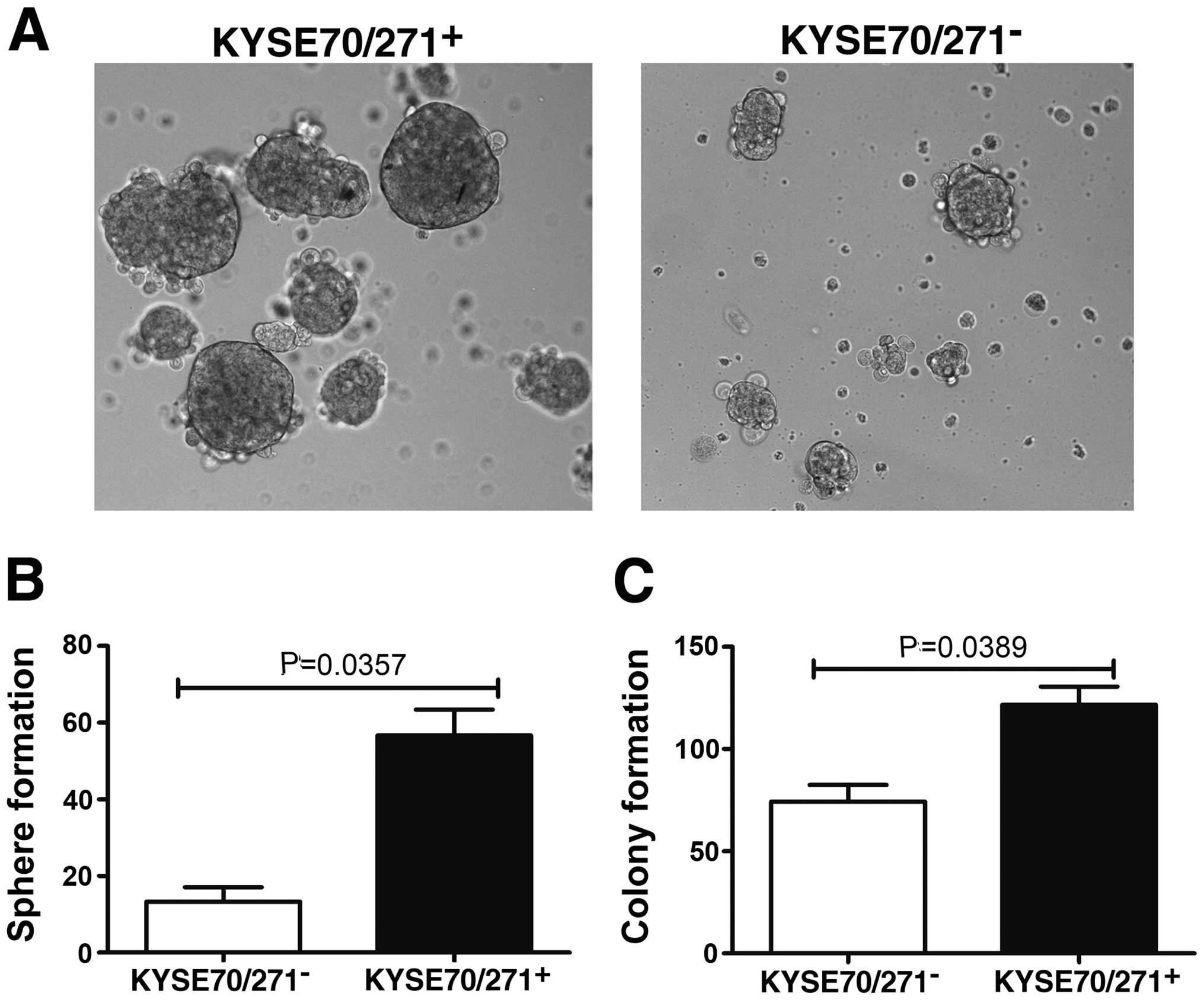
CD271+ cells form more
self-renewing spheres and promote anchorage-independent growth
One of the most important properties of GSCs is
self-renewal. We examined the tumor sphere formation ability, which
represents a self-renewal capacity. The sorted CD271+
and CD271− cells were seeded in 24-well ultra-low
cluster plates at a density of 5×10³ cells/ml in
DMEM/F12 medium supplemented with heparin, B27, EGF, and bFGF.
After culturing for 7 days, CD271+ cells exhibited a
marked ability for tumor sphere formation, compared with
CD271− cells (Fig. 3A and
B). In addition, CD271+ cells promoted
anchorage-independent growth (Fig.
3C), indicating that the CD271+ cells play an
important role in the maintenance of malignant growth of ESCC
cells. These results suggested that CD271+ ESCC cells
possess stem-like properties.
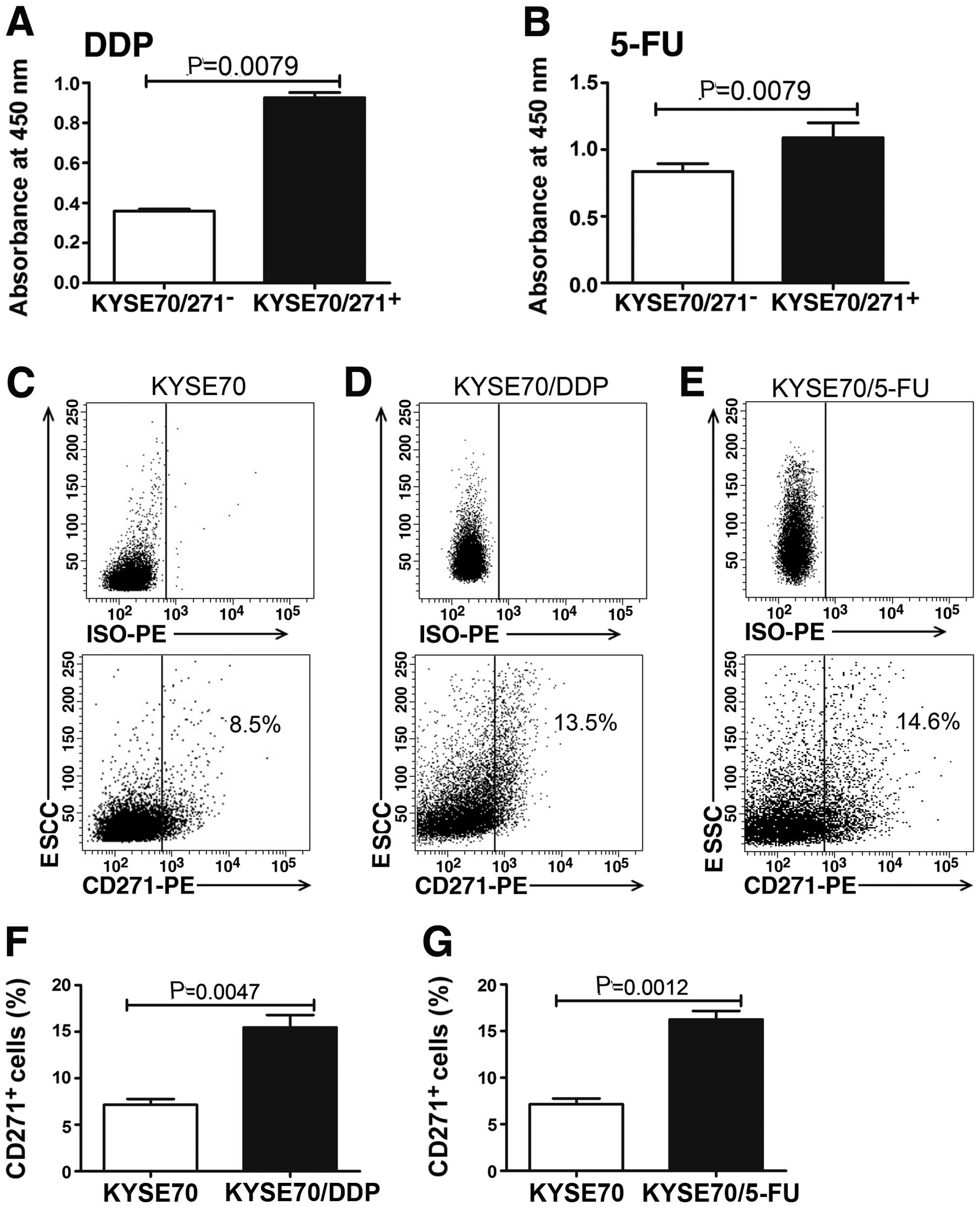
CD271+ cells possess the
ability to resist conventional chemotherapeutic reagents in
vitro
Cancer stem cells are more resistant to conventional
chemotherapeutic drugs. To examine whether the self-renewing
CD271+ cells possess the hypothesized CSC chemoresistant
ability, the sensitivity of the sorted CD271+ and
CD271− cells to DDP and 5-FU, respectively, was
analyzed. The survival rates of CD271+ cells were higher
under the treatment of DDP and 5-FU, compared with
CD271− cells (Fig. 4A and
B). We also identified CD271+ stem-like cells could
be enriched in DDP- and 5-FU-resistant cells (Fig. 4C–G). The results validated a role
for CD271+ cells in chemoresistance, which may explain
the failure of current therapies to eradicate progenitors and
prevent tumor recurrence.
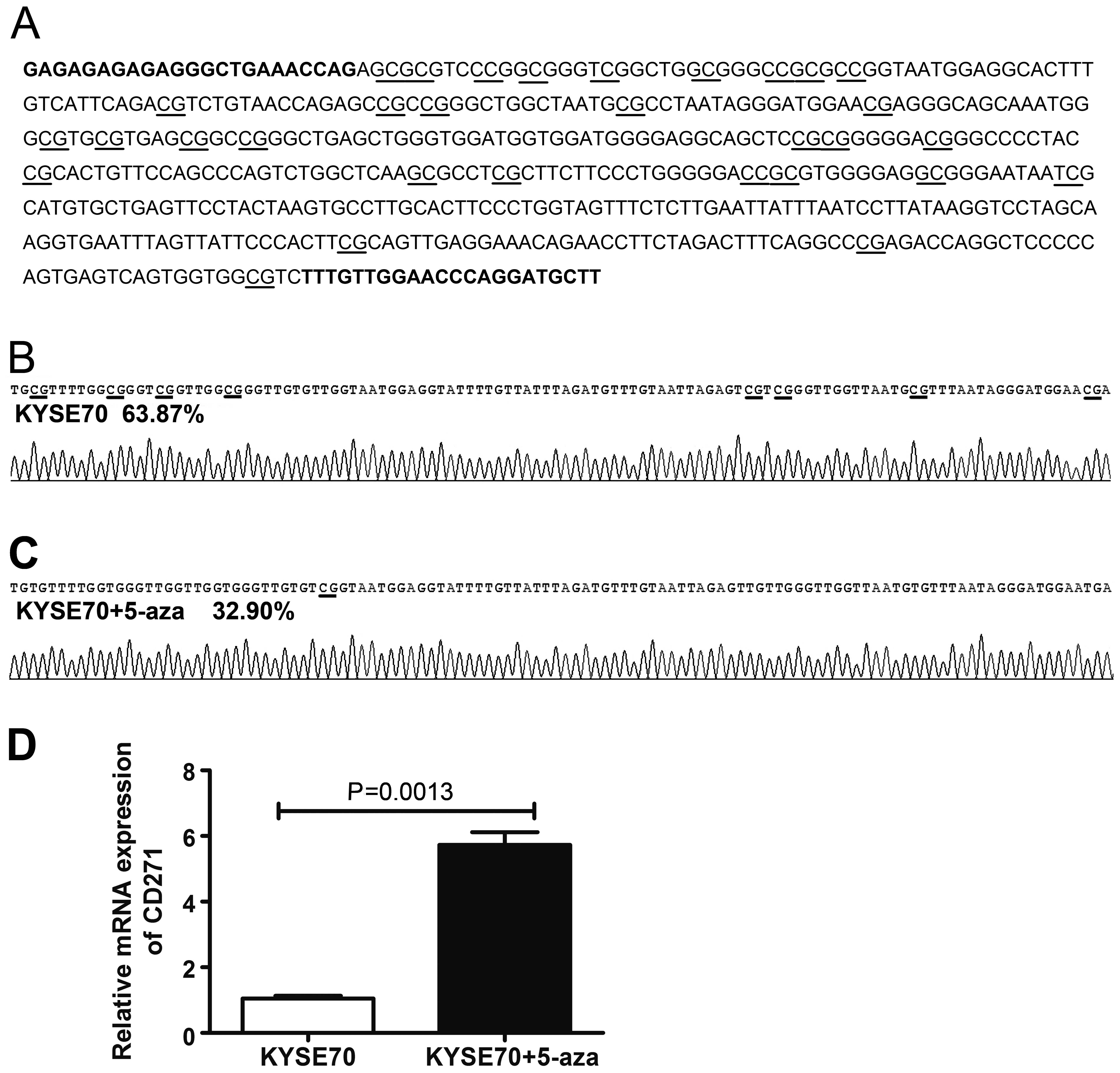
CD271 expression is regulated by DNA
methylation
Transcription factors and epigenetic modifications
often guide external signals to a specific genetic response. We
examined whether epigenetics including DNA methylation are involved
in regulating CD271 expression. To investigate promoter methylation
of CD271, BSP was performed. The area of the CpG-rich region
spanning 31 CpG sites, was sequenced (Fig. 5A). We found most CpG dinucleotides
were methylated in KYSE70 cells (Fig.
5B). To elucidate whether the methylation of CD271 was
associated with its expression, KYSE70 cells were treated with
5-aza (an inhibitor of the methylase enzyme, which can reactivate
mRNA expression suppressed by methylation) for 6 days and performed
BSP and qPCR to detect the promoter methylation and expression of
CD271. Following treatment with 5 μM 5-aza for 6 days, CD271 gene
exhibited an obvious induction and the promoter methylation level
was reduced (Fig. 5C and D),
suggesting that the expression of CD271 is regulated by DNA
methylation.
Discussion
The majority of ESCC patients present with an
advanced stage at the time of diagnosis, with poor prognosis, rapid
growth and spread due to late diagnosis. Accumulating evidence
suggested the CSCs theory in that tumorigenic potential is largely
restricted to CSCs (21,22). CSCs from tumor tissues or
established cancer cell lines can be isolated by cell-surface
markers expressed on CSCs (17,23).
In recent studies ALDH was identified as the CSC marker for various
types of cancer including ovarian (24), breast (25), lung (26) and prostate (27) cancer,
CD44+CD24− for breast (28) and ovarian (29) cancer, CD133 for non-small cell lung
(30), liver (31) and lung (32) cancer. However, Meng et al
reported that both CD133+ and CD133−
subpopulations contain similar numbers of CSCs (33). Therefore, identification of specific
cell-surface markers to define CSCs is important for the possible
establishment of target-specific therapies using small molecule
inhibitors or humanized antibodies.
CD271 knockdown was found to eliminate the capacity
of melanoma cells to form heterogeneous tumors most likely through
the downregulation of mediators for melanoma invasion and
metastasis (GLI-2, SOX2 and ERBB3), angiogenesis (IGFBP-2),
proliferation (FST and MITF) or chemoresistance (RHOJ) (34). CD271 was also identified as a
predominant molecule responsible for the proliferation,
tumorigenecity and plasticity of melanoma cells. Accumulating
evidence has shown that metastases develop when distant organs are
seeded with CSCs derived from a primary tumor (35). Boiko et al identified that
CD271+ melanoma cells lacked the expression of TYR,
MART1 and MAGE genes in 86, 69 and 68% of melanoma patients,
respectively, which may explain the reason for T-cell therapies
targeting these antigens usually resulting in only temporary tumor
shrinkage (16). Okumura et
al have shown that CD271 expression correlated with negative
lymph node metastasis and lower TNM staging. There was no
significant correlation between CD271 expression and distant
metastasis in ESCC (19). However,
in this study we show that the expression of CD271 was
significantly higher in ESCC tissues than adjacent non-cancerous
tissues. In contrast to findings of a previous study (19), we found that the overexpression of
CD271 was significantly associated with TNM stage and lymph node
metastasis. Taken together, these data strongly suggest CD271 as an
oncogene that plays an important role in ESCC progression.
Cell sorting yielded CD271+ and
CD271− cells and identified the overexpression of the
stem-related gene NANOG in CD271+ cells. Since EMT plays
a key role in tumor invasion and metastasis during tumor
progression, we tested the expression of EMT markers between the
two subpopulations. As expected, CD271+ cells highly
expressed mesenchymal markers, such as FIBRO and VIM. We also found
that CD271+ cells were able to form more tumor spheres
and colonies compared with CD271− cells. Our results
further showed that CD271+ cells exhibited general
resistance to DDP and 5-FU, with higher survival percentages
compared with CD271− cells. Taken together, these data
suggest that CD271 is a marker of stem-like cells in ESCC. In
addition, we observed increased CD271+ cells in DDP- and
5-FU-resistant cells. In other words, conventional chemotherapeutic
drugs, such as DDP and 5-FU, may selectively decrease the
CD271− population, resulting in a relative increase of
CD271+ cell frequency. Thus, CSCs can be responsible for
treatment failures of chemotherapy and poor clinical outcomes.
Accumulating evidence has suggested that CSCs are
not only governed by genetic alterations but also aberrant
epigenetic regulation. Sun et al reported that a unique set
of genes such as NANOG, OCT4 and SOX9, were demethylated in
invasive cancer cells but methylated in non-invasive cancer cells,
indicating that they may be biologically important in the invasive
population and upregulated during the EMT process (36). Schirmer et al (39) found that the gene L1CAM, which can
promote cell motility, invasion and metastasis formation in various
human cancers (37–39), could be induced by treatment with
the demethylation agent 5-aza in endometrial carcinoma cell lines
(40). We previously found that
tumor-suppressor gene SPINT2 was induced by a demethylation agent
(41). This shows that following
epigenetic drug treatment, hypermethylated tumor-suppressor genes
as well as oncogenes may be activated simultaneously. In the
present study, we firstly found CD271 was regulated epigenetically
and its expression could be induced by demethylation agent
treatment. Therefore, epigenetic therapy may be a ‘double-edge
sword’ and unexpected side effects may occur.
In conclusion, we have demonstrated that CD271, the
ESCC cancer stem cell marker, is a potential prognostic marker of
patients with ESCC and is regulated epigenetically.
Acknowledgements
This study was supported by grants from the China-US
(NFSC-NIH) Program for Biomedical Collaborative Research (Grant no.
812111102), the National Natural Science Foundation of China (Grant
no. 81171986), Research Grant from the Ministry of Public Health
(no. 20110110001), the Basic and Advanced Technology Research
Foundation from Science and Technology Department of Henan Province
(Grant no. 112300410153, Grant no. 122300410155), Funds for
Creative Research Team of Henan Province, Creative Research Team of
Higher Education of Henan Province and the Innovation Team of The
First Affiliated Hospital of Zhengzhou University.
References
|
1
|
Yuequan J, Shifeng C and Bing Z:
Prognostic factors and family history for survival of esophageal
squamous cell carcinoma patients after surgery. Ann Thorac Surg.
90:908–913. 2010. View Article : Google Scholar : PubMed/NCBI
|
|
2
|
Reya T, Morrison SJ, Clarke MF and
Weissman IL: Stem cells, cancer, and cancer stem cells. Nature.
414:105–111. 2001. View
Article : Google Scholar : PubMed/NCBI
|
|
3
|
Boman BM and Wicha MS: Cancer stem cells:
a step toward the cure. J Clin Oncol. 26:2795–2799. 2008.
View Article : Google Scholar : PubMed/NCBI
|
|
4
|
Wicha MS, Liu S and Dontu G: Cancer stem
cells: an old idea - a paradigm shift. Cancer Res. 66:1883–1890.
2006. View Article : Google Scholar
|
|
5
|
Hermann PC, Huber SL, Herrler T, et al:
Distinct populations of cancer stem cells determine tumor growth
and metastatic activity in human pancreatic cancer. Cell Stem Cell.
1:313–323. 2007. View Article : Google Scholar
|
|
6
|
Frank NY, Margaryan A, Huang Y, et al:
ABCB5-mediated doxorubicin transport and chemoresistance in human
malignant melanoma. Cancer Res. 65:4320–4333. 2005. View Article : Google Scholar : PubMed/NCBI
|
|
7
|
Zhang Y, Wang Z, Yu J, et al: Cancer
stem-like cells contribute to cisplatin resistance and progression
in bladder cancer. Cancer Lett. 322:70–77. 2012. View Article : Google Scholar : PubMed/NCBI
|
|
8
|
Cho RW and Clarke MF: Recent advances in
cancer stem cells. Curr Opin Genet Dev. 18:48–53. 2008. View Article : Google Scholar : PubMed/NCBI
|
|
9
|
Tilghman J, Wu H, Sang Y, et al: HMMR
maintains the stemness and tumorigenicity of glioblastoma stem-like
cells. Cancer Res. 74:3168–3179. 2014. View Article : Google Scholar : PubMed/NCBI
|
|
10
|
Hirsch D, Barker N, McNeil N, et al: LGR5
positivity defines stem-like cells in colorectal cancer.
Carcinogenesis. 35:849–858. 2014. View Article : Google Scholar
|
|
11
|
Ding K, Banerjee A, Tan S, et al: Artemin,
a member of the glial cell line-derived neurotrophic factor family
of ligands, is HER2 regulated and mediates acquired Trastuzumab
resistance by promoting cancer stem cell-like behaviour in mammary
carcinoma cells. J Biol Chem. 289:16057–16071. 2014. View Article : Google Scholar : PubMed/NCBI
|
|
12
|
Liepinsh E, Ilag LL, Otting G and Ibanez
CF: NMR structure of the death domain of the p75 neurotrophin
receptor. EMBO J. 16:4999–5005. 1997. View Article : Google Scholar : PubMed/NCBI
|
|
13
|
Rabizadeh S, Oh J, Zhong LT, et al:
Induction of apoptosis by the low-affinity NGF receptor. Science.
261:345–348. 1993. View Article : Google Scholar : PubMed/NCBI
|
|
14
|
Bibel M and Barde YA: Neurotrophins: key
regulators of cell fate and cell shape in the vertebrate nervous
system. Genes Dev. 14:2919–2937. 2000. View Article : Google Scholar : PubMed/NCBI
|
|
15
|
Civenni G, Walter A, Kobert N, et al:
Human CD271-positive melanoma stem cells associated with metastasis
establish tumor heterogeneity and long-term growth. Cancer Res.
71:3098–3109. 2011. View Article : Google Scholar : PubMed/NCBI
|
|
16
|
Boiko AD, Razorenova OV, van de Rijn M, et
al: Human melanoma-initiating cells express neural crest nerve
growth factor receptor CD271. Nature. 466:133–137. 2010. View Article : Google Scholar : PubMed/NCBI
|
|
17
|
Imai T, Tamai K, Oizumi S, et al: CD271
defines a stem cell-like population in hypopharyngeal cancer. PLoS
One. 8:e620022013. View Article : Google Scholar : PubMed/NCBI
|
|
18
|
Okumura T, Shimada Y, Imamura M and
Yasumoto S: Neurotrophin receptor p75(NTR) characterizes human
esophageal keratinocyte stem cells in vitro. Oncogene.
22:4017–4026. 2003. View Article : Google Scholar : PubMed/NCBI
|
|
19
|
Okumura T, Tsunoda S, Mori Y, et al: The
biological role of the low-affinity p75 neurotrophin receptor in
esophageal squamous cell carcinoma. Clin Cancer Res. 12:5096–5103.
2006. View Article : Google Scholar : PubMed/NCBI
|
|
20
|
Huang SD, Yuan Y, Liu XH, et al:
Self-renewal and chemotherapy resistance of p75NTR positive cells
in esophageal squamous cell carcinomas. BMC Cancer. 9:92009.
View Article : Google Scholar : PubMed/NCBI
|
|
21
|
Clarke MF, Dick JE, Dirks PB, et al:
Cancer stem cells -perspectives on current status and future
directions: AACR Workshop on cancer stem cells. Cancer Res.
66:9339–9344. 2006. View Article : Google Scholar : PubMed/NCBI
|
|
22
|
Eyler CE and Rich JN: Survival of the
fittest: cancer stem cells in therapeutic resistance and
angiogenesis. J Clin Oncol. 26:2839–2845. 2008. View Article : Google Scholar : PubMed/NCBI
|
|
23
|
Tirino V, Desiderio V, d‘Aquino R, et al:
Detection and characterization of CD133+ cancer stem
cells in human solid tumours. PLoS One. 3:e34692008. View Article : Google Scholar
|
|
24
|
Wang YC, Yo YT, Lee HY, et al:
ALDH1-bright epithelial ovarian cancer cells are associated with
CD44 expression, drug resistance, and poor clinical outcome. Am J
Pathol. 180:1159–1169. 2012. View Article : Google Scholar : PubMed/NCBI
|
|
25
|
Marcato P, Dean CA, Pan D, et al: Aldehyde
dehydrogenase activity of breast cancer stem cells is primarily due
to isoform ALDH1A3 and its expression is predictive of metastasis.
Stem Cells. 29:32–45. 2011. View Article : Google Scholar : PubMed/NCBI
|
|
26
|
Jiang F, Qiu Q, Khanna A, et al: Aldehyde
dehydrogenase 1 is a tumor stem cell-associated marker in lung
cancer. Mol Cancer Res. 7:330–338. 2009. View Article : Google Scholar : PubMed/NCBI
|
|
27
|
Hellsten R, Johansson M, Dahlman A,
Sterner O and Bjartell A: Galiellalactone inhibits stem cell-like
ALDH-positive prostate cancer cells. PLoS One. 6:e221182011.
View Article : Google Scholar : PubMed/NCBI
|
|
28
|
Sun H, Jia J, Wang X, et al:
CD44(+)/CD24(−) breast cancer cells isolated from MCF-7 cultures
exhibit enhanced angiogenic properties. Clin Transl Oncol.
15:46–54. 2012. View Article : Google Scholar
|
|
29
|
Meng E, Long B, Sullivan P, et al:
CD44+/CD24− ovarian cancer cells demonstrate
cancer stem cell properties and correlate to survival. Clin Exp
Metastasis. 29:939–948. 2012. View Article : Google Scholar : PubMed/NCBI
|
|
30
|
Tirino V, Camerlingo R, Franco R, et al:
The role of CD133 in the identification and characterisation of
tumour-initiating cells in non-small-cell lung cancer. Eur J
Cardiothorac Surg. 36:446–453. 2009. View Article : Google Scholar : PubMed/NCBI
|
|
31
|
Piao LS, Hur W, Kim TK, et al:
CD133+ liver cancer stem cells modulate radioresistance
in human hepatocellular carcinoma. Cancer Lett. 315:129–137. 2012.
View Article : Google Scholar
|
|
32
|
Bertolini G, Roz L, Perego P, et al:
Highly tumorigenic lung cancer CD133+ cells display
stem-like features and are spared by cisplatin treatment. Proc Natl
Acad Sci USA. 106:16281–16286. 2009. View Article : Google Scholar
|
|
33
|
Meng X, Li M, Wang X, Wang Y and Ma D:
Both CD133+ and CD133− subpopulations of A549
and H446 cells contain cancer-initiating cells. Cancer Sci.
100:1040–1046. 2009. View Article : Google Scholar : PubMed/NCBI
|
|
34
|
Redmer T, Welte Y, Behrens D, et al: The
nerve growth factor receptor CD271 is crucial to maintain
tumorigenicity and stem-like properties of melanoma cells. PLoS
One. 9:e925962014. View Article : Google Scholar : PubMed/NCBI
|
|
35
|
Balic M, Lin H, Young L, et al: Most early
disseminated cancer cells detected in bone marrow of breast cancer
patients have a putative breast cancer stem cell phenotype. Clin
Cancer Res. 12:5615–5621. 2006. View Article : Google Scholar : PubMed/NCBI
|
|
36
|
Sun L, Mathews LA, Cabarcas SM, et al:
Epigenetic regulation of SOX9 by the NF-κB signaling pathway in
pancreatic cancer stem cells. Stem Cells. 31:1454–1466. 2013.
View Article : Google Scholar : PubMed/NCBI
|
|
37
|
Chen DL, Zeng ZL, Yang J, et al: L1CAM
promotes tumor progression and metastasis and is an independent
unfavorable prognostic factor in gastric cancer. J Hematol Oncol.
6:432013. View Article : Google Scholar : PubMed/NCBI
|
|
38
|
Ben Q, An W, Fei J, et al: Downregulation
of L1CAM inhibits proliferation, invasion and arrests cell cycle
progression in pancreatic cancer cells. Exp Ther Med. 7:785–790.
2014.PubMed/NCBI
|
|
39
|
Schirmer U, Doberstein K, Rupp AK, et al:
Role of miR-34a as a suppressor of L1CAM in endometrial carcinoma.
Oncotarget. 5:462–472. 2014.PubMed/NCBI
|
|
40
|
Schirmer U, Fiegl H, Pfeifer M, et al:
Epigenetic regulation of L1CAM in endometrial carcinoma: comparison
to cancer-testis (CT-X) antigens. BMC Cancer. 13:1562013.
View Article : Google Scholar : PubMed/NCBI
|
|
41
|
Yue D, Fan Q, Chen X, et al: Epigenetic
inactivation of SPINT2 is associated with tumor suppressive
function in esophageal squamous cell carcinoma. Exp Cell Res.
322:149–158. 2014. View Article : Google Scholar
|