Introduction
The abscopal effect is a phenomenon noted in the
treatment of metastatic cancer where localized irradiation of a
tumor causes shrinking not only of the immediate target, but also
of tumors located far from the irradiated area. This phenomenon is
extremely rare, but when it does occur, its anticancer effects can
be dramatic, leading to the disappearance of malignant growths
throughout the body. Such success has been described for a variety
of malignancies, including melanoma, lymphoma, and lung metastases
of hepatocellular carcinoma (1–3). The
mechanism underlying the abscopal effect remains to be clarified,
although a variety of underlying biological events may be
responsible, particularly those induced by the immune system
(4). In mouse studies, the
observations support the immune hypothesis for the abscopal effect
(5–7).
The modulated electro-hyperthermia (mEHT) system is
a fast-developing complementary treatment method that is effective
against different types of tumors (8,9). The
principles involved are based on the classical method of
hyperthermia, but the aim, besides the absolute increase in
temperature, is to promote the direct absorption of electric-field
energy by the extracellular fluid, thereby destroying the membrane
of cancer cells.
Hyperthermia is usually applied as an adjunct to an
already established treatment modality, such as radiotherapy and
chemotherapy, in order to achieve tumor temperatures of 40–46°C.
Hyperthermia was originally controlled through modulating
temperature alone, whereas the more recently developed mEHT (also
described as ‘oncothermia’) is based on energy dose control,
replacing the single temperature concept (9).
Although hyperthermia has been extensively studied,
the effects of mEHT combined with dendritic cell (DC) treatment on
squamous cell carcinoma have not been addressed. The goal of this
study was to evaluate the antitumor effects of combined direct
intratumoral injection of DCs and mEHT treatment using a mouse
squamous cell carcinoma (SCCVII) cancer model. We showed that this
combined treatment causes both local and distant shrinkage of
SCCVII-derived tumors, and thereby offers hope to patients with
esophageal squamous cell carcinoma, particularly those who have
recurrent disease and are unable to undergo further radiotherapy.
Thus, this newly developed technique could represent a new strategy
for the treatment of squamous cell carcinoma, as well as other
cancers.
Materials and methods
Culture of SCCVII tumor cells
Mouse SCCVII cells, which have been previously
characterized (10), were used in
this study. Cells were cultured at 37°C in Dulbecco’s modified
Eagle’s medium (DMEM; D8062; Sigma Life Science) with 10%
heat-inactivated fetal bovine serum (FBS; 16140-071; Gibco,
Invitrogen) plus 1% glutamine-penicillin-streptomycin.
DC generation
DCs were generated using the method established by
Lutz et al (11). Cells were
prepared from the bone marrow cells of the femurs and tibias of
mice. On day 0, 2×10⁶ bone marrow cells in 10 ml of
RPMI-1640 medium (RPMI, 22400-089; Gibco, Invitrogen) with
penicillin (100 U/ml), streptomycin (100 μg/ml), L-glutamine (2
mM), 2-mercaptoethanol (50 μM), 10% FBS and 200 U/ml (20 ng/ml)
rmGM-CSF (G0282; Sigma, Japan) were placed into 100-mm diameter
Petri dishes (Falcon™ no. 1029; BD Biosciences, San Jose, CA, USA).
On day 3, another 10 ml of the medium with supplements was added to
the dishes. Non-adherent cells were harvested on day 6.
We determined whether the cells generated could be
used as DCs by performing flow cytometry analysis. Surface markers
such as MHC class II, CD11c, CD80 and CD86 were highly expressed by
70.6, 35.2, 63.4, and 72.0% of the cells, respectively, indicating
that they were indeed DCs.
Mice and the tumor model
Syngeneic 6- to 10-week-old female C3H/He mice,
purchased from Japan SLC (Shizuoka, Japan), were maintained in our
facility under specific pathogen-free conditions. SCCVII cancer
cells at a dose of 5×10⁵ per mouse were inoculated
subcutaneously into the left leg in order to seed the primary
treatment tumor, and the same number of SCCVII cells was
simultaneously inoculated subcutaneously into the chest wall to
seed a distant, non-treatment tumor. Mice were examined every two
or three days, and only those of a similar size were selected for
treatment. Animals that had developed palpable tumors 9 days after
inoculation prepared for treatment were divided into independent
experimental groups, each consisting of at least 4 mice. Animal
care was provided in accordance with the guidelines of Chiba
University. All animal experiments were conducted to the Guidelines
for the Welfare and Use of Animals in Cancer Research (12).
mEHT and DC treatment
We used the LAB-EHY device (Oncotherm Ltd. Hungary)
to induce mEHT treatment on the left leg tumors of the mice. The RF
power level was regulated using the fluoroptic temperature
measurement system (Luxtron m3300; Lumasense, Santa Clara, CA,
USA). Tumors on the left legs of the mice in the mEHT-only
treatment group and in the DC combined with mEHT treatment group
were treated on day 9, 11 and 13 after SCCVII cell inoculation. In
the DC-only treatment group and DC combined with mEHT treatment
group, 1×10⁶ DCs for each mouse were directly
administered only into the tumor on the left leg of the mice after
the mEHT treatment on day 9, 11 and 13.
Flow cytometric analysis for cytotoxic T
lymphocytes
After the mice were sacrificed on day 39 after
SCCVII cell inoculation, the tumor draining lymph nodes (TDLNs)
were collected and washed with PBS. After red blood cell reduction,
the cells were treated with mouse BD Fc Block (2.4G2; Pharmingen™,
BD) and then stained with antibodies conjugated with fluorescent
agents. For the investigation of cytotoxic T lymphocytes (CTLs) in
the TDLNs, CD3-PE (2134-0034; Biogenesis Inc., Kingston, NH, USA)
and CD8-FITC (2134-0083; Biogenesis Inc.) were used. Cell counting
was performed with a Coulter Epics XL cytometer (Beckman Coulter,
Miami, FL, USA), and cell populations were evaluated with gating
software, FlowJo for Windows (Tree Star Inc., Ashland, OR,
USA).
Immunostaining
After the mice were sacrificed on day 39 after
SCCVII cell inoculation, the tumor tissues on the chest were
harvested. The tissues were stained with anti-mouse CD8 (250596;
ABBIOTEC), S100 (GEX48819; Gene Tex), Foxp3 (NBP1-18319; Novus
Biologicals) and a fluorescein-conjugated secondary antibody
(k4003; Dako). The expression of CD8, S100 and Foxp3 was observed
under a microscope (Carl Zeiss, Oberkochen, Germany). According to
the method developed by Xavie et al (13), we quantified the results of the
immunostaining with the Imag Pro Plus 6.0 software (Media
Cybernetic, Silver Spring, MD, USA). The measurement parameter was
integrated optical density (IOD). The signal density of the tissue
areas from five randomly selected visions were counted blindly and
subjected to statistical analysis.
Real-time quantitative PCR (RT-PCR)
The RNA was extracted from tumor tissues on the
chest using TRIzol reagent (Invitrogen, Carlsbad, CA, USA) and 1 μg
of RNA was transcribed into cDNA using a High Capacity cDNA Reverse
Transcription kit (Applied Biosystems, Foster City, CA, USA). The
resulting 1 μg of cDNA was amplified via RT-PCR. RT-PCR was
conducted using SsoFast™ EvaGreen® Supermix (Bio-Rad,
Hercules, CA, USA) with the following primers (Operon,
Biotechnology, Tokyo, Japan): GP96, 5′-ACACGGC TTGCTAAACTTCT-3′ and
5′-ACTACAGTCTGCGGTCC AAA-3′. β-actin was used as an internal
control and the sequences used were 5′-TCATGAAGATCCTCACCGAG-3′ and
5′-TTGCCAATGGTGATTGACCTG-3′. The PCR was performed using MyiQ2
thermal cycler (Bio-Rad). The PCR conditions consisted of 40 cycles
of 95°C for 30 sec, 95°C for 5 sec and 60°C for 10 sec. This assay
was performed in triplicate.
Statistics
Results are expressed as the mean ± SE, and
statistical comparisons were performed using ANOVA. A P-value
<0.05 was considered to indicate a statistically significant
result. All data were analyzed by the SPSS software package (SPSS
version 19.0; SPSS Inc., USA).
Results
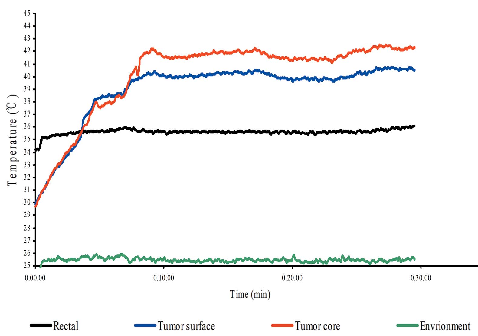
Temperature monitoring in local mEHT
treatment
By using the LAB-EHY device, it was possible to
increase the temperature within the tumor to 42°C within 10 min. By
controlling the energy input, we were able to maintain the
temperature within a range of 42–43°C. At the same time of
treatment, we found that the temperature on the surface of the
tumor was ~40°C, and the rectal temperature was maintained close to
35–37°C (Fig. 1).
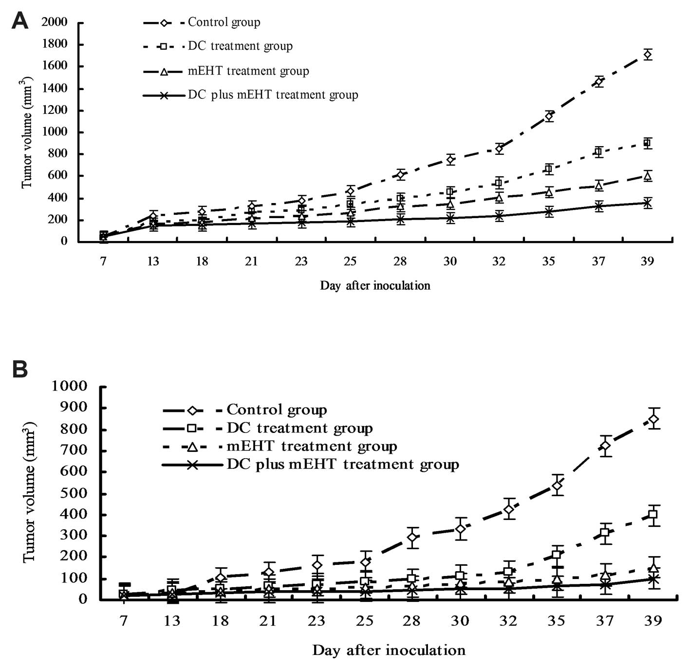
Efficacy of the combined therapy of DCs
and mEHT against the treated local tumor
We examined the antitumor effect by the combination
of DCs and mEHT treatment against the local tumor. The tumor on the
left leg of the mice in the control group grew to a mean volume of
1,709.25±2,557 4 mm3 by day 39. The mean volume of the
tumor on the left leg in the group treated with a combination of
DCs and mEHT was 359.92±62.01 mm3. The tumor growth rate
was obviously slow in the DC and mEHT treatment group compared to
that in the control group (P<0.05). The mean tumor volume in the
DC-only treatment group and the mEHT-only treatment group was
smaller compared to the control group (DC treatment group:
902.78±109.74 mm3, P<0.05; mEHT treatment group:
607.19±87.28 mm3, P<0.05) (Fig. 2A). Thus, the combination therapy of
DCs and mEHT showed the strongest antitumor effect in
vivo.
Efficacy of the combined therapy of DCs
and mEHT against the non-treated distant tumor
We examined the tumors on the chest wall of the mice
to investigate whether the combination treatment of DCs and mEHT
could elicit a systemic antitumor effect. On day 39, the mean
volume of the tumors on the chest wall of the mice in the control
group was 852.43±270.78 mm3, whereas the mean volume of
the tumors in the DC and the mEHT treatment group was only
99.84±9.21 mm3. The tumor growth rate was obviously
slower in the DC and mEHT treatment group compared to the control
group (P<0.05). The mean chest tumor volumes in the DC-only
treatment group and the mEHT-only treatment group were 396.32±58.52
and 152.78±19.12 mm3, respectively, both of which were
also significantly smaller (P<0.05) than the mean tumor volume
in the control group (Fig. 2B).
This phenomenon was thought to be the abscopal effect.
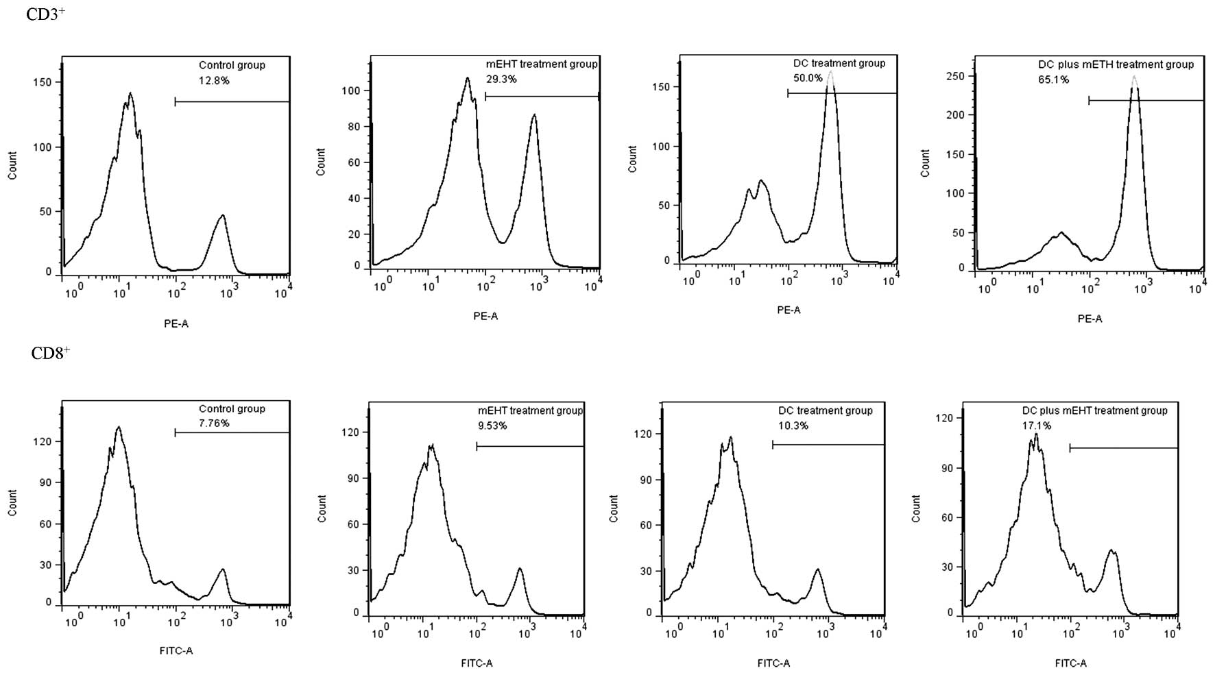
CD3+ and CD8+ T
cells in the TDLNs
We investigated whether the intratumoral injection
of DCs in addition to mEHT can elicit a systemic antitumor effect
in vivo by examining the populations of CD3+ and
CD8+ T cells in the TDLNs. As shown in Fig. 3, the CD3+ population in
the TDLNs of the mice treated with DCs plus mEHT was ~5 times
larger compared with this population in the TDLNs of the control
group, and the CD8+ population in the TDLNs of the mice
treated with DCs plus mEHT was ~2 times larger. This suggests that
the systemic antitumor activity that was evoked by DC plus mEHT
treatment can be a sensitizer of a DC-based immunoresponse.
Furthermore, we also evaluated whether mEHT alone has the
possibility of inducing CD3+ and CD8+ T
cells. We found that mEHT alone could increase that population.
This may be one of the pieces of evidence that can explain the
mechanism of the absocopal effects.
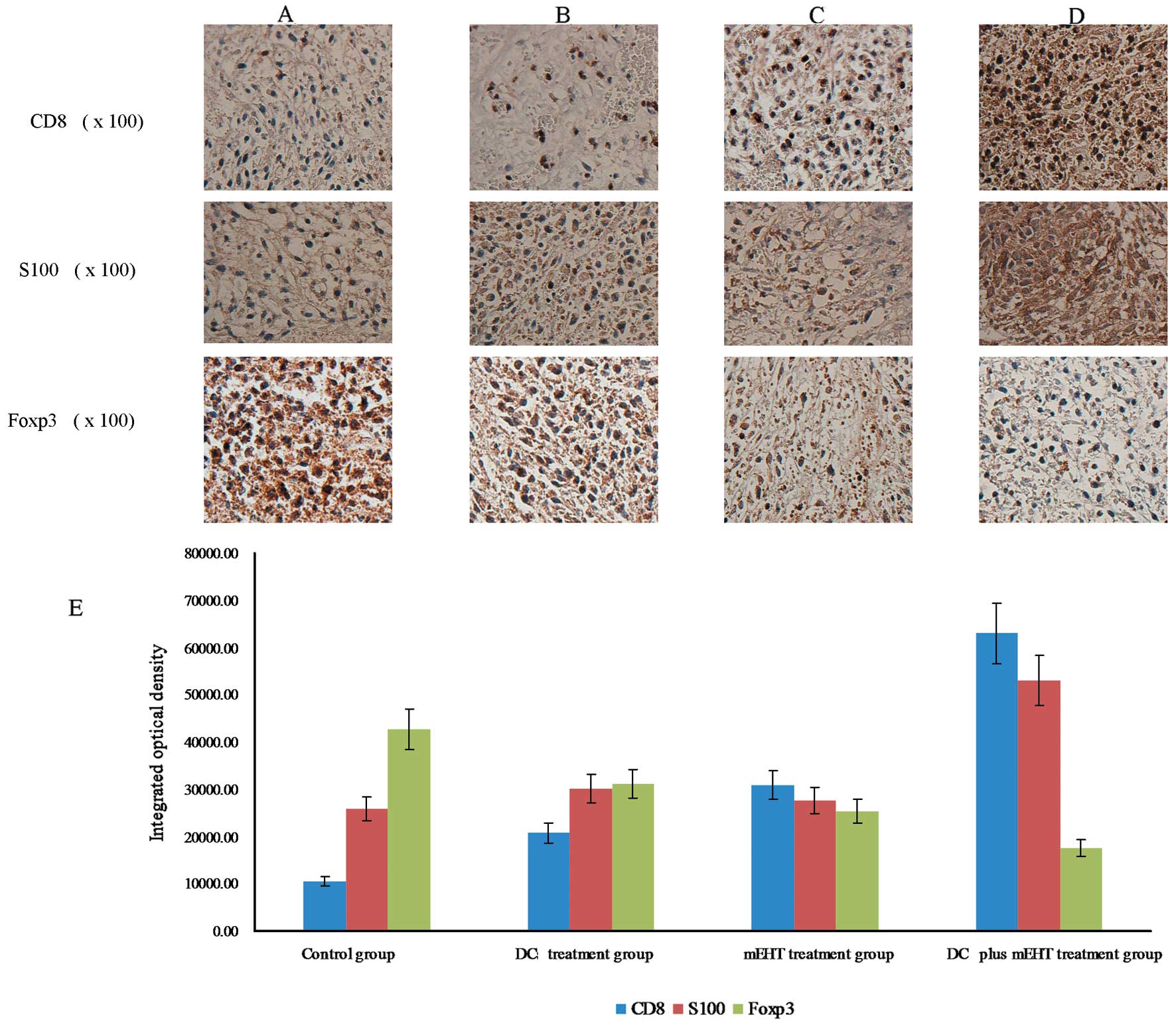
Immunostaining of the tumors
For the immunostaining, the tumor tissues were
examined for CD8, S100 and Foxp3 protein expression. The IOD was
calculated as the number of positive cells expressing the protein.
We found that in the DC plus mEHT treatment group, a greater number
of cells were positive for CD8 than the numbers in the other groups
(Fig. 4) (control group:
10,607.17±1,193.27; DC treatment group: 20,775.26±1,56.91; mEHT
treatment group: 30,893.19±2,067.52; DC and mEHT treatment group:
63,028.26±8,281.73; P<0.05). This indicates that a greater
number of cytotoxic lymphocytes were stimulated to attack the tumor
cells following the combined treatment. Similar findings were noted
for S100, a DC marker, with a significantly greater number of
S100-positive cells noted in the DC plus mEHT treatment group
compared with the other groups (Fig.
4) (control group: 25,944.49±2,084.24; DC treatment group:
30,108.26±1,827.21; mEHT treatment group: 27,685.48±1,373.36; DCs
and mEHT treatment group: 53,118.53±6,680.50, P<0.05). This
indicates that a greater number of DCs were induced to take part in
the immune reaction against the tumor. Unlike these other markers,
the extent of staining of Foxp3, a marker and determinant of
regulatory T (Treg) cells, was significantly higher in the control
group compared with the other 3 groups (Fig. 4) (control group: 42,773.29±1,461.24;
DC treatment group: 31,209.38±1,928.48; mEHT treatment group:
25,394.31±1,323.39; DC and mEHT treatment group:
17,496.13±1,018.26, P<0.05), suggesting that the
immunosuppression by Treg cells in the treated tissue was reduced
by mEHT.
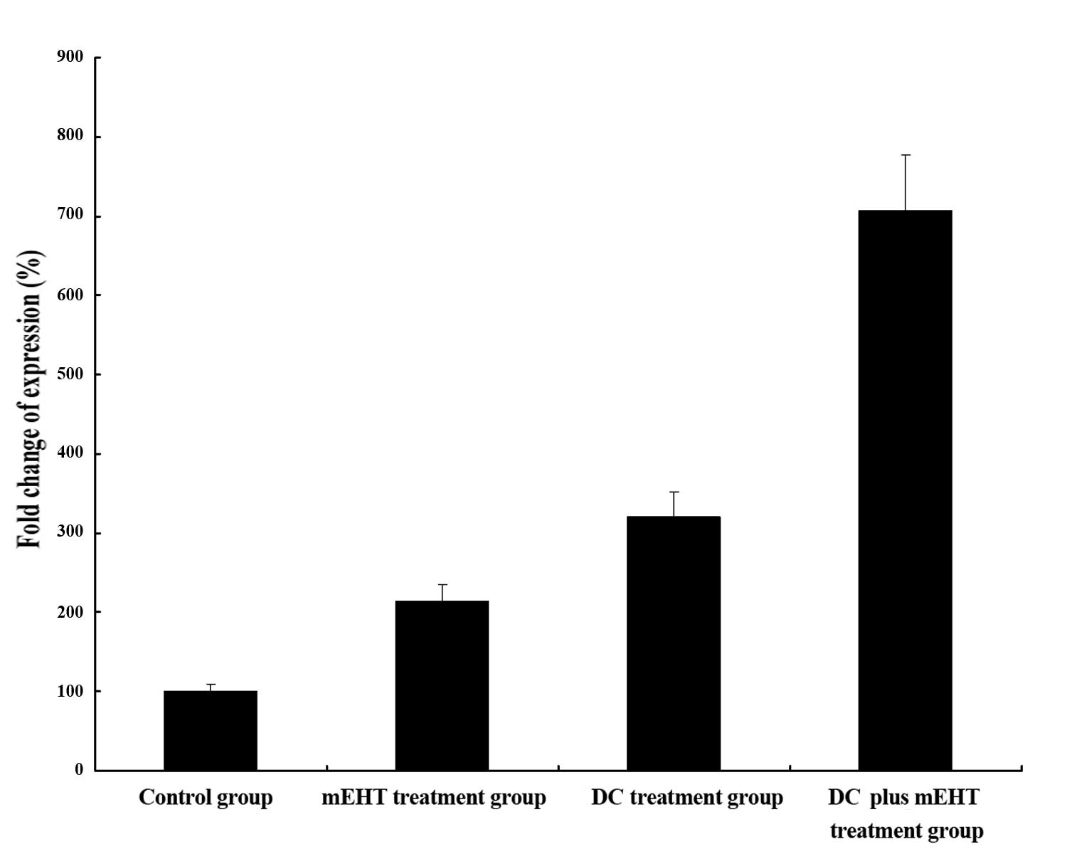
RT-PCR
RT-PCR was used to analyze the cDNA
reverse-transcribed RNA of the chest tumors from different groups.
As shown in Fig. 5, regarding the
expression of GP96, ~6-fold increase was observed in the DC plus
mEHT treatment group compared to the control group. In addition,
the expression of GP96 exhibited a 2-fold increase in the DC
treatment group and a 1-fold increased in the mEHT treatment group.
Based on the data, the GP96 gene was amplified in the treatment
groups compared to the control group. This may at least in part
explain the mechanism underlying the abscopal effect.
Discussion
Head and neck and esophageal cancer are still among
the most deadly malignant neoplasms, particularly in the Asian
populaton. These cancers have the tendency to metastasize easily to
lymph nodes (14,15). Their prognosis is poor and
unsatisfactory even with current treatments, as lymph node
metastases and distant organ metastases occur frequently. The
development of effective treatments is urgent.
Hyperthermia is a type of cancer treatment in which
tissue is exposed to a high temperature. Previous studies have
shown that high temperatures can damage and kill cancer cells,
usually with minimal injury to normal tissues. mEHT which is also
described as ‘oncothermia’ in Europe is a personalized treatment
using energy delivery to the targeted tumor (9). This treatment concentrates the
absorbed energy to intercellular electrolytes (8). According to clinical studies of mEHT,
the application of mEHT has good clinical outcome and makes a
stable basis for clinical applications in various advanced primary
and metastatic malignancies, such as bone (metastatic) (16), breast (17) and malignant gliomas (18). In the present study, as shown in
Fig. 1, only the tumor underwent an
increase in temperature during mEHT, without any increase in rectal
temperature. The temperature of the center of the tumor is higher
than the temperature of the tumor surface. This differential
temperature rise is achieved through the precise surface control
and moderate power usage in mEHT that enables the temperature to be
finely regulated (19).
In the present study, we found that treatment of the
tumors on the leg of mice resulted in the concurrent growth
inhibition of the distant tumors on the chest. This phenomenon is
known as the abscopal effect and is defined as a reaction following
irradiation but occurring outside the zone of actual radiation
absorption. It was first described more than 60 years ago by Dr
R.H. Mole, of Britain’s Medical Research Council (20). He was the first to use the term
‘abscopal effect,’ where ‘abscopal’ is derived from the Latin
prefix ab, meaning ‘away from,’ and the Greek word
skopos, meaning ‘target.’ Multiple case studies describing
an abscopal effect observed after radiotherapy have been published
for a variety of malignancies, including lymphoma, papillary
adenocarcinoma, melanoma, adenocarcinoma of the esophagus, chronic
lymphocytic leukemia and hepatocellular carcinoma (21–25).
The mechanism that underlies the abscopal effect
remains unclear, although studies in mice suggest that it may
depend upon activation of the immune system, and is mediated by T
cells (7). Our findings also
suggest that distant tumor regression was mediated by an immune
response (via CD3+ and CD8+ cells) activated
by mEHT and DCs. The combination of mEHT and DCs produced tumor
antigens by apoptosis and/or necrosis of tumor cells, and also
induced the abscopal effect against SCCVII cells. Moreover, direct
intratumoral administration of DCs are an important strategy. DCs
can be pulsed with a wide range of antigens and they are efficient
for the direct trafficking into tumors. This means that the
trafficking loss of DCs administered is minimized (26–28).
In the present research, we found that mEHT enhanced
the expression of GP96 in vivo. As shown in Fig. 5, mEHT treatment had the ability to
induce the expression of GP96. The product of this gene plays an
important role in the uptake of antigens by DCs. The tumor antigens
form a complex with GP96, which is then taken up by DCs through the
GP96 receptor in a process mediated by toll-like receptors 2 and 4.
This leads to DC maturation, enabling them in turn to induce
CD8+ CTL activation (29–33).
In the present study, we found that the upregulation of GP96 by
mEHT led to an increase in the number of CD8+ CTLs in
the TDLNs. This suggests that DCs pulsed with mEHT exerted a
stronger cytotoxic effect leading to tumor growth inhibition in
vivo, via an increase in the level of GP96 protein. Thus, one
key molecule, by which the abscopal effect induces systemic
antitumor immune reaction, may be GP96. As others have reported,
GP96 potentially boosts the activity of the systemic antitumor
effect by these CTLs, and this mechanism could be one explanation
for the abscopal effect (34–36).
In a case study from the Memorial Sloan-Kettering
Cancer Center in New York, changes in a metastatic melanoma
patient’s immune system were measured over the course of treatment
(1). Changes in tumor-directed
antibody levels and immune cell populations were found to coincide
with an abscopal effect. These findings support the idea that
radiation may help stimulate the immune system to fight cancer. We
believe that local hyperthermia induced both, direct and abscopal
antitumor effects, and that the latter may have been the result of
a systemic effect of hyperthermia. We found that there was a
significantly greater number of CD8- and S100-positive cells after
combined DC and mEHT treatment. This indicates that additional
cytotoxic lymphocytes were stimulated to attack the tumor cells.
Furthermore, there were fewer Foxp3-positive cells after this
treatment, suggesting that the immune-suppression mediated by Treg
cells in the treated tissue was reduced. Correspondingly, combined
treatment with DCs plus mEHT induced a more pronounced antitumor
effect against distant non-treated tumors compared with the leg
tumors treated with DCs alone. This is particularly important for
cancer treatment, as the combination of DCs and mEHT exerted a
strong abscopal effect against the distant non-treated tumors.
Our findings suggest that the combination of
hyperthermia with an intratumoral injection of DCs can induce whole
body antitumor effects. Both DC and mEHT therapy are non-invasive,
and therefore, this treatment model can be safely adapted for
treating patients. Based on our findings, the combination of direct
intratumoral administration of DCs and mEHT can be a feasible
future treatment, and warrants further studies, including human
clinical trials.
Acknowledgements
This study was supported by the JSPS KAKENHI (grant
no. 24501328).
References
|
1
|
Postow MA, Callahan MK, Barker CA, et al:
Immunologic correlates of the abscopal effect in a patient with
melanoma. N Engl J Med. 366:925–931. 2012. View Article : Google Scholar : PubMed/NCBI
|
|
2
|
Perego D and Faravelli A: Unexpected
consequence of splenectomy in composite lymphoma. The abscopal
effect. Haematologica. 85:2112000.PubMed/NCBI
|
|
3
|
Okuma K, Yamashita H, Niibe Y, Hayakawa K
and Nakagawa K: Abscopal effect of radiation on lung metastases of
hepatocellular carcinoma: a case report. J Med Case Rep. 5:1112011.
View Article : Google Scholar : PubMed/NCBI
|
|
4
|
Macklis RM, Mauch PM, Burakoff SJ and
Smith BR: Lymphoid irradiation results in long-term increases in
natural killer cells in patients treated for Hodgkin’s disease.
Cancer. 69:778–783. 1992.PubMed/NCBI
|
|
5
|
Lugade AA, Moran JP, Gerber SA, Rose RC,
Frelinger JG and Lord EM: Local radiation therapy of B16 melanoma
tumors increases the generation of tumor antigen-specific effector
cells that traffic to the tumor. J Immunol. 174:7516–7523. 2005.
View Article : Google Scholar
|
|
6
|
Lee Y, Auh SL, Wang Y, et al: Therapeutic
effects of ablative radiation on local tumor require
CD8+ T cells: changing strategies for cancer treatment.
Blood. 114:589–595. 2009. View Article : Google Scholar : PubMed/NCBI
|
|
7
|
Demaria S, Ng B, Devitt ML, et al:
Ionizing radiation inhibition of distant untreated tumors (abscopal
effect) is immune mediated. Int J Radiat Oncol Biol Phys.
58:862–870. 2004. View Article : Google Scholar : PubMed/NCBI
|
|
8
|
Andocs G, Renner H, Balogh L, Fonyad L,
Jakab C and Szasz A: Strong synergy of heat and modulated
electromagnetic field in tumor cell killing. Strahlenther Onkol.
185:120–126. 2009. View Article : Google Scholar : PubMed/NCBI
|
|
9
|
Andocs G, Szasz O and Szasz A: Oncothermia
treatment of cancer: from the laboratory to clinic. Electromagn
Biol Med. 28:148–165. 2009. View Article : Google Scholar : PubMed/NCBI
|
|
10
|
Khurana D, Martin EA, Kasperbauer JL, et
al: Characterization of a spontaneously arising murine squamous
cell carcinoma (SCC VII) as a prerequisite for head and neck cancer
immunotherapy. Head Neck. 23:899–906. 2001. View Article : Google Scholar : PubMed/NCBI
|
|
11
|
Lutz MB, Kukutsch N, Ogilvie AL, et al: An
advanced culture method for generating large quantities of highly
pure dendritic cells from mouse bone marrow. J Immunol Methods.
223:77–92. 1999. View Article : Google Scholar : PubMed/NCBI
|
|
12
|
Workman P, Aboagye EO, Balkwill F, et al:
Guidelines for the Welfare and Use of Animals in Cancer Research.
Br J Cancer. 102:1555–1577. 2010. View Article : Google Scholar : PubMed/NCBI
|
|
13
|
Xavier LL, Viola GG, Ferraz AC, et al: A
simple and fast densitometric method for the analysis of tyrosine
hydroxylase immunoreactivity in the substantia nigra pars compacta
and in the ventral tegmental area. Brain Res Brain Res Protoc.
16:58–64. 2005. View Article : Google Scholar
|
|
14
|
Bollschweiler E, Baldus SE, Schroder W, et
al: High rate of lymph-node metastasis in submucosal esophageal
squamous-cell carcinomas and adenocarcinomas. Endoscopy.
38:149–156. 2006. View Article : Google Scholar : PubMed/NCBI
|
|
15
|
Mayinger B, Horner P, Jordan M, et al:
Light-induced autofluorescence spectroscopy for the endoscopic
detection of esophageal cancer. Gastrointest Endosc. 54:195–201.
2001. View Article : Google Scholar : PubMed/NCBI
|
|
16
|
Bogovic J, Douwes F, Muravjov G and
Istomin J: Posttreatment histology and microcirculation status of
osteogenic sarcoma after a neoadjuvant chemo- and radiotherapy in
combination with local electromagnetic hyperthermia. Onkologie.
24:55–58. 2001. View Article : Google Scholar
|
|
17
|
Feyerabend T, Wiedemann GJ, Jager B,
Vesely H, Mahlmann B and Richter E: Local hyperthermia, radiation,
and chemotherapy in recurrent breast cancer is feasible and
effective except for inflammatory disease. Int J Radiat Oncol Biol
Phys. 49:1317–1325. 2001. View Article : Google Scholar : PubMed/NCBI
|
|
18
|
Fiorentini G, Giovanis P, Rossi S, et al:
A phase II clinical study on relapsed malignant gliomas treated
with electro-hyperthermia. In Vivo. 20:721–724. 2006.PubMed/NCBI
|
|
19
|
Hegyi G, Szigeti GP and Szasz A:
Hyperthermia versus oncothermia: cellular effects in complementary
cancer therapy. Evid Based Complement Alternat Med.
2013:6728732013.PubMed/NCBI
|
|
20
|
Mole RH: Whole body irradiation;
radiobiology or medicine? Br J Radiol. 26:234–241. 1953. View Article : Google Scholar : PubMed/NCBI
|
|
21
|
Nobler M: The abscopal effect in malignant
lymphoma and its relationship to lymphocyte circulation. Radiology.
93:410–412. 1969. View
Article : Google Scholar : PubMed/NCBI
|
|
22
|
Rees GJ: Abscopal regression in lymphoma:
a mechanism in common with total body irradiation? Clin Radiol.
32:475–480. 1981. View Article : Google Scholar : PubMed/NCBI
|
|
23
|
Rees GJ and Ross CM: Abscopal regression
following radiotherapy for adenocarcinoma. Br J Radiol. 56:63–66.
1983. View Article : Google Scholar : PubMed/NCBI
|
|
24
|
Sham RL: The abscopal effect and chronic
lymphocytic leukemia. Am J Med. 98:307–308. 1995. View Article : Google Scholar : PubMed/NCBI
|
|
25
|
Ohba K, Omagari K, Nakamura T, et al:
Abscopal regression of hepatocellular carcinoma after radiotherapy
for bone metastasis. Gut. 43:575–577. 1998. View Article : Google Scholar : PubMed/NCBI
|
|
26
|
Schmidt T, Ziske C, Marten A, et al:
Intratumoral immunization with tumor RNA-pulsed dendritic cells
confers antitumor immunity in a C57BL/6 pancreatic murine tumor
model. Cancer Res. 63:8962–8967. 2003.PubMed/NCBI
|
|
27
|
Candido KA, Shimizu K, McLaughlin JC, et
al: Local administration of dendritic cells inhibits established
breast tumor growth: implications for apoptosis-inducing agents.
Cancer Res. 61:228–236. 2001.PubMed/NCBI
|
|
28
|
Triozzi PL, Khurram R, Aldrich WA, Walker
MJ, Kim JA and Jaynes S: Intratumoral injection of dendritic cells
derived in vitro in patients with metastatic cancer. Cancer.
89:2646–2654. 2000. View Article : Google Scholar : PubMed/NCBI
|
|
29
|
Binder RJ and Srivastava PK: Essential
role of CD91 in re-presentation of gp96-chaperoned peptides. Proc
Natl Acad Sci USA. 101:6128–6133. 2004. View Article : Google Scholar : PubMed/NCBI
|
|
30
|
Facciponte JG, Wang XY, MacDonald IJ, et
al: Heat shock proteins HSP70 and GP96: Structural insights. Cancer
Immunol Immunother. 55:339–346. 2006. View Article : Google Scholar : PubMed/NCBI
|
|
31
|
Demine R and Walden P: Testing the role of
gp96 as peptide chaperone in antigen processing. J Biol Chem.
280:17573–17578. 2005. View Article : Google Scholar : PubMed/NCBI
|
|
32
|
Ramirez SR, Singh-Jasuja H, Warger T, et
al: Glycoprotein 96-activated dendritic cells induce a CD8-biased T
cell response. Cell Stress Chaperones. 10:221–229. 2005. View Article : Google Scholar : PubMed/NCBI
|
|
33
|
Segal BH, Wang XY, Dennis CG, et al: Heat
shock proteins as vaccine adjuvants in infections and cancer. Drug
Discov Today. 11:534–540. 2006. View Article : Google Scholar : PubMed/NCBI
|
|
34
|
Akutsu Y, Matsubara H, Urashima T, et al:
Combination of direct intratumoral administration of dendritic
cells and irradiation induces strong systemic antitumor effect
mediated by GRP94/gp96 against squamous cell carcinoma in mice. Int
J Oncol. 31:509–515. 2007.
|
|
35
|
Wang XH, Qin Y, Hu MH and Xie Y: Dendritic
cells pulsed with gp96-peptide complexes derived from human
hepatocellular carcinoma (HCC) induce specific cytotoxic T
lymphocytes. Cancer Immunol Immunother. 54:971–980. 2005.
View Article : Google Scholar : PubMed/NCBI
|
|
36
|
Liu S, Wang H, Yang Z, et al: Enhancement
of cancer radiation therapy by use of adenovirus-mediated
secretable glucose-regulated protein 94/gp96 expression. Cancer
Res. 65:9126–9131. 2005. View Article : Google Scholar : PubMed/NCBI
|