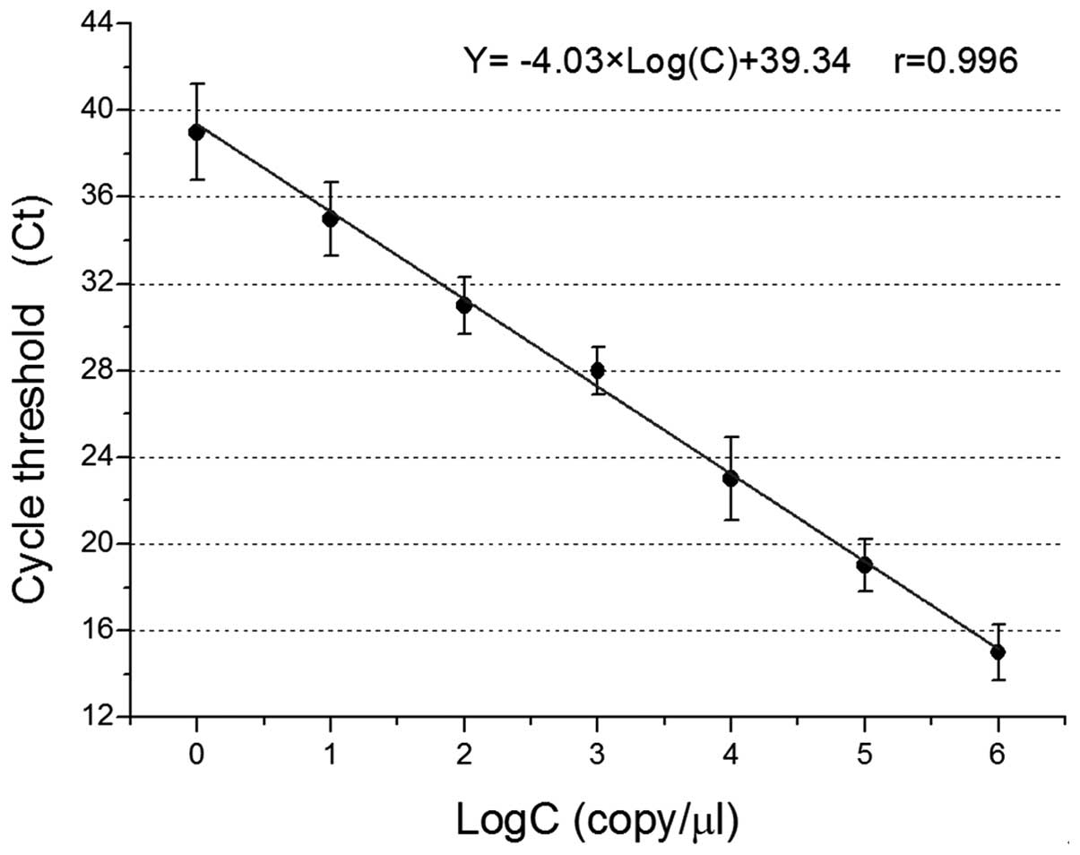
|
1
|
Lavaissiere L, Jia S, Nishiyama M, et al:
Overexpression of human aspartyl(asparaginyl)beta-hydroxylase in
hepatocellular carcinoma and cholangiocarcinoma. J Clin Invest.
98:1313–1323. 1996. View Article : Google Scholar : PubMed/NCBI
|
|
2
|
Korioth F, Gieffers C and Frey J: Cloning
and characterization of the human gene encoding aspartyl
beta-hydroxylase. Gene. 150:395–399. 1994. View Article : Google Scholar : PubMed/NCBI
|
|
3
|
Jones LR, Zhang L, Sanborn K, Jorgensen AO
and Kelley J: Purification, primary structure, and immunological
characterization of the 26-kDa calsequestrin binding protein
(junctin) from cardiac junctional sarcoplasmic reticulum. J Biol
Chem. 270:30787–30796. 1995. View Article : Google Scholar : PubMed/NCBI
|
|
4
|
Treves S, Feriotto G, Moccagatta L,
Gambari R and Zorzato F: Molecular cloning, expression, functional
characterization, chromosomal localization, and gene structure of
junctate, a novel integral calcium binding protein of
sarco(endo)plasmic reticulum membrane. J Biol Chem.
275:39555–39568. 2000. View Article : Google Scholar : PubMed/NCBI
|
|
5
|
Lee JH: Overexpression of humbug promotes
malignant progression in human gastric cancer cells. Oncol Rep.
19:795–800. 2008.PubMed/NCBI
|
|
6
|
Dinchuk JE, Henderson NL, Burn TC, et al:
Aspartyl beta-hydroxylase (Asph) and an evolutionarily conserved
isoform of Asph missing the catalytic domain share exons with
junctin. J Biol Chem. 275:39543–39554. 2000. View Article : Google Scholar : PubMed/NCBI
|
|
7
|
Ince N, de la Monte SM and Wands JR:
Overexpression of human aspartyl (asparaginyl) beta-hydroxylase is
associated with malignant transformation. Cancer Res. 60:1261–1266.
2000.PubMed/NCBI
|
|
8
|
Maeda T, Taguchi K, Aishima S, et al:
Clinicopathological correlates of aspartyl (asparaginyl)
beta-hydroxylase overexpression in cholangiocarcinoma. Cancer
Detect Prev. 28:313–318. 2004. View Article : Google Scholar
|
|
9
|
De la Monte SM, Tamaki S, Cantarini MC, et
al: Aspartyl-(asparaginyl)-beta-hydroxylase regulates
hepatocellular carcinoma invasiveness. J Hepatol. 44:971–983. 2006.
View Article : Google Scholar : PubMed/NCBI
|
|
10
|
Luu M, Sabo E, de la Monte SM, et al:
Prognostic value of aspartyl (asparaginyl)-beta-hydroxylase/humbug
expression in non-small cell lung carcinoma. Hum Pathol.
40:639–644. 2009. View Article : Google Scholar : PubMed/NCBI
|
|
11
|
Wang J, de la Monte SM, Sabo E, et al:
Prognostic value of humbug gene overexpression in stage II colon
cancer. Hum Pathol. 38:17–25. 2007. View Article : Google Scholar
|
|
12
|
Moshiri M, Lebowitz MS and Roberts SF:
Cancer biomarker, haah (human aspartyl (asparaginyl)
beta-hydroxylase), a companion diagnostic strategy. In: Proceedings
of the Fifth Oncology Biomarker Conference; Zurich. pp. 2332012
|
|
13
|
Silbermann E, Moskal P, Bowling N, Tong M
and de la Monte SM: Role of aspartyl-(asparaginyl)-beta-hydroxylase
mediated notch signaling in cerebellar development and function.
Behav Brain Funct. 6:682010. View Article : Google Scholar
|
|
14
|
Yang H, Song K, Xue T, et al: The
distribution and expression profiles of human Aspartyl/Asparaginyl
β-hydroxylase in tumor cell lines and human tissues. Oncol Rep.
24:1257–1264. 2010.PubMed/NCBI
|
|
15
|
Xue T, Xue XP, Huang QS, Wei L and Sun K:
Monoclonal antibodies against human aspartyl (asparaginyl)
beta-hydroxylase developed by DNA immunization. Hybridoma.
28:251–257. 2009. View Article : Google Scholar : PubMed/NCBI
|
|
16
|
Xian ZH, Zhang SH, Cong WM, Yan HX, Wang K
and Wu MC: Expression of aspartyl beta-hydroxylase and its
clinicopathological significance in hepatocellular carcinoma. Mod
Pathol. 19:280–286. 2006. View Article : Google Scholar
|
|
17
|
Lander ES, Linton LM, Birren B, et al:
Initial sequencing and analysis of the human genome. Nature.
409:860–921. 2001. View
Article : Google Scholar : PubMed/NCBI
|
|
18
|
Sepe PS, Lahousse SA, Gemelli B, et al:
Role of the aspartyl-asparaginyl-beta-hydroxylase gene in
neuroblastoma cell motility. Lab Invest. 82:881–891. 2002.
View Article : Google Scholar : PubMed/NCBI
|
|
19
|
Davila JA, Morgan RO, Shaib Y, McGlynn KA
and El Serag HB: Hepatitis C infection and the increasing incidence
of hepatocellular carcinoma: a population-based study.
Gastroenterology. 127:1372–1380. 2004. View Article : Google Scholar : PubMed/NCBI
|
|
20
|
Ho SP, Scully MS, Krauthauser CM, et al:
Antisense oligonucleotides selectively regulate aspartyl
beta-hydroxylase and its truncated protein isoform in vitro but
distribute poorly into A549 tumors in vivo. J Pharmacol Exp Ther.
302:795–803. 2002. View Article : Google Scholar : PubMed/NCBI
|
|
21
|
Fuller S, Stewart S, Lebowitz M, et al:
Immunogenicity of a lambda phage-based anti-cancer vaccine
targeting HAAH. J Immunother Cancer. 1:P2102013. View Article : Google Scholar
|
|
22
|
Wang K, Liu J, Yan ZL, et al:
Overexpression of aspartyl-(asparaginyl)-beta-hydroxylase in
hepatocellular carcinoma is associated with worse surgical outcome.
Hepatology. 52:164–173. 2010. View Article : Google Scholar : PubMed/NCBI
|
|
23
|
Tyagi S and Kramer FR: Molecular beacons:
probes that fluoresce upon hybridization. Nat Biotechnol.
14:303–308. 1996. View Article : Google Scholar : PubMed/NCBI
|
|
24
|
Huang Z, Zhou M and Wang L: Study on the
geographic distribution of liver cancer mortality and HBsAg carrier
rate in China. Disease Surveillance. 22:242–245. 2007.(In
Chinese).
|
|
25
|
Fan X, Zhao H and Zhang L: A 1:2 matched
case-control study on risk factors of hepatocellular carcinoma in
North Shaanxi. J Fourth Military Medical University. 23:891–895.
2002.(In Chinese).
|
|
26
|
Cantarini MC, De la Monte SM, Pang M, Tong
M, D’Errico A, Trevisani F and Wands JR: Aspartyl-asparagyl beta
hydroxylase over-expression in human hepatoma is linked to
activation of insulin-like growth factor and notch signaling
mechanisms. Hepatology. 44:446–457. 2006. View Article : Google Scholar : PubMed/NCBI
|