1. Introduction to nanotechnology
Nanotechnology is the management of material
properties at nanometer levels. The concept of nanotechnology was
first introduced by the American physicist Richard Feynman who gave
a talk entitled 'There's Plenty of Room at the Bottom' at an
American Physical Society meeting at Caltech in 1959. Feynman did
not mention 'nanotechnology', yet considered the potential to
manipulate individual atoms at the molecular level. This talk
inspired the conceptual beginning for nanotechnology science
through creating the concept of 'molecular manufacturing' (1). The prefix 'nano' derives from the
Greek word 'nanos' which means 'a dwarf'. A nanometer (nm) measures
one-billionth of a meter. Many biological structures are within the
nanometer size. While a single RBC is 2,500 nm, a DNA molecule is
2.5 nm, and while a protein molecule is 1–20 nm, the biggest amino
acid (tryptophan) is 1.2 nm (2).
Nanotechnology is defined as the creation and use of
structures, materials, devices and systems with completely novel
properties and functions obtained from their size which ranges from
1 to 100 nm. It is applied in many fields including electronics,
information technology, material developments and biomedicine
(3). Nanobiotechnology represents a
specific category of nanotechnology, based on biological molecules
and processes that involve the creation of devices and systems at
the nanolevel to examine or control biological processes (4,5).
Cancer nanotechnology is a striving field (6), current methods for cancer diagnosis
have limitations regarding the sensitivity and the ability to
detect cancer at early stages (7).
In addition, conventional treatment options have considerable
side-effects due to non-target tissue toxicity and their success is
limited in advanced disease stages due to the development of drug
resistance. An additional diagnostic and follow-up problem is
related to the detection of minimal residual diseases (MRD) after
treatment (7). Therefore, there is
an urgent need to develop novel therapies and cell type-specific
delivery systems to deliver anticancer therapies more effectively.
Hematological malignancies representing cancers that affect blood,
bone marrow and lymph nodes, are associated with these same
problems encountered with other types of cancer. Nanotechnology may
find novel solutions to all of these problems. New nanodevices can
perform different key functions such as detecting cancer cells at
the initial stages and determining their location in the body.
Nanotechnology, either alone or in combination with traditional
diagnostic methods, provides new sensitive, specific, reproducible
and cost-effective methods for diagnosis of different types of
cancers including hematological malignancies. In the field of
cancer therapy, nanodevices are used to carry drugs to specific
target cells and tumor sites, with nanoparticle-based drug delivery
approaches making remarkable success in site-specific release of
different therapies due to their unique physical, chemical and
biological features.
The advantages that nanotechnology offers include
targeted delivery of drugs, reduced dosage, reduced frequency of
dosing, better solubility, reduced immunogenicity and better
half-life of these drugs when used in vivo. Multiple types
of nanomaterials can be used to generate the nanoscale drug
delivery systems including lipid-based nanocapsules or liposomes
and natural or synthetic polymers. Liposomes are biocompatible
vesicles formed of amphipathic lipid bilayer membranes. Polymers
could be derived from biocompatible drug conjugates or micelles
that self-assemble to form a hydrophilic outer corona and a
hydrophobic inner core for drug encapsulation. The choices of
different nanoscale systems are based on the components,
applications, advantages and limitations of each preparation. These
novel nanotherapeutics are rapidly progressing as they offer
solutions to many limitations of conventional drug delivery systems
such as nonspecific targeting, limited biodistribution, poor
bioavailability and low therapeutic indices. Factors that determine
nanomaterial design and characterization include size and shape,
blood half-life, controlled drug release and active targeting of
nanoparticles. Efficient cell uptakes were reported with
nanoparticles with a size range of 40–50 nm (8). Studies examining the effects of
nanoparticle shapes demonstrated that rod-shaped micelles continue
in the circulation 10 times longer than spherical micelles
(9), while these spherical micelles
bind more tightly to circulating and leukemic cells (10). Moreover, nanoscale formulations can
be rendered multifunctional with the concept of combination therapy
in practice. In the following section, a number of applications of
nanotechnology in hematological malignancies will be discussed with
a particular emphasis on the utilization of nanotechnology in
diagnosis, treatment and follow-up of different types of leukemias
and lymphomas.
2. Nanotechnology applications in different
types of leukemia
Nanotechnology applications in acute
leukemias
Acute leukemias represent a group of cancers caused
by malignant transformation of hematopoietic cells and usually
associated with maintaining a partial capacity of differentiation
of the progenitor tumor initiating cells (11). In general, acute leukemias are
divided into acute myeloid (also called non-lymphocytic) leukemia
(AML) (12) and acute lymphoid
leukemia (ALL) (lymphoblastic leukemia/lymphoma) (13). ALL has a higher incidence in
children, while AML usually predominates in adults, yet age
profiles overlap. Sex distribution for AML is almost equal until
the age of 60, after which a considerable male predominance occurs.
The following section will discuss the different diagnostic and
therapeutic applications of nanotechnology in acute leukemia.
Aptamer-conjugated nanoparticles for
selective detection of leukemic cells
Diagnosis and classification of acute leukemias
depend on integration of information from morphologic and
cytochemical examination along with data obtained from
immunophenotyping analysis using flow cytometry (FCM) and
cytogenetic or molecular analyses of diagnostic genetic mutations
associated with prognosis (14). As
antibodies used in FCM may not detect all the molecular events
associated with the development of malignancy or other factors that
may be useful for clinical management, the study of cell surface
proteins with nanotechnology may improve treatment strategies.
Creating probes to detect surface proteins can be used to classify
tumors depending on the molecular features of these cells rather
than their tissues of origin (15).
In this regard, using molecular aptamers is a new strategy of
identifying novel biomarkers that can be used to increase
effectiveness of therapy and survival of leukemic patients
(16). Aptamers are synthetic
nucleic acid ligands that can be produced against many targets
including proteins, drugs and amino acids (17). Compared with antibodies, aptamers
have several advantages. To name a few, aptamers have a higher
affinity and are easily synthesized with limited toxicity. In
addition, aptamers can also fold into three-dimensional
conformations with unique ability to bind biomolecular ligands
(18). Making use of these
advantages, Herr et al (19)
have chosen an aptamer for acute leukemic cells with a specific
sequence. In this study, they created an assay using
aptamer-conjugated nanoparticles for quick recognition of acute
leukemic cells using high-affinity DNA aptamers for signal
detection. An oligonucleotide of 88 base pairs, which has the
capability of binding to acute leukemic cells was attached to
magnetic and fluorescent nanoparticles. The fluorescent
nanoparticles then provided a method of detection, while the use of
magnetic nanoparticles eliminated the need for centrifugation and
helped the removal of unwanted particles.
Enhanced leukemic cell detection using a
novel magnetic needle and nanoparticles
Detection of MRD in leukemia is essential for
follow-up of patients in order to change treatment strategies.
Although the utilization of morphological examination of blood and
bone marrow for detection of MRD has decreased, being replaced by
more sensitive techniques, it is still being used as one of the
easiest methods for detection of MRD (20). Morphological examination of
peripheral blood and bone marrow has been used for years for
detection of MRD. However, microscopic diagnosis is subject to the
examiner's interpretations and depends on the experience of the
hematologist, therefore distinguishing leukemic cells from normal
lympho-hematopoietic progenitors can be challenging (21). Traditional methods for detection of
MRD include FCM, polymerase chain reaction (PCR) as well as
immunoglobulin (Ig) and T-cell receptor (TCR) gene rearrangements.
These methods have a detection limit of 0.001% leukemic cells
therefore decreasing dependence on morphological examination for
detection of MRD (22). In order to
improve the sensitivity of detection of leukemic cells in the bone
marrow, Jaetao et al (23)
developed a new device that uses antibodies coupled to
superparamagnetic iron oxide nanoparticles (SPION) that were
directed against the acute leukemia antigen CD34 linked with a
magnetic needle biopsy. CD34-conjugated nanoparticles bound highly
to CD34 expressing cell lines. In addition, the magnetic needle
allowed the recognition of cells from cell lines and patient
leukemic cells diluted into normal blood at low concentrations.
They also observed that the magnetic needle improved the percentage
of blasts visible by light microscopy by 10-fold indicating that
using this needle may improve the detection levels for MRD
(23).
Therapeutic nanotechnology approaches in
acute leukemia
After approval of Doxil, a pegylated liposomal
doxorubicin by the US Food and Drug Administration (FDA) in 1995
for treatment of metastatic ovarian cancer and AIDS-related Kaposi
sarcoma, several other drugs with similar features have been
developed and were either approved or in different phases of
clinical trials (24).
Nanotechnology has a high potential for improving current
therapeutic approaches in leukemia. In 2012, vincristine sulfate
liposome injection (VSLI) was approved by FDA for treatment of
adult patients with Philadelphia chromosome-negative ALL when those
patients are in relapse or have progressive disease after two or
more anti-leukemia therapies (25).
Several other approaches have also been investigated. One of the
approaches tested by Krishnan et al (26) was the incorporation of
dexamethasone, which is commonly used for treatment of childhood
leukemia, in polymeric NPs and testing the ability of the conjugate
against leukemic cells in vitro and in vivo. The
cytotoxic activity of the complex was similar to that of free
dexamethasone. In vivo models have shown that the complex
enhanced the quality of life of mice in comparison with
dexamethasone alone and that NPs were cleared from the body with
time, providing evidence for the potential use of this method to
deliver dexamethasone in lower doses and more efficient forms.
Another approach is incorporating different compounds into lipid
nanoparticles. Lipid nanoparticles have many advantages as drug
carriers being biocompatible, biodegradable, with a low toxicity
and able to incorporate different drugs types. This approach was
tested by Rahman et al (27)
who loaded zerumbone, a natural dietary compound with an antitumor
effect and a low water solubility, into nano-structured lipid
carriers. The activity of the formulation was tested in human
T-cell ALL cell lines and has shown a sustained release and a
highly effective killing effect against these ALL cells.
Conjugation of drugs with colloids was also
investigated. Cosco et al (28) investigated the effect of linking
cytarabine (Ara-C) with one product of squalene which is a natural
product used as a precursor in the synthesis of sterols. The
product was assessed against different cancer cell lines (L1210,
K562 and MCF-7) and has shown an effect against resistant leukemic
cells (L1210R) compared with the naked compound as the conjugate
protected the drug from cellular metabolic degradation. These
results were also confirmed by an in vivo leukemic model in
which mice treated with the conjugate had a better survival rate,
than those treated with Ara-C alone.
Nanotechnology for reversal of multidrug
resistance (MDR) in leukemia
A major problem that represents an obstacle to the
success of treatment of leukemia is the development of MDR, which
is responsible for ~90% of cancer treatment failure (29). As MDR develops, cancer cells become
resistant to the cytotoxic effects of chemotherapeutic agents
(29). Using higher doses of these
chemotherapies does not overcome the effects of MDR. On the
contrary, it is frequently associated with several toxic effects
creating a need for the development of a new approach combining
conventional methods with new strategies to increase the delivery
and concentration of drugs in target tissues (30). Numerous mechanisms have been
implicated in the development of drug resistance in patients with
AML. One of the common mechanisms is the development of efflux
pumps, such as P-glycoprotein and MDR-associated protein (MRP)
(31). An approach that may be used
to overcome MDR is utilizing antibody drug conjugates (ADCs) by
linking an antibody (or an antibody fragment) to a cytotoxic drug
to improve the anticancer effect of antibodies and decrease the
toxicity of the conjugated drugs (32).
Chen et al (33) prepared
Fe3O4-MNPs (magnetic nanoparticles) loaded
with adriamycin and tetrandrine to interfere with MDR of K562/A02
cells. Measurement of several biological parameters has shown that
adriamycin- and tetrandrine-loaded Fe3O4-MNPs
can help in counteracting the effects of MDR in K562/A02 cells, due
to the buildup of drugs caused by its polymerization with
Fe3O4-MNPs. In another study, Chen et
al (34) synthesized
tetraheptylammonium-capped MNPs-Fe3O4 causing
an increase in the concentration of daunorubicin in MDR leukemia
K562/A02 cells and augmenting the therapeutic effect of
daunorubicin in MDR leukemic K562/A02 cells. This study also showed
that utilizing daunorubicin only is less sensitive than
daunorubicin associated with MNPs-Fe3O4.
In vitro cytotoxicity assays provided evidence that
MNPs-Fe3O4s are biocompatible materials.
Combination of Fe3O4-MNP with other
therapeutic forms can also represent an alternative strategy to
overcome MDR in leukemia. Magnetic drug targeting (MDT) is a novel
approach developed to increase the concentration of anticancer
drugs in the tumor and decrease the amount of leakage into the
surrounding healthy tissues (35).
Ren et al (3) investigated
this approach and combined Fe3O4-MNP with
hyperthermia and chemotherapy in nude mice harboring tumor
xenografts. Pathological examination of the tumors and markers of
apoptosis were assessed. According to their results, tumors became
smaller in size and apoptosis was observed in cells from the
treated group, providing evidence for the benefits of using this
approach to reverse MDR in leukemia.
Another promising approach to overcome MDR is the
use of nanodiamonds (NDs). Several advantages of NDs favor their
use in the field of medicine such as being inert, transparent,
having high surface area and being biocompatible (36). NDs can be used as carriers for drugs
or therapeutic nucleic acids. Man et al (37) adjusted different parameters such as
pH and concentration for loading daunorubicin into NDs. The
resultant complex was tested in K562 human leukemic cells, with MDR
induced by gradual exposure to daunorubicin. The results showed
that NDs could help the release of daunorubicin and overcome
mechanisms of drug efflux, which induce resistance, therefore
adding further evidence for the potential use of NDs to overcome
MDR. In another study, Ghoneum et al (38) examined the effects of using a
mixture of NDs and nanoplatinum (NP) known as DPV576 to overcome
MDR in MDR human myeloid leukemic cells (HL60/AR) and MDR-sensitive
cells (HL60). According to their results, the combination of
daunorubicin with DPV576 was associated with higher incidence of
the development of cell membrane holes in the MDR cells in
comparison with the control cells that allowed better delivery of
the drugs inside such cells.
Modifications of all-trans retinoic acid
for treatment of acute promyelocytic leukemia
Acute promyelocytic leukemia (APL) is a specific
subtype of AML. The hallmark of APL is the presence of specific
t(15;17) chromosomal translocation. The translocation occurs
between retinoic acid receptor-α (RARα) gene and the PML gene on
chromosomes 17 and 15, respectively (39). APL cells are induced by
all-trans retinoic acid (ATRA) to differentiate into mature
myeloid cells and ATRA may induce complete remission in APL
patients (40). However, several
complications appear to be associated with the use of ATRA
including hypertriglyceridermia, mucocutaneous dryness and
headache. In addition, ATRA resistance may develop due to lower
plasma concentration of the drug leading to recurrence of the
disease. Kim et al (41)
developed new nanoparticles based on creating a complex between low
molecular weight water-soluble chitosan (LMWSC) and ATRA. LMWSC has
many advantages, in addition to being water soluble, including ease
of modification and potential use as a gene or drug carrier. In
their study, Kim et al (41)
observed that ATRA was released from the nanoparticles for 10 days
and had superior effects on CT-26 colon carcinoma cells on day 1,
but the same cytotoxicity on day 2. These results provide
proof-of-concept of using LMWSC nanoparticles in the drug delivery
field. However, overcoming treatment resistance in APL is still a
challenge. Understanding the molecular mechanisms associated with
resistance and development of targeted therapy could be the
ultimate solution to this problem (42).
Nanotechnology applications in chronic
myeloid leukemia (CML)
Due to their unique properties, gold nanoparticles
(AuNPs) are among the interesting materials for use in the field of
nanotechnology. AuNPs have many optical properties that make them
suitable for bioimaging applications (43). AuNPs have superior absorption and
scattering properties than traditional organic dyes. The size and
shape of AuNPs affect their optical properties where they can
absorb light from visible to the near-infrared (NIR) region raising
interest for their medicinal use (44). Light absorption, scattering and
emission are enhanced in AuNPs due to the interaction of light with
the free electrons in the particles. Accordingly, the field of
light causes oscillation of the conduction band electrons at the
surface of the particles. This oscillation of free electrons in
resonance with the electro-magnetic field is called the surface
plasmon resonance (SPR), which is responsible for the unique
properties of AuNPs (43).
AuNPs have also proven to be biocompatible, have
limited toxicity and low susceptibility to photobleaching making
them suitable for use in studies with human cells (45). These exceptional optical properties
together with easy synthesis and functionality of AuNPs with
different biomolecules allow the use of AuNPs for molecular assays
and permit the detection of very low concentrations of nucleic
acids, therefore providing five times higher sensitivity than
fluorescence based techniques (46). Using gold nanoparticles in molecular
assays can improve current technique performances or help in the
development of completely new assays, which can be faster, more
sensitive and less expensive. This is of great importance
particularly for use in developing countries (45,47).
CML is one of the examples in which AuNPs have been
used for molecular diagnosis. CML is one of the most common
myeloproliferative disorders. It is a clonal disorder of
hematopoietic stem cells that passes through three stages. The
first phase is the chronic phase characterized by a marked increase
in the total leukocytic count with predominance of progenitors and
mature cells of myeloid nature. With time, the disease progresses
into an accelerated and finally a blastic phase characterized by
maturation arrest concomitant with the appearance of an increased
number of blasts (48). Most CML
patients, ~90%, have an acquired translocation between the long
arms of chromosomes 9 and 22 t(9;22 q34;q11) resulting in what is
known as the Philadelphia chromosome (Ph). The translocation
results in the formation of fusion gene between BCR and ABL genes
with production of fusion protein that has a greater tyrosine
kinase activity than normal. The new gene encodes p210 BCR-ABL1
protein, instead of the p145 Abelson protein causing growth factor
independence and unlimited growth of leukemic cells (49). Common procedures for the diagnosis
of Philadelphia chromosome include karyotype analysis, fluorescence
in-situ hybridization (FISH) as well as reverse
transcriptase polymerase chain reaction analyses (RT-PCR). However,
those methods are costly and time-consuming creating the need for
developing methods that are sensitive and cost effective at the
same time (50).
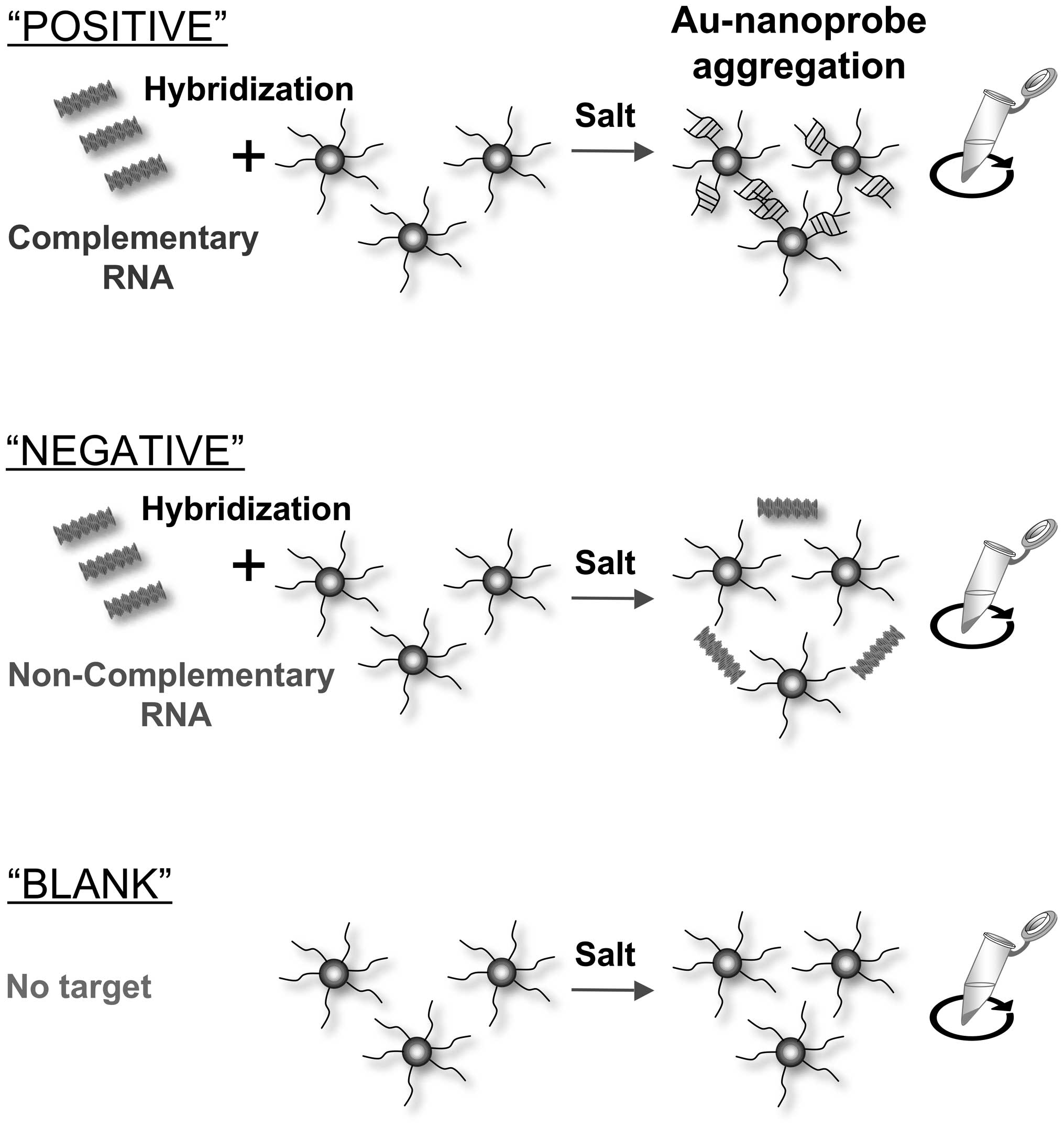
Conde et al (51) presented an Au-nanoparticle-based
method for the diagnosis and quantification of the BCR-ABL fusion
transcript associated with CML. The technique depends on the
optical properties of AuNPs. According to the size, Au-nanoprobes
absorb light in different regions of the spectrum. Au-nanoprobes of
13 nm are red and have a narrow SPR band ~520 nm. On the other
hand, when aggregated Au-nanoprobes are present, the solution is
blue. The colors of the solutions are compared before and after
aggregation induced by salt either visually and/or using
spectroscopy. As shown in Fig. 1,
the existence of a complementary target does not allow aggregation
and the solution appears red (SPR peak at ±520 nm), while the
absence of this target allows aggregation of Au-nanoprobes,
associated with alteration of color from red into blue (red-shift
of the SPR peak to 600–650 nm).
The same team worked on another idea within the same
scope. Baptista et al (52)
have developed nanoparticles in the form of an alloy with different
metal compositions and have linked them with different
thiol-modified single stranded DNA (nanoprobes). This was used for
simultaneous detection of multiple targets depending on the
different colors detected by the different alloy composition. In
this study, the targets detected were various gene products in CML;
ABL, BCR and BCR-ABL fusion product allowing identification of
multiple targets at the same time. The presence of BCR-ABL fusion
gene transcript also provided the base for targeted therapy for
CML. Imatinib mesylate (IM), a tyrosine kinase inhibitor which was
introduced in 1998, acts selectively against the oncoprotein
BCR-ABL with a high success rate (53). Despite major hematological and
cytogenetic responses achieved by IM, resistance to IM develops in
a significant number of patients, resulting in disease relapse
(54). Palamà et al
(55) developed new polyelectrolyte
complexes to act as carriers for IM in KU812 CML cell line and
CD34-positive cells collected from patients. The results of these
studies have shown that the used complexes allowed longer BCR-ABL
kinase inactivation even at lower doses of IM compared with the
previously formed microscale formulation polyelectrolyte
microcapsules, giving hope that such complexes can be used with
success to overcome drug resistance and prevent relapse in CML
patients (55). A major limitation
to this approach is that failure of therapy in most CML cases is
due to persistence of leukemic stem cells (LSCS) in which tyrosine
kinase therapy (TKI) is not effective. A better understanding of
the pathways regulating LSC survival and the mechanisms involved in
disease progression is a major need to overcome resistance of
therapy and relapse (56).
Nanotechnology applications in B-chronic
lymphocytic leukemia (B-CLL)
Chronic lymphocytic leukemia (CLL) is a clonal
disorder characterized by accumulation of mature-appearing
lymphocytes in the peripheral blood, bone marrow, lymph nodes as
well as the spleen. CLL cells are frequently monoclonal
B-lymphocytes that express CD19, CD5 and CD23, with weak or no
expression of surface immunoglobulin (Ig), CD20, CD79b and FMC7
(57). CLL cells are also
characterized by resistance to apoptosis. Although most CLL cells
are non-replicating cells in G0 phase of the cell cycle, a small
percentage of cells are replicating and these account for disease
progression. The arrest of cells at this stage might explain the
resistance of these CLL cells to cytotoxic drugs, which work
through induction of apoptosis. Bcl-2 overexpression is one of the
specific genes that is overexpressed in CLL and may induce such
resistance (58).
The other finding that can affect growth and
development of CLL is the presence of abnormal vascularization. CLL
cells manufacture and release vascular endothelial growth factor
(VEGF) and express VEGF membrane receptors (VEGF-R1 and VEGF-R2).
VEGF also plays a significant role in CLL cells resistance to
apoptosis (59). These findings can
create the base for using targeted therapy with anti-VEGF
antibodies (avastin; bevacizumab) to induce apoptosis in CLL cells.
It has been shown that avastin selectively induces apoptosis in CLL
cell through the mitochondrial pathway of caspase activation while
sparing normal lymphocytes (60).
However, a limitation to such an approach is the high concentration
of the antibody necessary to achieve reasonable effects (58). Mukherjee et al (58) studied the functionality of gold
nanoparticles with anti-VEGF antibodies on the release of the drug.
In their study, all samples examined have shown a response to
gold-AbVF that was considerably superior to CLL cells exposed to
only AbVF or GNP with downregulation of anti apoptotic proteins.
GNP alone has shown some response with induction of apoptosis in
some of these CLL cells. Phase II clinical studies conducted on 46
CLL patients concluded that while anti-VEGF therapy, including
avastin, remains a viable therapy for CLL, using a single agent
anti-VEGF monotherapy had limited activity in CLL patients, and
combination therapy is a more feasible approach particularly for
patients with relapsed/refractory CLL (61). The combination of monoclonal
antibodies, such as anti-CD20 or rituximab, with other types of
chemotherapy is another promising approach for treatment in CLL
(62).
Another strategy, developed later by Yu et al
(63), recognized that anti-CD37
monoclonal antibody immunoliposomes could be used as carriers for
specific targeting of B-CLL cells. Anti-CD19 or anti-CD20 was
combined with anti-CD37 to form dual immunoliposomes that were used
to induce apoptosis in B-CLL cells. Their results have proven that
using the dual liposomes have higher effects regarding induction of
apoptosis than using either anti-CD19 immunoliposome or anti-CD20
immunoliposome suggesting that this strategy can be favorable for
personalized treatment of B-CLL and B-cell malignancies in
general.
3. Nanotechnology applications in
lymphomas
Nanotechnology applications in Hodgkin
lymphoma (HL)
CD30 is an antigen that is expressed on T-cells,
activated B-cells as well as natural killer cells and has a very
low expression on normal cells. Therefore, CD30 is an ideal target
in classical HL. Brentuximab vedotin (BV) is a CD30-directed
antibody-drug conjugate that received approval from FDA for the
treatment of patients with HL after relapse (64). CD30 has also been investigated as a
target for photothermal therapy using gold nanoparticles. Gold
nanoparticles have several advantages that enhance their potential
as important agents in nanotechnology. In addition to their
absorption and scattering properties, gold nanoparticles can absorb
light and switch it into heat. This property can be used to induce
killing of cancer cells through protein denaturation and induction
of apoptosis. Photothermal therapy also allows for monitoring of
the process that eventually will lead to the death of the cancer
cells (65). Accordingly, Zharov
et al made use of this property for photothermal therapy of
HL. They developed two gold nanoparticle-antibody conjugates, one
of them was combined with an anti-CD30 receptor which binds to CD30
on the surface of L-428 Hodgkin cells and the other with an
anti-CD25-receptor as a control. High killing power was achieved
using appropriate doses of laser irradiation and gold concentration
for gold-targeted L-428 cells with little to no effect on
neighboring non-targeted cancer cells. These data further support
previous findings for the potential use of gold nanoparticles as a
safe modality for treatment of cancer.
Nanotechnology applications in anaplastic
large cell lymphoma (ALCL)
ALCL is a type of T-cell lymphoma with aggressive
features and poor prognosis. One of the main features that
characterize ALCL cells is abnormal expression of the anaplastic
lymphoma kinase (ALK) together with the surface expression of CD30.
Based on the presence or the absence of ALK translocation, the
tumor is classified into either ALK-positive or -negative-ALCL. The
presence of these unique and specific features can provide the base
of targeted therapy for treatment of ALCL (66). For example, BV has been approved, in
addition to its role in treatment of HL, for treatment of relapsed
systemic ALCL (67). Crizotinib, an
ALK inhibitor, is another FDA-approved drug for treatment of
ALK-positive anaplastic large-cell lymphoma. However, resistance to
crizotinib may develop limiting its use for long-term therapy
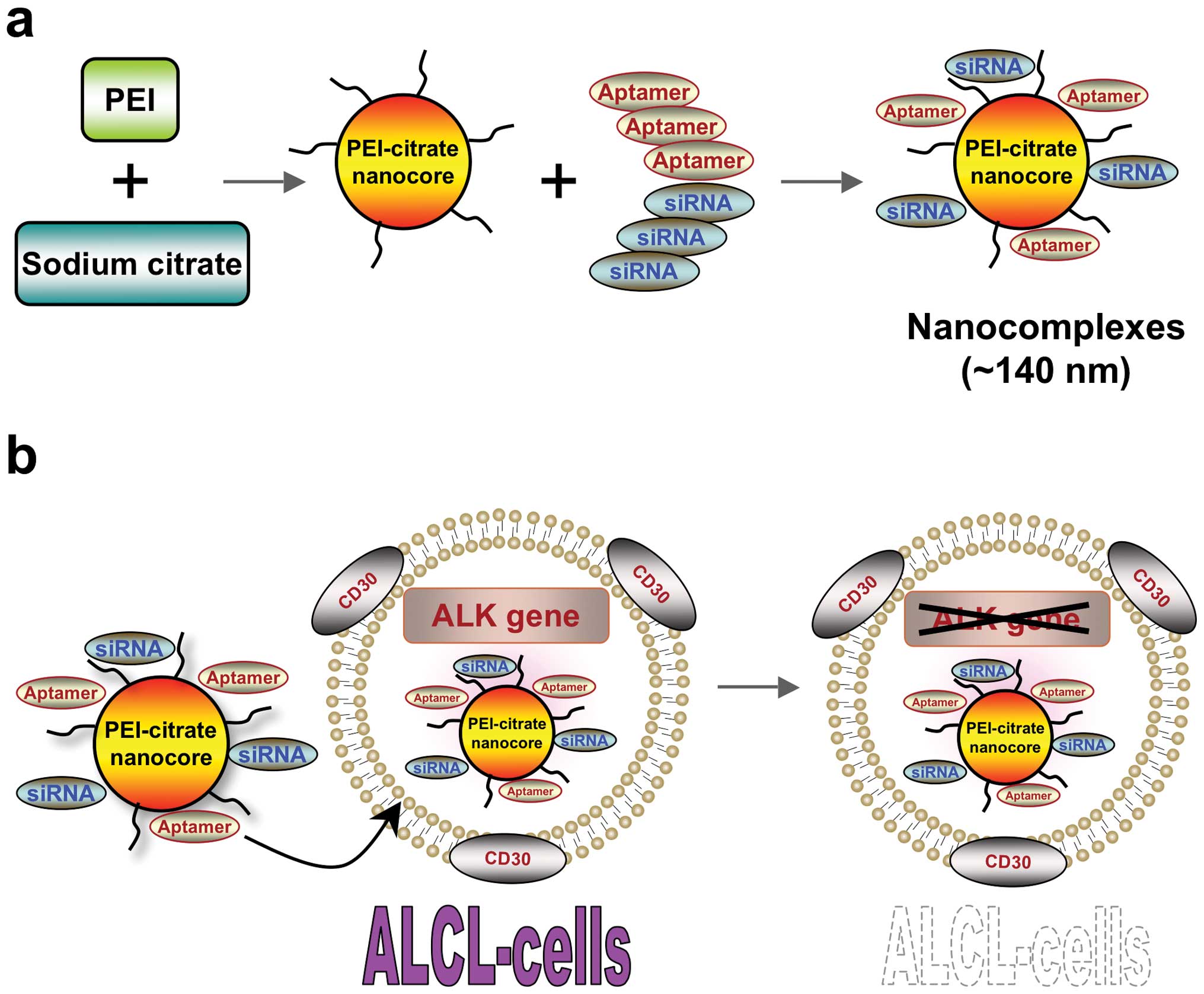
(68). Using an approach that
combines both characteristics, Zhao et al (69) have developed an RNA aptamer, which
binds only to CD30 protein in solution. The complex produced was
formed by inserting an RNA-based CD30 aptamer probe and ALK siRNA
into polyethyleneimine-citrate carriers in nanosize (Fig. 2). According to their results, this
complex allowed silencing of the expression of ALK gene in ALCL
cells, stopping the growth of these cells with induction of
apoptosis.
Nanotechnology applications in mantle
cell lymphoma
Mantle cell lymphoma (MCL) is a high-risk subtype of
NHL characterized by an aggressive clinical course and by having
the worst prognosis among B-cell NHL (70). These characters create a need for
development of new agents and targets against this type of cancer.
One of the agents that has attracted attention is lenalidomide, an
immunomodulatory agent, that has been tested with some success for
treatment of refractory MCL (71).
Using high-throughput techniques can provide a better understanding
of the mechanisms underlying excessive proliferation and apoptotic
resistance in MCL, thus allowing the identification of new targets
for therapy (72). One of these
potential targets is SYK, which is one of the factors that control
apoptosis. Overexpression of SYK is associated with several B-cell
malignancies including MCL. Cely et al (73) designed a nanotechnology platform
targeting specifically a SYK inhibitor in the MCL cells. According
to their results, the developed liposomal nanoparticles (NP) loaded
with a product called compound 61 (C-61) were able to induce
apoptosis of the MCL cells in one day offering the base for using
this therapeutic innovation against a large spectrum of lymphoid
malignancies, including MCL.
4. Conclusions and future prospects
Nanotechnology represents a promising technology
that can be of great value in the war against cancer. However,
attention should be put towards safety issues and ethical concerns.
Since particles at the nanosize have unique physical and chemical
properties, care must be taken when dealing with particles or
products for in vivo applications. Possible changes in the
reactions of the particles with cells of the body may occur and
changes in the absorption and secretion properties of such
particles should be well investigated. FDA has recommended that
meticulous research should be conducted in areas related to these
novel properties that may cause toxicity. FDA also recommended that
further research for development of devices and methods that can be
used for measurement of nanoscale materials should be developed.
Another important issue that is related to nanotechnology, being an
emerging technology, is the ethical concern. In many cases, as
science leaps forward, ethics lag behind. Part of the funding
should be directed towards ethical, legal and social issues related
to nanotechnology. One of the most famous examples for this
readiness was during the Human Genome Project when James Watson
suggested that 3–5% of the budget be devoted for study of ethical,
legal and social issues.
The future prospects of nanotechnology and
nanomedicine are very promising. Nanotechnology development for
targeted therapy of cancer is one of the most interesting areas in
research currently underway where drugs are delivered specifically
to the diseased tissues or cells of interest without affecting
nearby normal cells. Development of biomarkers for early detection
and imaging of cancer cells is another important topic for research
using nanotechnology. Scientists hope that progress in proteomics
and bioinformatics can be used by nanotechnology to identify and
kill malignant cells with the highest efficacy and the least
possible side-effects, therefore bringing us closer to the goal of
more effective cancer care.
Acknowledgments
We would like to thank Dr Joseph Bertino and Dr
Robert DiPaola (Rutgers Cancer Institute of New Jersey) and Mervat
El Ansary (Cairo University School of Medicine) for their helpful
discussions. We also appreciate the excellent work of all the
personnel at the Central Hematology Laboratory, Department of
Clinical Pathology, Faculty of Medicine and National Cancer
Institute, Cairo University. The authors of this review are
generously supported by Rutgers Cancer Institute of New Jersey, New
Jersey Health Foundation, Cairo University, and the Egyptian
Academy for Scientific Research and Technology. H.E.S. is a founder
and stockholder, and H.G. is a collaborator of Celvive, Inc., a
company with a profile focused on stem cell therapy technologies.
No products or off label use of any products of Celvive have been
discussed in this review.
References
|
1
|
Feynman R: There's plenty of room at the
bottom: An invitation to enter a new field of physics. Caltech
Engineering and Science. 23(5): 22–36. 1959.
|
|
2
|
Jain KK: Introduction to nanomedicine. The
handbook of nanomedicine. Jain KK: Hamana press; pp. 1–5. 2008,
View Article : Google Scholar
|
|
3
|
Ren Y, Zhang H, Chen B, Cheng J, Cai X,
Liu R, Xia G, Wu W, Wang S, Ding J, et al: Multifunctional magnetic
Fe3O4 nanoparticles combined with
chemotherapy and hyperthermia to overcome multidrug resistance. Int
J Nanomed. 7:2261–2269. 2012.
|
|
4
|
Fakruddin M, Hossain Z and Afroz H:
Prospects and applications of nanobiotechnology: A medical
perspective. J Nanobiotechnol. 10:312012. View Article : Google Scholar
|
|
5
|
Kim ES, Ahn EH, Chung E and Kim DH: Recent
advances in nanobiotechnology and high-throughput molecular
techniques for systems biomedicine. Mol Cells. 36:477–484. 2013.
View Article : Google Scholar : PubMed/NCBI
|
|
6
|
Cai W, Gao T, Hong H and Sun J:
Applications of gold nanoparticles in cancer nanotechnology.
Nanotechnol Sci Appl. 1:17–32. 2008.PubMed/NCBI
|
|
7
|
Bharali DJ and Mousa SA: Emerging
nanomedicines for early cancer detection and improved treatment:
Current perspective and future promise. Pharmacol Ther.
128:324–335. 2010. View Article : Google Scholar : PubMed/NCBI
|
|
8
|
Jiang W, Kim BY, Rutka JT and Chan WC:
Nanoparticle-mediated cellular response is size-dependent. Nat
Nanotechnol. 3:145–150. 2008. View Article : Google Scholar : PubMed/NCBI
|
|
9
|
Geng Y, Dalhaimer P, Cai S, Tsai R, Tewari
M, Minko T and Discher DE: Shape effects of filaments versus
spherical particles in flow and drug delivery. Nat Nanotechnol.
2:249–255. 2007. View Article : Google Scholar
|
|
10
|
Verma A and Stellacci F: Effect of surface
properties on nanoparticle-cell interactions. Small. 6:12–21. 2010.
View Article : Google Scholar
|
|
11
|
Olsen RJ, Chang CC, Herrick JL, Zu Y and
Ehsan A: Acute leukemia immunohistochemistry: A systematic
diagnostic approach. Arch Pathol Lab Med. 132:462–475.
2008.PubMed/NCBI
|
|
12
|
Ferrara F and Schiffer CA: Acute myeloid
leukaemia in adults. Lancet. 381:484–495. 2013. View Article : Google Scholar : PubMed/NCBI
|
|
13
|
Inaba H, Greaves M and Mullighan CG: Acute
lymphoblastic leukaemia. Lancet. 381:1943–1955. 2013. View Article : Google Scholar : PubMed/NCBI
|
|
14
|
Salem DA and Abd El-Aziz SM:
Flowcytometric immunopheno-typic profile of acute leukemia:
Mansoura experience. Indian J Hematol Blood Transfus. 28:89–96.
2012. View Article : Google Scholar :
|
|
15
|
Sefah K, Tang ZW, Shangguan DH, Chen H,
Lopez-Colon D, Li Y, Parekh P, Martin J, Meng L, Phillips JA, et
al: Molecular recognition of acute myeloid leukemia using aptamers.
Leukemia. 23:235–244. 2009. View Article : Google Scholar : PubMed/NCBI
|
|
16
|
Yang M, Jiang G, Li W, Qiu K, Zhang M,
Carter CM, Al-Quran SZ and Li Y: Developing aptamer probes for
acute myelogenous leukemia detection and surface protein biomarker
discovery. J Hematol Oncol. 7:52014. View Article : Google Scholar : PubMed/NCBI
|
|
17
|
Mallikaratchy PR, Ruggiero A, Gardner JR,
Kuryavyi V, Maguire WF, Heaney ML, McDevitt MR, Patel DJ and
Scheinberg DA: A multivalent DNA aptamer specific for the B-cell
receptor on human lymphoma and leukemia. Nucleic Acids Res.
39:2458–2469. 2011. View Article : Google Scholar :
|
|
18
|
Lakhin AV, Tarantul VZ and Gening LV:
Aptamers: Problems, solutions and prospects. Acta Naturae. 5:34–43.
2013.
|
|
19
|
Herr JK, Smith JE, Medley CD, Shangguan D
and Tan W: Aptamer-conjugated nanoparticles for selective
collection and detection of cancer cells. Anal Chem. 78:2918–2924.
2006. View Article : Google Scholar : PubMed/NCBI
|
|
20
|
Shook D, Coustan-Smith E, Ribeiro RC,
Rubnitz JE and Campana D: Minimal residual disease quantitation in
acute myeloid leukemia. Clin Lymphoma Myeloma. 9(Suppl 3):
S281–S285. 2009. View Article : Google Scholar : PubMed/NCBI
|
|
21
|
Campana D and Coustan-Smith E:
Measurements of treatment response in childhood acute leukemia.
Korean J Hematol. 47:245–254. 2012. View Article : Google Scholar
|
|
22
|
Campana D: Minimal residual disease in
acute lymphoblastic leukemia. Hematology Am Soc Hematol Educ
Program. 2010:7–12. 2010. View Article : Google Scholar
|
|
23
|
Jaetao JE, Butler KS, Adolphi NL, Lovato
DM, Bryant HC, Rabinowitz I, Winter SS, Tessier TE, Hathaway HJ,
Bergemann C, et al: Enhanced leukemia cell detection using a novel
magnetic needle and nanoparticles. Cancer Res. 69:8310–8316. 2009.
View Article : Google Scholar : PubMed/NCBI
|
|
24
|
Pillai JJ, Thulasidasan AK, Anto RJ,
Chithralekha DN, Narayanan A and Kumar GS: Folic acid conjugated
cross-linked acrylic polymer (FA-CLAP) hydrogel for site specific
delivery of hydrophobic drugs to cancer cells. J Nanobiotechnol.
12:252014. View Article : Google Scholar
|
|
25
|
Davis T and Farag SS: Treating relapsed or
refractory Philadelphia chromosome-negative acute lymphoblastic
leukemia: Liposome-encapsulated vincristine. Int J Nanomed.
8:3479–3488. 2013.
|
|
26
|
Krishnan V, Xu X, Barwe SP, Yang X,
Czymmek K, Waldman SA, Mason RW, Jia X and Rajasekaran AK:
Dexamethasone-loaded block copolymer nanoparticles induce leukemia
cell death and enhance therapeutic efficacy: A novel application in
pediatric nanomedicine. Mol Pharm. 10:2199–2210. 2013. View Article : Google Scholar
|
|
27
|
Rahman HS, Rasedee A, How CW, Abdul AB,
Zeenathul NA, Othman HH, Saeed MI and Yeap SK: Zerumbone-loaded
nanostructured lipid carriers: Preparation, characterization, and
antileukemic effect. Int J Nanomed. 8:2769–2781. 2013. View Article : Google Scholar
|
|
28
|
Cosco D, Rocco F, Ceruti M, Vono M, Fresta
M and Paolino D: Self-assembled squalenoyl-cytarabine
nanostructures as a potent nanomedicine for treatment of leukemic
diseases. Int J Nanomed. 7:2535–2546. 2012.
|
|
29
|
Lai BB, Chen BA, Cheng J, Gao F, Xu WL,
Ding JH, Gao C, Sun XC, Li GH, Chen WJ, et al: Daunorubicin-loaded
magnetic nanoparticles of Fe3O4 greatly
enhance the responses of multi-drug-resistant K562 leukemic cells
in a nude mouse xenograft model to chemotherapy. Zhongguo Shi Yan
Xue Ye Xue Za Zhi. 17:345–351. 2009.PubMed/NCBI
|
|
30
|
He Q, Gao Y, Zhang L, Zhang Z, Gao F, Ji
X, Li Y and Shi J: A pH-responsive mesoporous silica
nanoparticles-based multi-drug delivery system for overcoming
multi-drug resistance. Biomaterials. 32:7711–7720. 2011. View Article : Google Scholar : PubMed/NCBI
|
|
31
|
De Boer AB, De Lange EL, Van der Sandt IC
and Breimer DD: Transporters and the blood-brain barrier (BBB). Int
J Clin Pharmacol Ther. 36:14–15. 1998.PubMed/NCBI
|
|
32
|
Cianfriglia M: Targeting
MDR1-P-glycoprotein (MDR1-Pgp) in immunochemotherapy of acute
myeloid leukemia (AML). Ann Ist Super Sanita. 49:190–208.
2013.PubMed/NCBI
|
|
33
|
Chen B, Sun Q, Wang X, Gao F, Dai Y, Yin
Y, Ding J, Gao C, Cheng J, Li J, et al: Reversal in multidrug
resistance by magnetic nanoparticle of Fe3O4
loaded with adriamycin and tetrandrine in K562/A02 leukemic cells.
Int J Nanomed. 3:277–286. 2008.
|
|
34
|
Chen BA, Lai BB, Cheng J, Xia GH, Gao F,
Xu WL, Ding JH, Gao C, Sun XC, Xu CR, et al: Daunorubicin-loaded
magnetic nanoparticles of Fe3O4 overcome
multidrug resistance and induce apoptosis of K562-n/VCR cells in
vivo. Int J Nanomed. 4:201–208. 2009. View Article : Google Scholar
|
|
35
|
Janko C, Dürr S, Munoz LE, Lyer S, Chaurio
R, Tietze R, Löhneysen S, Schorn C, Herrmann M and Alexiou C:
Magnetic drug targeting reduces the chemotherapeutic burden on
circulating leukocytes. Int J Mol Sci. 14:7341–7355. 2013.
View Article : Google Scholar : PubMed/NCBI
|
|
36
|
Zhu Y, Li J, Li W, Zhang Y, Yang X, Chen
N, Sun Y, Zhao Y, Fan C and Huang Q: The biocompatibility of
nanodiamonds and their application in drug delivery systems.
Theranostics. 2:302–312. 2012. View Article : Google Scholar : PubMed/NCBI
|
|
37
|
Man HB, Kim H, Kim HJ, Robinson E, Liu WK,
Chow EK and Ho D: Synthesis of nanodiamond-daunorubicin conjugates
to overcome multidrug chemoresistance in leukemia. Nanomedicine.
10:359–369. 2014. View Article : Google Scholar :
|
|
38
|
Ghoneum A, Sharma S and Gimzewski J:
Nano-hole induction by nanodiamond and nanoplatinum liquid, DPV576,
reverses multidrug resistance in human myeloid leukemia (HL60/AR).
Int J Nanomed. 8:2567–2573. 2013. View Article : Google Scholar
|
|
39
|
Grignani F, Testa U, Fagioli M, Barberi T,
Masciulli R, Mariani G, Peschle C and Pelicci PG: Promyelocytic
leukemia-specific PML-retinoic acid alpha receptor fusion protein
interferes with erythroid differentiation of human erythroleukemia
K562 cells. Cancer Res. 55:440–443. 1995.PubMed/NCBI
|
|
40
|
Imaizumi M, Suzuki H, Yoshinari M, Sato A,
Saito T, Sugawara A, Tsuchiya S, Hatae Y, Fujimoto T, Kakizuka A,
et al: Mutations in the E-domain of RARα portion of the PML/RARα
chimeric gene may confer clinical resistance to all-trans retinoic
acid in acute promyelocytic leukemia. Blood. 92:374–382.
1998.PubMed/NCBI
|
|
41
|
Kim DG, Jeong YI, Choi C, Roh SH, Kang SK,
Jang MK and Nah JW: Retinol-encapsulated low molecular
water-soluble chitosan nanoparticles. Int J Pharm. 319:130–138.
2006. View Article : Google Scholar : PubMed/NCBI
|
|
42
|
Fung TK and So CW: Overcoming treatment
resistance in acute promyelocytic leukemia and beyond. Oncotarget.
4:1128–1129. 2013.PubMed/NCBI
|
|
43
|
Huang X, Jain PK, El-Sayed IH and El-Sayed
MA: Gold nanoparticles: Interesting optical properties and recent
applications in cancer diagnostics and therapy. Nanomedicine.
2:681–693. 2007. View Article : Google Scholar : PubMed/NCBI
|
|
44
|
Choi YE, Kwak JW and Park JW:
Nanotechnology for early cancer detection. Sens Basel. 10:428–455.
2010. View Article : Google Scholar
|
|
45
|
Radwan SH and Azzazy HM: Gold
nanoparticles for molecular diagnostics. Expert Rev Mol Diagn.
9:511–524. 2009. View Article : Google Scholar : PubMed/NCBI
|
|
46
|
Hwang SH, Im SG, Hah SS, Cong VT, Lee EJ,
Lee YS, Lee GK, Lee DH and Son SJ: Effects of upconversion
nanoparticles on polymerase chain reaction. PLoS One. 8:e734082013.
View Article : Google Scholar : PubMed/NCBI
|
|
47
|
Gormally E, Vineis P, Matullo G, Veglia F,
Caboux E, Le Roux E, Peluso M, Garte S, Guarrera S, Munnia A, et
al: TP53 and KRAS2 mutations in plasma DNA of healthy subjects and
subsequent cancer occurrence: A prospective study. Cancer Res.
66:6871–6876. 2006. View Article : Google Scholar : PubMed/NCBI
|
|
48
|
Silver RT, Woolf SH, Hehlmann R, Appelbaum
FR, Anderson J, Bennett C, Goldman JM, Guilhot F, Kantarjian HM,
Lichtin AE, et al: An evidence-based analysis of the effect of
busulfan, hydroxyurea, interferon, and allogeneic bone marrow
transplantation in treating the chronic phase of chronic myeloid
leukemia: Developed for the American Society of Hematology. Blood.
94:1517–1536. 1999.PubMed/NCBI
|
|
49
|
Shet AS, Jahagirdar BN and Verfaillie CM:
Chronic myelogenous leukemia: Mechanisms underlying disease
progression. Leukemia. 16:1402–1411. 2002. View Article : Google Scholar : PubMed/NCBI
|
|
50
|
Gabert J, Beillard E, van der Velden VH,
Bi W, Grimwade D, Pallisgaard N, Barbany G, Cazzaniga G, Cayuela
JM, Cavé H, et al: Standardization and quality control studies of
'real-time' quantitative reverse transcriptase polymerase chain
reaction of fusion gene transcripts for residual disease detection
in leukemia - a Europe Against Cancer program. Leukemia.
17:2318–2357. 2003. View Article : Google Scholar : PubMed/NCBI
|
|
51
|
Conde J, Doria G and Baptista P: Noble
metal nanoparticles applications in cancer. J Drug Deliv.
2012:7510752012. View Article : Google Scholar
|
|
52
|
Baptista PV, Doria G, Quaresma P, Cavadas
M, Neves CS, Gomes I, Eaton P, Pereira E and Franco R:
Nanoparticles in molecular diagnostics. Prog Mol Biol Transl Sci.
104:427–488. 2011. View Article : Google Scholar : PubMed/NCBI
|
|
53
|
El-Metnawy WH, Mattar MM, El-Nahass YH,
Samra MA, Abdelhamid HM, Abdlfattah RM and Hamed AR: Predictive
value of pretreatment BCR-ABL (IS) transcript level on response to
imatinib therapy in Egyptian patients with chronic phase chronic
myeloid leukemia (CPCML). Int J Biomed Sci. 9:48–53.
2013.PubMed/NCBI
|
|
54
|
Kang Y, Hodges A, Ong E, Roberts W,
Piermarocchi C and Paternostro G: Identification of drug
combinations containing imatinib for treatment of BCR-ABL+
leukemias. PLoS One. 9:e1022212014. View Article : Google Scholar : PubMed/NCBI
|
|
55
|
Palamà IE, Coluccia AM and Gigli G: Uptake
of imatinib-loaded polyelectrolyte complexes by BCR-ABL+
cells: A long-acting drug-delivery strategy for targeting
oncoprotein activity. Nanomedicine. 9:2087–2098. 2014. View Article : Google Scholar
|
|
56
|
Jamieson CH: Chronic myeloid leukemia stem
cells. Hematology Am Soc Hematol Educ Program. 2008:436–442. 2008.
View Article : Google Scholar
|
|
57
|
Vilpo J, Hulkkonen J, Hurme M and Vilpo L:
Surface membrane antigen expression changes induced in vitro by
exogenous growth factors in chronic lymphocytic leukemia cells.
Leukemia. 16:1691–1698. 2002. View Article : Google Scholar : PubMed/NCBI
|
|
58
|
Mukherjee P, Bhattacharya R, Bone N, Lee
YK, Patra CR, Wang S, Lu L, Secreto C, Banerjee PC, Yaszemski MJ,
et al: Potential therapeutic application of gold nanoparticles in
B-chronic lymphocytic leukemia (BCLL): Enhancing apoptosis. J
Nanobiotechnol. 5:42007. View Article : Google Scholar
|
|
59
|
Lee YK, Bone ND, Strege AK, Shanafelt TD,
Jelinek DF and Kay NE: VEGF receptor phosphorylation status and
apoptosis is modulated by a green tea component,
epigallocatechin-3-gallate (EGCG), in B-cell chronic lymphocytic
leukemia. Blood. 104:788–794. 2004. View Article : Google Scholar : PubMed/NCBI
|
|
60
|
Bogusz J, Majchrzak A, Mędra A,
Cebula-Obrzut B, Robak T and Smolewski P: Mechanisms of action of
the anti-VEGF monoclonal antibody bevacizumab on chronic
lymphocytic leukemia cells. Postepy Hig Med Dosw Online.
67:107–118. 2013. View Article : Google Scholar : PubMed/NCBI
|
|
61
|
Shanafelt TD, Gunderson H and Call TG:
Commentary: Chronic lymphocytic leukemia - the price of progress.
Oncologist. 15:601–602. 2010. View Article : Google Scholar
|
|
62
|
Żołnierczyk JD, Borowiak A, Błoński JZ,
Cebula-Obrzut B, Rogalińska M, Kotkowska A, Wawrzyniak E, Smolewski
P, Robak T and Kiliańska ZM: In vivo and ex vivo responses of CLL
cells to purine analogs combined with alkylating agent. Pharmacol
Rep. 65:460–475. 2013. View Article : Google Scholar : PubMed/NCBI
|
|
63
|
Yu B, Mao Y, Yuan Y, Yue C, Wang X, Mo X,
Jarjoura D, Paulaitis ME, Lee RJ and Byrd JC: Targeted drug
delivery and cross-linking induced apoptosis with anti-CD37 based
dual-ligand immunoliposomes in B chronic lymphocytic leukemia
cells. Biomaterials. 34:6185–6193. 2013. View Article : Google Scholar : PubMed/NCBI
|
|
64
|
Siddiqi T, Thomas SH and Chen R: Role of
brentuximab vedotin in the treatment of relapsed or refractory
Hodgkin lymphoma. Pharmgenomics Pers Med. 7:79–85. 2014.PubMed/NCBI
|
|
65
|
Zharov VP, Mercer KE, Galitovskaya EN and
Smeltzer MS: Photothermal nanotherapeutics and nanodiagnostics for
selective killing of bacteria targeted with gold nanoparticles.
Biophys J. 90:619–627. 2006. View Article : Google Scholar
|
|
66
|
Kutok JL and Aster JC: Molecular biology
of anaplastic lymphoma kinase-positive anaplastic large-cell
lymphoma. J Clin Oncol. 20:3691–3702. 2002. View Article : Google Scholar : PubMed/NCBI
|
|
67
|
Heidegger S, Beer AJ, Geissinger E,
Rosenwald A, Peschel C, Ringshausen I and Keller U: Combination
therapy with brentuximab vedotin and cisplatin/cytarabine in a
patient with primarily refractory anaplastic lymphoma kinase
positive anaplastic large cell lymphoma. Onco Targets Ther.
7:1123–1127. 2014. View Article : Google Scholar : PubMed/NCBI
|
|
68
|
Zdzalik D, Dymek B, Grygielewicz P,
Gunerka P, Bujak A, Lamparska-Przybysz M, Wieczorek M and Dzwonek
K: Activating mutations in ALK kinase domain confer resistance to
structurally unrelated ALK inhibitors in NPM-ALK-positive
anaplastic large-cell lymphoma. J Cancer Res Clin Oncol.
140:589–598. 2014. View Article : Google Scholar : PubMed/NCBI
|
|
69
|
Zhao N, Bagaria HG, Wong MS and Zu Y: A
nanocomplex that is both tumor cell-selective and cancer
gene-specific for anaplastic large cell lymphoma. J Nanobiotechnol.
9:22011. View Article : Google Scholar
|
|
70
|
Zinzani PL, Vose JM, Czuczman MS, Reeder
CB, Haioun C, Polikoff J, Tilly H, Zhang L, Prandi K, Li J, et al:
Long-term follow-up of lenalidomide in relapsed/refractory mantle
cell lymphoma: Subset analysis of the NHL-003 study. Ann Oncol.
24:2892–2897. 2013. View Article : Google Scholar : PubMed/NCBI
|
|
71
|
Desai M, Newberry K, Ou Z, Wang M and
Zhang L: Lenalidomide in relapsed or refractory mantle cell
lymphoma: Overview and perspective. Ther Adv Hematol. 5:91–101.
2014. View Article : Google Scholar : PubMed/NCBI
|
|
72
|
Pighi C, Gu TL, Dalai I, Barbi S, Parolini
C, Bertolaso A, Pedron S, Parisi A, Ren J, Cecconi D, et al:
Phospho-proteomic analysis of mantle cell lymphoma cells suggests a
pro-survival role of B-cell receptor signaling. Cell Oncol.
34:141–153. 2011. View Article : Google Scholar
|
|
73
|
Cely I, Yiv S, Yin Q, Shahidzadeh A, Tang
L, Cheng J and Uckun FM: Targeting mantle cell lymphoma with
anti-SYK nanoparticles. J Anal Oncol. 1:1–9. 2012.
|