Introduction
Lung cancer is one of the most refractory
malignancies and the leading cause of cancer-related mortality
worldwide (1–3). Lung cancer is mainly classified into two
categories, small-cell lung cancer (SCLC) and non-SCLC (NSCLC).
Recent advances in lung cancer research have identified several
novel therapeutic agents, such as pemetrexed and bevacizumab
(4), which target non-squamous cell
carcinomas, i.e., mainly adenocarcinomas; thus, an accurate
subclassification of NSCLC is required. Furthermore, the use of
molecular-targeted agents, such as gefitinib and erlotinib,
necessitated the subclassification of adenocarcinomas from the
aspect of molecular characteristics (5,6). Thus, the
existing classifications of the World Health Organization (WHO) in
2004 required a revision. The new international, multidisciplinary
classification of lung adenocarcinoma, was proposed by the
International Association for the Study of Lung Cancer (IASLC), the
American Thoracic Society (ATC) and the European Respiratory
Society (ERS) (7). In this
classification, pulmonary adenocarcinoma (PA) with a micropapillary
component (PA-MPC) was recommended as a new subtype of PA in
addition to the lepidic, acinar, papillary and solid subtypes
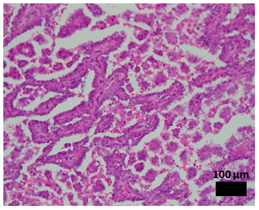
defined in the 2004 WHO classification (8). MPC was defined as tumor cells growing in
papillary tufts lacking fibrovascular cores that may float within
alveolar spaces (Fig. 1) (9,10). PA-MPC
has been associated with an aggressive clinical course compared
with traditional papillary adenocarcinoma and bronchioloalveolar
carcinoma (9,11–13).
PA-MPC is frequently encountered in non-smokers, with intralobar
satellites, and frequently metastasizes to the contralateral lung,
mediastinal lymph nodes, bone and adrenal glands, with a high
mortality rate (11–14). Although PA-MPC represents a unique
form of PA, its molecular profile is yet to be elucidated. In the
present study, PA-MPC was analyzed for the common genetic mutations
in pa, including endothelial growth factor receptor gene
(EGFR), Kirsten rat sarcoma viral oncogene homolog
(KRAS) and echinoderm microtubule-associated protein-like
4-anaplastic lymphoma kinase fusion gene (EML4-ALK) to
determine whether a distinct genetic profile was associated with
this histopathological growth pattern.
Patients and methods
Patients
The pathological reports of patients who underwent
surgical resection for lung cancer between April, 2004 and May,
2012 at the Okayama University Hospital (Okayama, Japan) were
reviewed. Of the 674 patients diagnosed with PA, 28 were found to
have MPC. The ratio of MPC varied widely (3–80%) among these 28
patients. A total of 138 resected PAs without MPC were randomly
selected in the same period to serve as age-, gender- and smoking
status-matched controls to the PA-MPC cases (Table I). Our institutional review board
approved this study's protocol and informed consent was obtained
from all the patients.
 | Table I.Patient characteristics. |
Table I.
Patient characteristics.
|
| MPC |
|
|---|
|
|
|
|
|---|
|
Characteristics | Positive
(n=28) | Negative
(n=138) | P-value |
|---|
| Age (years) | 65.3±11.0 | 66.0±9.8 | NS |
| Gender |
|
| NS |
|
Male | 21 | 101 |
|
|
Female | 7 | 37 |
|
| Smoking
history |
|
| NS |
|
Smoker | 20 | 100 |
|
|
Non-smoker | 8 | 38 |
|
| Pathological
stage |
|
|
<0.001a |
| I | 13 | 104 |
|
| II | 7 | 12 |
|
|
III | 4 | 18 |
|
| IV | 4 | 4 |
|
DNA and RNA extraction
Genomic DNA was obtained from primary tumors by
standard phenol-chloroform (1:1) extraction followed by ethanol
precipitation, or by using the DNeasy Tissue kit (Qiagen, Valencia,
CA, USA). Total RNA was extracted from primary tumors using the
RNeasy Mini kit (Qiagen) according to the manufacturer's protocol.
Oligo(dT)-primed cDNA was synthesized using the High-Capacity cDNA
Reverse Transcription kit (Applied Biosystems, Foster City, CA,
USA) with DNase treatment.
Genotype screening
Using DNA derived from frozen tumor specimens,
genotyping was performed by SNaPshot, a targeted mutational
analysis assay designed by Su et al (15). The platform involves two methods: a
screen (SNaPshot) based on multiplex polymerase chain reaction
(PCR), primer extension and capillary electrophoresis that was
designed to assess 38 somatic mutations in 8 genes [(AKT1, BRAF,
EGFR, KRAS, mitogen-activated protein kinase kinase 1
(MEK1), neuroblastoma RAS viral oncogene homolog
(NRAS), phosphatidylinositol-4,5-bisphosphate 3-kinase,
catalytic subunit α (PIK3CA) and phosphatase and tensin
homolog (PTEN)]; and a PCR-based sizing assay that assesses
EGFR exon 19 (deletions), EGFR exon 20 (insertions)
and human epidermal growth factor receptor 2 (HER2) exon 20
(insertions).
Detection of EGFR and KRAS
mutations
PCR-based assays or direct sequencing were performed
to confirm the results if mutations of EGFR and KRAS
were detected by the SNaPshot assay.
The EGFR mutational status was determined
using a PCR-based length polymorphism and restriction fragment
length polymorphism assay, as previously reported (16). Briefly, the common deletions of exon
19 were distinguished from the wild-type based on PCR product
length polymorphisms using 12% polyacrylamide gel electrophoresis
(PAGE) via ethidium bromide staining. For the exon 21 L858R
mutation, Sau96I digestion, which specifically digests the
mutant type, was performed prior to 12% PAGE.
The KRAS mutations in codons 12 and 13 were
examined using PCR-based direct sequencing on an ABI PRISM 3130xl
Genetic Analyzer (Applied Biosystems), as previously reported
(17–19).
Detection of EML4-ALK fusion
events
EML4-ALK fusion was screened by ALK
immunohistochemistry (IHC) and confirmed by reverse transcription
(RT)-PCR assay and fluorescence in situ hybridization (FISH)
analysis.
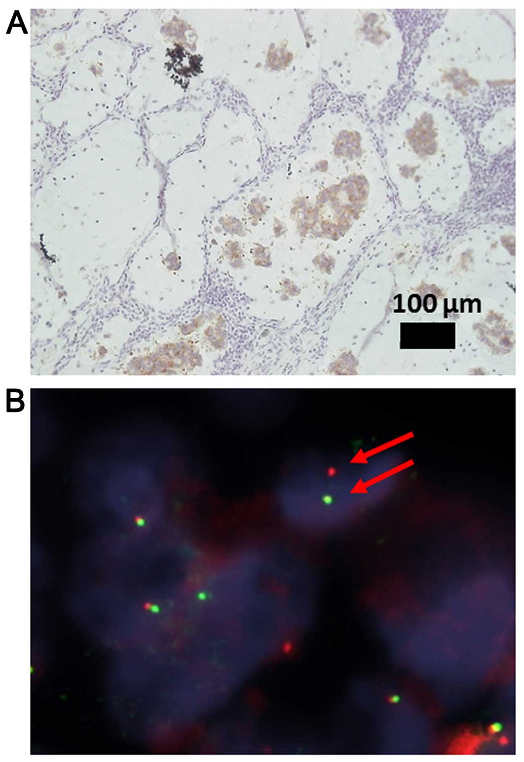
ALK IHC
Unstained paraffin-embedded sections were
deparaffinised in xylene, hydrated through, and rinsed in distilled
water. Heat-induced epitope retrieval was performed with EnVision
FLEX Target Retrieval Solution, High pH (Dako, Carpinteria,
California, USA). The slides were then incubated at room
temperature with mouse anti-ALK monoclonal antibody (clone 5A4;
dilution, 1:100; cat. no. ab17127; Abcam) for 30 min. The slides
were incubated at room temperature with EnVision FLEX+Mouse Linker
(Dako) for 15 min. The immune complexes were then detected with the
dextran polymer reagent (Fig. 2A)
(20,21).
RT-PCR
The primers used to identify the EML4-ALK
fusion transcript were selected to enable the detection of all
possible in-frame fusions of EML4 to exon 20 of ALK,
in which the kinase domain of ALK would be preserved. The
forward primers used were EML4 72F
(5′-GTCAGCTCTTGAGTCACGAGTT-3′) and fusion-RT-S
(5′-GTGCAGTGTTTAGCATTCTTGGGG-3′); the reverse primer was ALK 3078RR
(5′-ATCCAGTTCGTCCTGTTCAGAGC-3′) (22). PCR was performed for EML4-ALK under
the following conditions: 94°C for 10 min, followed by 35 cycles of
denaturation at 94°C for 1 min, annealing at 64°C for 1 min and
polymerization at 72°C for 1 min, with a final extension step at
72°C for 7 min. RT-PCR for GAPDH expression as an internal control
was performed under the same conditions in each tumor sample.
FISH
FISH was performed on formalin-fixed,
paraffin-embedded tumor tissues using a break-apart probe to the
ALK gene (Vysis LSI ALK Dual Color, Break Apart
Rearrangement Probe; Abbott Molecular, Abbott Park, IL, USA) as per
the manufacturer's instructions. cases were defined as
FISH-positive when there was a >15% split signal in the tumor
cells (Fig. 2B) (23,24).
Statistical analysis
Differences in statistical significance among the
categorized groups were compared using the Chi-square test or the
Student's t-test. An analysis of overall survival and disease-free
survival was performed using the Kaplan-Meier method with the
log-rank test. The data were analyzed using SPSS v22.0 software
(IBM SPSS, Armonk, NY, USA). P<0.05 was considered to indicate a
statistically significant difference for each analysis.
Results
Genetic alterations in clinical
samples
Regarding genetic alterations, 13 (46.4%) of the 28
PA-MPCs harbored mutually exclusive mutations: 9 (32.1%)
EGFR mutations, 1 (3.6%) KRAS mutation and 3 (10.7%)
EML4-ALK fusion genes. PAs without MPC harbored 42 (30.4%)
EGFR mutations, 17 (12.3%) KRAS mutations, 1 (0.7%)
PIK3CA mutation and 3 (2.2%) EML4-ALK fusion genes in
a mutually exclusive manner, except that 1 case had EGFR
G719A and L861Q mutations. There were no mutations in the AKT1,
BRAF, MEK1, NRAS, PTEN or HER2 genes in either group.
EML4-ALK fusion genes appeared to occur significantly more
frequently in PA-MPCs compared with PA without MPC (P=0.027)
(Table II). Regarding the
EGFR and KRAS mutational status, the results of the
SNaPshot assay were consistent with those of PCR-based assays or
direct sequencing.
| Table II.Association between MPC and genetic
alterations. |
Table II.
Association between MPC and genetic
alterations.
|
| MPC, patient no.
(%) |
|
|---|
|
|
|
|
|---|
| Genes | Positive
(n=28) |
Negative(n=138) | P-value |
|---|
| EGFR | 9
(32.1) | 42 (30.4) | 0.9 |
| KRAS | 1 (3.6) | 17 (12.3) | 0.2 |
|
EML4-ALK | 3
(10.7) | 3 (2.2) |
0.027 |
Effect of PA-MPCs on clinical
outcome
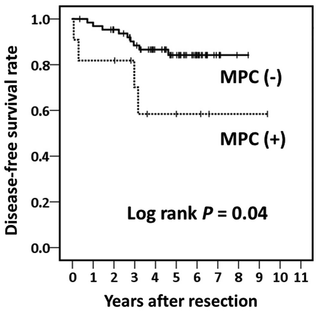
To confirm the clinical outcome of PA-MPC, 11 cases
with pathological stage IA PA-MPC were compared with 65 cases with
pathological stage IA PA without MPC (Table III). As of December, 2013, 13
(17.1%) patients had succumbed to the disease, with a median
follow-up period of 62.0 months; 2 (18.2%) of the patients with
PA-MPC had succumbed to PA during follow-up; 11 (16.9%) patients
with PA without MPC had succumbed (7 to PA and 4 to other causes).
The 5-year overall survival rates of all pathological stage IA
patients (n=76), patients with PA-MPC (n=11), and patients with PA
without MPC (n=65) were 82.0, 87.5 and 81.0%, respectively. A total
of 13 (17.1%) patients developed disease relapse (4 patients with
PA-MPC and 7 patients with PA without MPC). The 5-year disease-free
survival rates of all pathological stage IA patients, patients with
PA-MPC, and patients with PA without MPC were 80.7, 58.4 and 84.2%,
respectively. Patients with PA-MPC exhibited a significantly poorer
disease-free survival rate compared with those with PA without MPC
(log-rank test, P=0.04) (Fig. 3).
| Table III.Association among MPC, patient
characteristics and genetic alterations in pathological stage IA
lung adenocarcinoma. |
Table III.
Association among MPC, patient
characteristics and genetic alterations in pathological stage IA
lung adenocarcinoma.
|
| MPC |
|
|---|
|
|
|
|
|---|
|
Characteristics |
Positive(n=11) |
Negative (n=65) |
P-value |
|---|
| Age (years) | 67.3±9.2 | 67.2±9.5 | NS |
| Gender |
|
| NS |
|
Male | 7 | 44 |
|
|
Female | 4 | 21 |
|
| Smoking
history |
|
| NS |
|
Smoker | 6 | 42 |
|
|
Non-smoker | 5 | 23 |
|
| Tumor size
(cm) | 1.8±0.5 | 1.7±0.7 | NS |
| EGFR
mutations |
|
| NS |
|
Positive | 3 | 19 |
|
|
Wild-type | 8 | 46 |
|
| KRAS
mutations |
|
| NS |
|
Positive | 0 | 6 |
|
|
Wild-type | 11 | 59 |
|
|
EML4-ALK |
|
| NS |
|
Positive | 1 | 1 |
|
|
Negative | 10 | 64 |
|
Discussion
The IASLC/ATS/ERS classification is the result of
the advances in the research of PA. Although PA-MPC is newly
classified in it, its detailed molecular characteristics, including
EML4-ALK fusion, have not been determined. In this regard,
this is a unique report describing a comprehensive gene mutational
analysis, including EML4-ALK fusion gene, in patients with
PA-MPC. In the present study, EML4-ALK fusion genes appeared
to occur significantly more frequently in PA-MPC compared with PA
without MPC. According to previous reports, the frequency of
EML4-ALK fusion genes in PA is low (4.3–6%) (23,25,26).
Furthermore, ALK-positive lung cancers tend to be more common among
younger patients and marginally more common in women, which may be
associated with the difference in smoking rates between the two
genders (27). In our study, the 6
ALK-positive patients included 2 smoking men, 1 non-smoking woman
with PA-MPC, and 3 non-smoking women with PA without MPC. The
ALK-positive patients were younger than ALK-negative patients (56.0
vs. 66.3 years, respectively; P=0.013). Inamura et al
reported that adenocarcinomas with EML4-ALK fusion were
predominantly classified as the acinar or papillary subtypes
(26). Although our sample size was
small, our data suggest that the EML4-ALK fusion gene is one
of the most common genetic alterations in PA-MPCs.
As regards the association between PA-MPC and gene
status, De Oliveira Duarte Achcar et al reported the genetic
alterations of 15 micropapillary-dominant cases, namely 5 (33%)
Kras, 3 (20%) EGFR and 3 (20%) BRAF mutations
(28). A number of studies regarding
patients with PA-MPC reported that they often harbored EGFR
mutations (13,28,29). As
previously reported, EGFR mutations are common in Asian,
female, non-smoker PA patients (30,31) and
the frequency of EGFR mutations in Japanese PA patients is
~44% (32). Conversely, in our
series, the prevalence of EGFR mutations in PA was lower (51
of 166, 30.7%) compared with that previously reported, likely
because the population of this study included several smokers (120
of 166, 72.3%) and men (122 of 166, 73.5%). In never-smoker
patients, the prevalence of the EGFR mutation (24 of 46,
52.2%) was similar to our previous report (32).
Similar to previous studies (9,11–13), our data suggest a poorer prognosis for
PA-MPC compared with that for PA without MPC. Miyoshi et al
reported that a higher ratio (6–100%) of MPC was associated with a
poorer prognosis compared with a lower ratio (1–5%) (12). The ratio of MPC in the 28 PA-MPC cases
varied widely (3–80%) in this study. Our results indicated that
PA-MPC had a tendency for relapse, even if the ratio of MPC was
low. Thus, clinicians should bear in mind the possibility for
metastasis when MPC is present in PA.
In conclusion, our study suggests that the molecular
pathogenesis of PA-MPC may differ from that of other
adenocarcinomas, which is associated with its aggressive clinical
behavior. Further investigation is required to elucidate the
characteristics of PA-MPC and lead to the development of new
therapeutic strategies.
Acknowledgements
We would like to thank Ms. Fumiko Isobe (Department
of Thoracic, Breast and Endocrinological Surgery, Okayama
University Graduate School of Medicine, Dentistry and
Pharmaceutical Sciences, Okayama, Japan) for the preparation of the
pathological materials. We would also like to thank Yukinari
Isomoto (Central Research Laboratory, Okayama University Medical
School, Okayama, Japan) for technical support for the SNaPshot
assay. This study was supported by a Grant-in-Aid for Scientific
Research from the Japan Society for the Promotion of Science (JSPS
KAKENHI grant nos. 22700916 to J.S. and 25293302 to S.T.).
References
|
1
|
Siegel RL, Miller KD and Jemal A: Cancer
statistics, 2015. CA Cancer J Clin. 65:5–29. 2015. View Article : Google Scholar : PubMed/NCBI
|
|
2
|
Malvezzi M, Bertuccio P, Rosso T, Rota M
and Levi F: LaV ecchia C and Negri E: European cancer mortality
predictions for the year 2015: Does lung cancer have the highest
death rate in EU women? Ann Oncol. 26:779–786. 2015. View Article : Google Scholar : PubMed/NCBI
|
|
3
|
Katanoda K, Hori M, Matsuda T, Shibata A,
Nishino Y, Hattori M, Soda M, Ioka A, Sobue T and Nishimoto H: An
updated report on the trends in cancer incidence and mortality in
Japan, 1958-2013. Jpn J Clin Oncol. 45:390–401. 2015. View Article : Google Scholar : PubMed/NCBI
|
|
4
|
Barlesi F, Scherpereel A, Gorbunova V,
Gervais R, Vikström A, Chouaid C, Chella A, Kim JH, Ahn MJ, Reck M,
et al: Maintenance bevacizumab-pemetrexed after first-line
cisplatin-pemetrexed-bevacizumab for advanced nonsquamous
nonsmall-cell lung cancer: Updated survival analysis of the AVAPERL
(MO22089) randomized phase III trial. Ann Oncol. 25:1044–1052.
2014. View Article : Google Scholar : PubMed/NCBI
|
|
5
|
Lynch TJ, Bell DW, Sordella R,
Gurubhagavatula S, Okimoto RA, Brannigan BW, Harris PL, Haserlat
SM, Supko JG, Haluska FG, et al: Activating mutations in the
epidermal growth factor receptor underlying responsiveness of
non-small-cell lung cancer to gefitinib. N Engl J Med.
350:2129–2139. 2004. View Article : Google Scholar : PubMed/NCBI
|
|
6
|
Cadranel J, Mauguen A, Faller M, Zalcman
G, Buisine MP, Westeel V, Longchampt E, Wislez M, Coudert B, Daniel
C, et al: Impact of systematic EGFR and KRAS mutation evaluation on
progression-free survival and overall survival in patients with
advanced non-small-cell lung cancer treated by erlotinib in a
French prospective cohort (ERMETIC project-part 2). J Thorac Oncol.
7:1490–1502. 2012. View Article : Google Scholar : PubMed/NCBI
|
|
7
|
Travis WD, Brambilla E, Noguchi M,
Nicholson AG, Geisinger KR, Yatabe Y, Beer DG, Powell CA, Riely GJ,
Van Schil PE, et al: International association for the study of
lung cancer/american thoracic society/european respiratory society
international multidisciplinary classification of lung
adenocarcinoma. J Thorac Oncol. 6:244–285. 2011. View Article : Google Scholar : PubMed/NCBI
|
|
8
|
Yoshizawa A, Motoi N, Riely GJ, Sima CS,
Gerald WL, Kris MG, Park BJ, Rusch VW and Travis WD: Impact of
proposed IASLC/ATS/ERS classification of lung adenocarcinoma,
Prognostic subgroups and implications for further revision of
staging based on analysis of 514 stage I cases. Mod Pathol.
24:653–664. 2011. View Article : Google Scholar : PubMed/NCBI
|
|
9
|
Amin MB, Tamboli P, Merchant SH, Ordóñez
NG, Ro J, Ayala AG and Ro JY: Micropapillary component in lung
adenocarcinoma, A distinctive histologic feature with possible
prognostic significance. Am J Surg Pathol. 26:358–364. 2002.
View Article : Google Scholar : PubMed/NCBI
|
|
10
|
Sánchez-Mora N, Presmanes MC, Monroy V,
Moreno N, Lara-Martínez JM, Aladro MH and Alvarez-Fernández E:
Micropapillary lung adenocarcinoma A distinctive histologic subtype
with prognostic significance. Histopathology. Hum Pathol.
39:324–330. 2008. View Article : Google Scholar : PubMed/NCBI
|
|
11
|
Makimoto Y, Nabeshima K, Iwasaki H,
Miyoshi T, Enatsu S, Shiraishi T, Iwasaki A, Shirakusa T and
Kikuchi M: Micropapillary pattern, A distinct pathological marker
to subclassify tumours with a significantly poor prognosis within
small peripheral lung adenocarcinoma (</=20 mm) with mixed
bronchioloalveolar and invasive subtypes (Noguchi's type C
tumours). Histopathology. 46:677–684. 2005. View Article : Google Scholar : PubMed/NCBI
|
|
12
|
Miyoshi T, Satoh Y, Okumura S, Nakagawa K,
Shirakusa T, Tsuchiya E and Ishikawa Y: Early-stage lung
adenocarcinomas with a micropapillary pattern, a distinct
pathologic marker for a significantly poor prognosis. Am J Surg
Pathol. 27:101–109. 2003. View Article : Google Scholar : PubMed/NCBI
|
|
13
|
Motoi N, Szoke J, Riely GJ, Seshan VE,
Kris MG, Rusch VW, Gerald WL and Travis WD: Lung adenocarcinoma,
Modification of the 2004 WHO mixed subtype to include the major
histologic subtype suggests correlations between papillary and
micropapillary adenocarcinoma subtypes, EGFR mutations and gene
expression analysis. Am J Surg Pathol. 32:810–827. 2008. View Article : Google Scholar : PubMed/NCBI
|
|
14
|
Roh MS, Lee JI, Choi PJ and Hong YS:
Relationship between micropapillary component and micrometastasis
in the regional lymph nodes of patients with stage I lung
adenocarcinoma. Histopathology. 45:580–586. 2004. View Article : Google Scholar : PubMed/NCBI
|
|
15
|
Su Z, Dias-Santagata D, Duke M, Hutchinson
K, Lin YL, Borger DR, Chung CH, Massion PP, Vnencak-Jones CL,
Iafrate AJ and Pao W: A platform for rapid detection of multiple
oncogenic mutations with relevance to targeted therapy in
non-small-cell lung cancer. J Mol Diagn. 13:74–84. 2011. View Article : Google Scholar : PubMed/NCBI
|
|
16
|
Asano H, Toyooka S, Tokumo M, Ichimura K,
Aoe K, Ito S, Tsukuda K, Ouchida M, Aoe M, Katayama H, et al:
Detection of EGFR gene mutation in lung cancer by mutant-enriched
polymerase chain reaction assay. Clin Cancer Res. 12:43–48. 2006.
View Article : Google Scholar : PubMed/NCBI
|
|
17
|
Tokumo M, Toyooka S, Ichihara S, Ohashi K,
Tsukuda K, Ichimura K, Tabata M, Kiura K, Aoe M, Sano Y, et al:
Double mutation and gene copy number of EGFR in gefitinib
refractory non-small-cell lung cancer. Lung Cancer. 53:117–121.
2006. View Article : Google Scholar : PubMed/NCBI
|
|
18
|
Toyooka S, Matsuo K, Shigematsu H, Kosaka
T, Tokumo M, Yatabe Y, Ichihara S, Inukai M, Suehisa H, Soh J, et
al: The impact of sex and smoking status on the mutational spectrum
of epidermal growth factor receptor gene in non small cell lung
cancer. Clin Cancer Res. 13:5763–5768. 2007. View Article : Google Scholar : PubMed/NCBI
|
|
19
|
Toyooka S, Tsukuda K, Ouchida M, Tanino M,
Inaki Y, Kobayashi K, Yano M, Soh J, Kobatake T, Shimizu N and
Shimizu K: Detection of codon 61 point mutations of the K-ras gene
in lung and colorectal cancers by enriched PCR. Oncol Rep.
10:1455–1459. 2003.PubMed/NCBI
|
|
20
|
Jokoji R, Yamasaki T, Minami S, Komuta K,
Sakamaki Y, Takeuchi K and Tsujimoto M: Combination of
morphological feature analysis and immunohistochemistry is useful
for screening of EML4-ALK-positive lung adenocarcinoma. J Clin
Pathol. 63:1066–1070. 2010. View Article : Google Scholar : PubMed/NCBI
|
|
21
|
Yoshida A, Tsuta K, Nakamura H, Kohno T,
Takahashi F, Asamura H, Sekine I, Fukayama M, Shibata T, Furuta K
and Tsuda H: Comprehensive histologic analysis of ALK-rearranged
lung carcinomas. Am J Surg Pathol. 35:1226–1234. 2011. View Article : Google Scholar : PubMed/NCBI
|
|
22
|
Takeuchi K, Choi YL, Soda M, Inamura K,
Togashi Y, Hatano S, Enomoto M, Takada S, Yamashita Y, Satoh Y, et
al: Multiplex reverse transcription-PCR screening for EML4-ALK
fusion transcripts. Clin Cancer Res. 14:6618–6624. 2008. View Article : Google Scholar : PubMed/NCBI
|
|
23
|
Rodig SJ, Mino-Kenudson M, Dacic S, Yeap
BY, Shaw A, Barletta JA, Stubbs H, Law K, Lindeman N, Mark E, et
al: Unique clinicopathologic features characterize ALK-rearranged
lung adenocarcinoma in the western population. Clin Cancer Res.
15:5216–5223. 2009. View Article : Google Scholar : PubMed/NCBI
|
|
24
|
Kwak EL, Bang YJ, Camidge DR, Shaw AT,
Solomon B, Maki RG, Ou SH, Dezube BJ, Jänne PA, Costa DB, et al:
Anaplastic lymphoma kinase inhibition in non-small-cell lung
cancer. N Engl J Med. 363:1693–1703. 2010. View Article : Google Scholar : PubMed/NCBI
|
|
25
|
Paik JH, Choe G, Kim H, Choe JY, Lee HJ,
Lee CT, Lee JS, Jheon S and Chung JH: Screening of anaplastic
lymphoma kinase rearrangement by immunohistochemistry in non-small
cell lung cancer, Correlation with fluorescence in situ
hybridization. J Thorac Oncol. 6:466–472. 2011. View Article : Google Scholar : PubMed/NCBI
|
|
26
|
Inamura K, Takeuchi K, Togashi Y, Hatano
S, Ninomiya H, Motoi N, Mun MY, Sakao Y, Okumura S, Nakagawa K, et
al: EML4-ALK lung cancers are characterized by rare other
mutations, a TTF-1 cell lineage, an acinar histology and young
onset. Mod Pathol. 22:508–515. 2009. View Article : Google Scholar : PubMed/NCBI
|
|
27
|
Mitsudomi T, Yatabe Y, Akita H, Genma A,
Soda M, Toyooka T, Nakagawa K, Nishio K and Hagiwara K: Guidance
for ALK gene testing in lung cancer patients. Biomarker Committee,
the Japan Lung Cancer Society. 2011.
|
|
28
|
De Oliveira Duarte Achcar R, Nikiforova MN
and Yousem SA: Micropapillary lung adenocarcinoma: EGFR, K-ras and
BRAF mutational profile. Am J Clin Pathol. 131:694–700. 2009.
View Article : Google Scholar : PubMed/NCBI
|
|
29
|
Ninomiya H, Hiramatsu M, Inamura K, Nomura
K, Okui M, Miyoshi T, Okumura S, Satoh Y, Nakagawa K, Nishio M, et
al: Correlation between morphology and EGFR mutations in lung
adenocarcinomas. Significance of the micropapillary pattern and the
hobnail cell type. Lung Cancer. 63:235–240. 2009. View Article : Google Scholar : PubMed/NCBI
|
|
30
|
Tokumo M, Toyooka S, Kiura K, Shigematsu
H, Tomii K, Aoe M, Ichimura K, Tsuda T, Yano M, Tsukuda K, et al:
The relationship between epidermal growth factor receptor mutations
and clinicopathologic features in non-small cell lung cancers. Clin
Cancer Res. 11:1167–1173. 2005.PubMed/NCBI
|
|
31
|
Mitsudomi T: Advances in target therapy
for lung cancer. Jpn J Clin Oncol. 40:101–106. 2010. View Article : Google Scholar : PubMed/NCBI
|
|
32
|
Shigematsu H, Lin L, Takahashi T, Nomura
M, Suzuki M, Wistuba II, Fong KM, Lee H, Toyooka S, Shimizu N, et
al: Clinical and biological features associated with epidermal
growth factor receptor gene mutations in lung cancers. J Natl
Cancer Inst. 97:339–346. 2005. View Article : Google Scholar : PubMed/NCBI
|