Spandidos Publications Logo
  • About
    • About Spandidos
    • Aims and Scopes
    • Abstracting and Indexing
    • Editorial Policies
    • Reprints and Permissions
    • Job Opportunities
    • Terms and Conditions
    • Contact
  • Journals
    • All Journals
    • Oncology Letters
      • Oncology Letters
      • Information for Authors
      • Editorial Policies
      • Editorial Board
      • Aims and Scope
      • Abstracting and Indexing
      • Bibliographic Information
      • Archive
    • International Journal of Oncology
      • International Journal of Oncology
      • Information for Authors
      • Editorial Policies
      • Editorial Board
      • Aims and Scope
      • Abstracting and Indexing
      • Bibliographic Information
      • Archive
    • Molecular and Clinical Oncology
      • Molecular and Clinical Oncology
      • Information for Authors
      • Editorial Policies
      • Editorial Board
      • Aims and Scope
      • Abstracting and Indexing
      • Bibliographic Information
      • Archive
    • Experimental and Therapeutic Medicine
      • Experimental and Therapeutic Medicine
      • Information for Authors
      • Editorial Policies
      • Editorial Board
      • Aims and Scope
      • Abstracting and Indexing
      • Bibliographic Information
      • Archive
    • International Journal of Molecular Medicine
      • International Journal of Molecular Medicine
      • Information for Authors
      • Editorial Policies
      • Editorial Board
      • Aims and Scope
      • Abstracting and Indexing
      • Bibliographic Information
      • Archive
    • Biomedical Reports
      • Biomedical Reports
      • Information for Authors
      • Editorial Policies
      • Editorial Board
      • Aims and Scope
      • Abstracting and Indexing
      • Bibliographic Information
      • Archive
    • Oncology Reports
      • Oncology Reports
      • Information for Authors
      • Editorial Policies
      • Editorial Board
      • Aims and Scope
      • Abstracting and Indexing
      • Bibliographic Information
      • Archive
    • Molecular Medicine Reports
      • Molecular Medicine Reports
      • Information for Authors
      • Editorial Policies
      • Editorial Board
      • Aims and Scope
      • Abstracting and Indexing
      • Bibliographic Information
      • Archive
    • World Academy of Sciences Journal
      • World Academy of Sciences Journal
      • Information for Authors
      • Editorial Policies
      • Editorial Board
      • Aims and Scope
      • Abstracting and Indexing
      • Bibliographic Information
      • Archive
    • International Journal of Functional Nutrition
      • International Journal of Functional Nutrition
      • Information for Authors
      • Editorial Policies
      • Editorial Board
      • Aims and Scope
      • Abstracting and Indexing
      • Bibliographic Information
      • Archive
    • International Journal of Epigenetics
      • International Journal of Epigenetics
      • Information for Authors
      • Editorial Policies
      • Editorial Board
      • Aims and Scope
      • Abstracting and Indexing
      • Bibliographic Information
      • Archive
    • Medicine International
      • Medicine International
      • Information for Authors
      • Editorial Policies
      • Editorial Board
      • Aims and Scope
      • Abstracting and Indexing
      • Bibliographic Information
      • Archive
  • Articles
  • Information
    • Information for Authors
    • Information for Reviewers
    • Information for Librarians
    • Information for Advertisers
    • Conferences
  • Language Editing
Spandidos Publications Logo
  • About
    • About Spandidos
    • Aims and Scopes
    • Abstracting and Indexing
    • Editorial Policies
    • Reprints and Permissions
    • Job Opportunities
    • Terms and Conditions
    • Contact
  • Journals
    • All Journals
    • Biomedical Reports
      • Information for Authors
      • Editorial Policies
      • Editorial Board
      • Aims and Scope
      • Abstracting and Indexing
      • Bibliographic Information
      • Archive
    • Experimental and Therapeutic Medicine
      • Information for Authors
      • Editorial Policies
      • Editorial Board
      • Aims and Scope
      • Abstracting and Indexing
      • Bibliographic Information
      • Archive
    • International Journal of Epigenetics
      • Information for Authors
      • Editorial Policies
      • Editorial Board
      • Aims and Scope
      • Abstracting and Indexing
      • Bibliographic Information
      • Archive
    • International Journal of Functional Nutrition
      • Information for Authors
      • Editorial Policies
      • Editorial Board
      • Aims and Scope
      • Abstracting and Indexing
      • Bibliographic Information
      • Archive
    • International Journal of Molecular Medicine
      • Information for Authors
      • Editorial Policies
      • Editorial Board
      • Aims and Scope
      • Abstracting and Indexing
      • Bibliographic Information
      • Archive
    • International Journal of Oncology
      • Information for Authors
      • Editorial Policies
      • Editorial Board
      • Aims and Scope
      • Abstracting and Indexing
      • Bibliographic Information
      • Archive
    • Medicine International
      • Information for Authors
      • Editorial Policies
      • Editorial Board
      • Aims and Scope
      • Abstracting and Indexing
      • Bibliographic Information
      • Archive
    • Molecular and Clinical Oncology
      • Information for Authors
      • Editorial Policies
      • Editorial Board
      • Aims and Scope
      • Abstracting and Indexing
      • Bibliographic Information
      • Archive
    • Molecular Medicine Reports
      • Information for Authors
      • Editorial Policies
      • Editorial Board
      • Aims and Scope
      • Abstracting and Indexing
      • Bibliographic Information
      • Archive
    • Oncology Letters
      • Information for Authors
      • Editorial Policies
      • Editorial Board
      • Aims and Scope
      • Abstracting and Indexing
      • Bibliographic Information
      • Archive
    • Oncology Reports
      • Information for Authors
      • Editorial Policies
      • Editorial Board
      • Aims and Scope
      • Abstracting and Indexing
      • Bibliographic Information
      • Archive
    • World Academy of Sciences Journal
      • Information for Authors
      • Editorial Policies
      • Editorial Board
      • Aims and Scope
      • Abstracting and Indexing
      • Bibliographic Information
      • Archive
  • Articles
  • Information
    • For Authors
    • For Reviewers
    • For Librarians
    • For Advertisers
    • Conferences
  • Language Editing
Login Register Submit
  • This site uses cookies
  • You can change your cookie settings at any time by following the instructions in our Cookie Policy. To find out more, you may read our Privacy Policy.

    I agree
Search articles by DOI, keyword, author or affiliation
Search
Advanced Search
presentation
Molecular and Clinical Oncology
Join Editorial Board Propose a Special Issue
Print ISSN: 2049-9450 Online ISSN: 2049-9469
Journal Cover
November-2018 Volume 9 Issue 5

Full Size Image

Sign up for eToc alerts
Recommend to Library

Journals

International Journal of Molecular Medicine

International Journal of Molecular Medicine

International Journal of Molecular Medicine is an international journal devoted to molecular mechanisms of human disease.

International Journal of Oncology

International Journal of Oncology

International Journal of Oncology is an international journal devoted to oncology research and cancer treatment.

Molecular Medicine Reports

Molecular Medicine Reports

Covers molecular medicine topics such as pharmacology, pathology, genetics, neuroscience, infectious diseases, molecular cardiology, and molecular surgery.

Oncology Reports

Oncology Reports

Oncology Reports is an international journal devoted to fundamental and applied research in Oncology.

Experimental and Therapeutic Medicine

Experimental and Therapeutic Medicine

Experimental and Therapeutic Medicine is an international journal devoted to laboratory and clinical medicine.

Oncology Letters

Oncology Letters

Oncology Letters is an international journal devoted to Experimental and Clinical Oncology.

Biomedical Reports

Biomedical Reports

Explores a wide range of biological and medical fields, including pharmacology, genetics, microbiology, neuroscience, and molecular cardiology.

Molecular and Clinical Oncology

Molecular and Clinical Oncology

International journal addressing all aspects of oncology research, from tumorigenesis and oncogenes to chemotherapy and metastasis.

World Academy of Sciences Journal

World Academy of Sciences Journal

Multidisciplinary open-access journal spanning biochemistry, genetics, neuroscience, environmental health, and synthetic biology.

International Journal of Functional Nutrition

International Journal of Functional Nutrition

Open-access journal combining biochemistry, pharmacology, immunology, and genetics to advance health through functional nutrition.

International Journal of Epigenetics

International Journal of Epigenetics

Publishes open-access research on using epigenetics to advance understanding and treatment of human disease.

Medicine International

Medicine International

An International Open Access Journal Devoted to General Medicine.

Journal Cover
November-2018 Volume 9 Issue 5

Full Size Image

Sign up for eToc alerts
Recommend to Library

  • Article
  • Citations
    • Cite This Article
    • Download Citation
    • Create Citation Alert
    • Remove Citation Alert
    • Cited By
  • Similar Articles
    • Related Articles (in Spandidos Publications)
    • Similar Articles (Google Scholar)
    • Similar Articles (PubMed)
  • Download PDF
  • Download XML
  • View XML
Case Report

Ultrasonography findings of proximal‑type epithelioid sarcoma of the vulva: A case report

  • Authors:
    • Yuanyuan Yue
    • Ya Lu
    • Xiaojuan Ma
    • Zhongping Tang
    • Yinrong Cheng
  • View Affiliations / Copyright

    Affiliations: Department of Ultrasound, Chengdu First People's Hospital, Chengdu, Sichuan 610041, P.R. China, Department of Pathology, West China Hospital, Sichuan University, Chengdu, Sichuan 610065, P.R. China, Department of Pathology, Chengdu First People's Hospital, Chengdu, Sichuan 610041, P.R. China
  • Pages: 507-510
    |
    Published online on: September 3, 2018
       https://doi.org/10.3892/mco.2018.1708
  • Expand metrics +
Metrics: Total Views: 0 (Spandidos Publications: | PMC Statistics: )
Metrics: Total PDF Downloads: 0 (Spandidos Publications: | PMC Statistics: )
Cited By (CrossRef): 0 citations Loading Articles...

This article is mentioned in:


Abstract

Proximal-type epithelioid sarcoma (PES) of the vulva is an exceedingly rare malignant soft tissue tumor. We herein present the case of a 41‑year‑old female patient who presented to our hospital with complaints of a painless mass in the right mons pubis that she had first noticed 3 years prior. Ultrasonographic (US) and color Doppler ultrasonographic (CDUS) examination revealed a solid mass with well‑defined, homogeneous hypoechoic structure and quite hypervascular on CDUS. The results of the immunohistochemical examination confirmed the diagnosis of vulvar PES (myxoid variant). The patient was treated with wide local excision and remained recurrence‑ and metastasis‑free at 9 months postoperatively. Although cases of PES in the pelvic region had been previously reported, to the best of our knowledge, the US or CDUS findings of PES of the vulva have not been described to date.

Introduction

Proximal-type epithelioid sarcoma (PES) of the vulva is an exceedingly rare malignant soft tissue tumor. The first case of vulvar PES was described in 1972 (1), and <60 cases have been reported in the literature to date. Compared with other soft tissue tumors, accurate diagnosis of this neoplasm is generally challenging due to its rarity and the likelihood of misdiagnosis as a benign lesion, including Bartholin's gland cyst, Bartholin's gland abscess, inguinal or femoral hernias or other benign soft tissue tumors (2).

Several imaging modalities have been used to assess soft tissue tumors, including ultrasonography (US), computed tomography (CT) and magnetic resonance imaging (MRI), plain radiography and positron emission tomography (PET). US in particular is the most common first-line examination in vulvar lesions due to its real-time and radiation-free properties. As PES lesions may present with various and non-specific signs and symptoms, the US findings of vulvar PES may lack specificity compared with other solid masses. However, US can assess the mass size, shape, echogenicity, margin, composition and vascularity (3). Furthermore, to the best of our knowledge, US and CDUS findings of vulvar PES have not been described in the literature to date. In the present study, the case of a patient with vulvar PES is presented and the US and CDUS findings are discussed.

Case report

A 41-year old female patient presented to the Department of Ultrasonography of Chengdu First People's Hospital (Chengdu, China) in May 2017, with complaints of a painless mass in the right mons pubis that she had first noticed 3 years prior. The patient had no other significant medical history and her family history did not include malignancies in first-degree relatives. Gynecological examination of the vagina, cervix and uterus appeared normal. Inspection of the vulva revealed a mass 2 cm in greatest diameter in the right mons pubis. Physical examination of the mass revealed a non-tender nodule with a smooth surface and hard consistency. No other abnormalities were observed on physical 
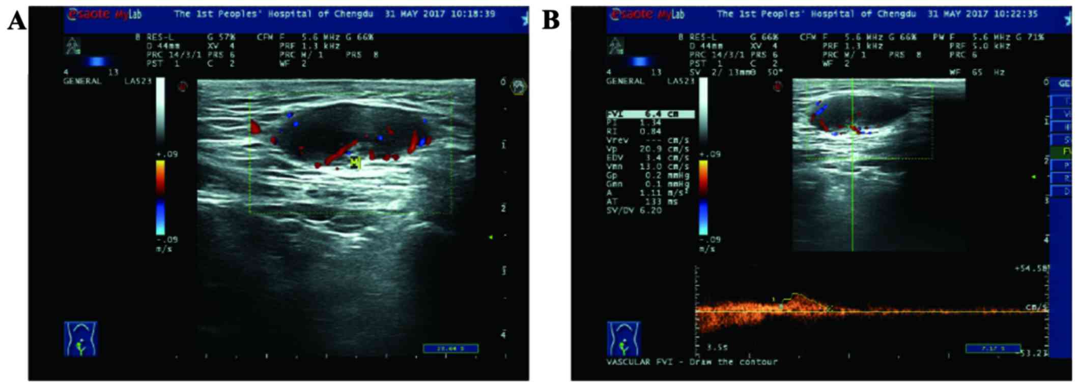
examination. Gray-scale US and CDUS of the vulva were performed with a MyLab Twice ultrasound scanner (Esaote, Genova, Italy) and a 5–10 MHZ multifrequency linear-array transducer. A prominent homogenous hypoechoic and ovoid solid mass with a well-defined border, sized 27×11 mm, was detected in the subcutaneous tissue in the right mons pubis. CDUS and pulse Doppler US revealed arterial blood flow with low resistivity index and low systolic peaks (20.9 cm/sec) in the mass (Fig. 1). The mass was clinically considered to be a benign lesion and was removed by excisional biopsy.

Figure 1.

Real-time ultrasonogram combining Color Doppler with Pulse Doppler findings of the lesion in the subcutaneous region of the right mons pubis. (A) The lesion was a homogenous hypoechoic and ovoid solid mass with a well-defined border and moderate blood flow. (B) The lesion exhibited arterial blood flow with low resistivity index and low systolic peaks (20.9 cm/sec).

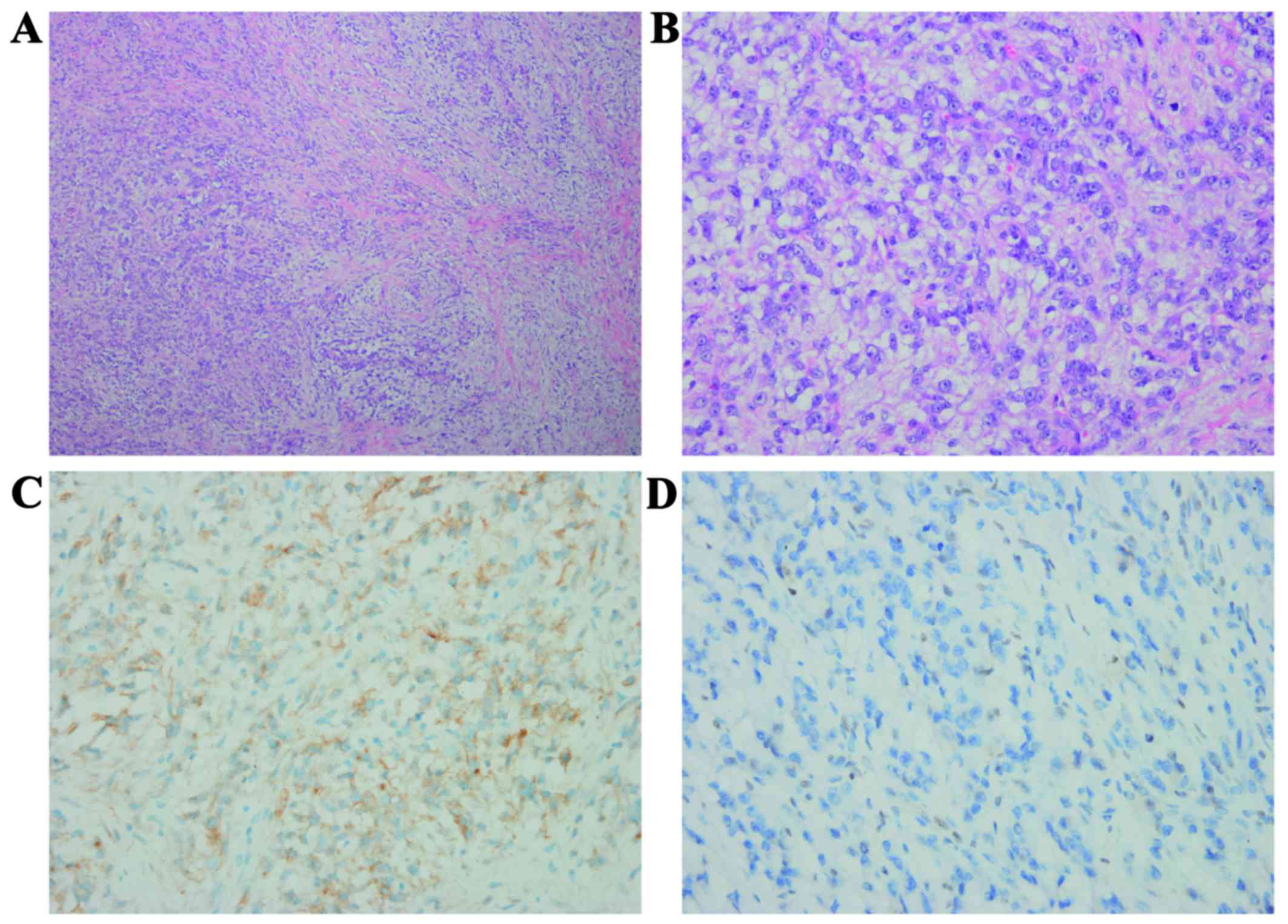
The initial pathology report suggested that the mass was rich in myxoid stroma. Subsequently, the surgical specimen was sent to the West China Hospital for consultation. The results of the immunohistochemical staining of the specimen were as follows: Negative for S100 protein, desmin, CD31, CD34, P40, P63, erythroblast transformation-specific-related gene, anaplastic lymphoma kinase-1, cytokeratin (CK)18, PAN-CK, smooth muscle actin and myogenin; the Ki-67 (MIB-1) index was positive (15% of the nuclei); the specimen was positive for epithelial membrane antigen (EMA) (Fig. 2). Additional INI1 staining revealed a complete loss of expression in tumor cells. These immunohistochemical results confirmed the diagnosis of vulvar PES (myxoid variant) in the right mons pubis. The patient underwent a second excision at the Sichuan Cancer Hospital. There were no residual tumor cells found in the first surgical area. The patient is currently well, without evidence of recurrence or metastasis, 9 months after the operation. During the follow-up period, the patient did not receive any adjuvant therapy.

Figure 2.

Hematoxylin-eosin staining and immunohistochemical expression pattern of the lesion in the mons pubis. (A) The lesion was composed of a mixed population of proliferating epithelioid and spindle cells. The transition between the two cell types is gradual and intercellular collagen deposition may be observed (magnification, ×100). (B) The lesion was rich in myxoid stroma and the tumor cells displayed abundant cytoplasm, enlarged vesicular nuclei and prominent nucleoli (magnification, ×400). (C) The tumor cells exhibited membranous positivity for epithelial membrane antigen (magnification, ×400). (D) The tumor cells exhibited a complete loss of INI1 expression (magnification, ×400).

Discussion

Epithelioid sarcoma (ES), first described by Enzinger in 1970 (4), as an independent entity, is a rare neoplasm accounting for <1% of adult soft tissue sarcomas. Two histological variants have been identified: Classic ES (also referred to as distal-type ES) and proximal-type ES. The former generally occurs in the distal extremities (mostly the hand or wrist) of adolescents and young adults, and it may appear as solitary or multinodular lesions involving either the deep or superficial soft tissues. The latter is rarer, generally occurring in middle-aged or older patients. It often develops in the axilla and the genital area (groin, mons pubis, clitoris and vulva) (5). The most common initial symptom of vulvar PES is a slowly growing, relatively painless mass in the labia majora. Due to its benign appearance as a painless subcutaneous nodule, diagnosis and treatment may often be delayed (6). In the present case, PES occurred in the mons pubis as a slow-growing, painless mass. The mass was clinically considered to be a benign lesion and was removed by excisional biopsy.

US, CT and MRI, plain radiography and PET have been used to assess soft tissue tumors. As US is cost-effective, non-invasive, real-time and easy to perform, it has been used as the main first-line examination method for evaluating soft tissue tumors over several years (7). Due to the technological advances, US can be more sensitive to the detection of minute lesions. Furthermore, the combination of CDUS and spectral analysis may depict the vascularity in malignant tumors, whereas other imaging modalities cannot obtain this type of information (8). In addition, together with clinical examination, US-guided needle biopsy is a more cost-effective method for confirming the final diagnosis (3). Compared with other benign masses, soft tissue sarcomas are more likely to recur and metastasize after surgery. Fornage et al (9) reported that US may not definitively distinguish between recurrent tumor and postsurgical changes in the early postoperative period (3–6 months); however, the sensitivity and specificity of US for identification of recurrent tumors was comparable to that of routine MRI for long-term follow-up.

Immunohistochemistry is useful for differentiating epithelioid sarcomas from other tumors, including other sarcomas with epithelioid cells, malignant melanoma, metastatic carcinoma and malignant extrarenal rhabdoid tumor (MERT) (10). Generally, vulvar PES expresses epithelioid and mesenchymal markers, such as vimentin, EMA, CKs and CD34. Staining for S100 or HMB45 to exclude malignant melanoma is a particularly useful differential diagnosis for PES and MERT. The innovative molecular marker INI 1 may also be used in the differential diagnosis of soft tissue tumors (11).

Optimal treatment for PES of the vulva has not been established due to its rarity. There is no universally accepted treatment for vulvar epithelioid sarcoma (12). However, the cornerstone of initial management is wide surgical excision with an adequate margin (≥2 cm) (13). The role and selection of adjuvant therapy also remain unclear due to the rarity of this disease. Adjuvant radiotherapy is advocated in high-grade tumors or cases with inadequate surgical margins (14), and also due to the high incidence of local recurrence and distant metastasis (15). However, the results of radiotherapy are controversial and show no statistically significant reduction in mortality (16). The role of chemotherapy in the adjuvant setting appears marginally effective at best for the treatment of metastatic disease (17).

In conclusion, US and CDUS may be used to evaluate vulvar PES, a rare soft tissue sarcoma, by location, echogenicity, margin, shape, composition, sound transmission and vascularity, although vulvar PES generally presents as a non-specific solid mass. Combination with other imaging modalities may aid the diagnosis. However, definite diagnosis relies on the histopathological examination.

Acknowledgements

The authors gratefully acknowledge the assistance of Dr Buyun Ma, Department of Ultrasonography, West China Hospital, Sichuan University (Chengdu, China).

Funding

No funding was received.

Availability of materials and data

Not applicable.

Authors' contributions

YY analyzed the clinical data and drafted the manuscript. YL provided the pathological results. YC provided ultrasound examination results and conducted follow-up on the patient in the outpatient clinic. ZT provided professional guidance on pathological results. XM contributed to critical review and supervised the entire study. All the authors have read and approved the final version of this manuscript.

Ethics approval and consent to participate

Not applicable.

Patient consent for publication

Written informed consent was obtained from the patient for publication of this case report and any accompanying images.

Competing interests

The authors declare no potential conflicts of interest with respect to the research, authorship and/or publication of this article.

References

1 

Piver MS, Tsukada Y and Barlow J: Epithelioid sarcoma of the vulva. Obstet Gynecol. 40:839–842. 1972.PubMed/NCBI

2 

Altundag K, Dikbas O, Oyan B, Usubutun A and Turker A: Epithelioid sarcoma of vulva: A case report and review of the literature. Med Oncol. 21:367–372. 2004. View Article : Google Scholar : PubMed/NCBI

3 

Fornage BD: Soft-tissue masses: The case for increased utilization of sonography. Ald Radiol. 29:8–22. 2000.

4 

Enzinger FM: Epithelioid sarcoma. A sarcoma simulating a granulomas or a carcinom. Cancer. 26:1029–1041. 1970. View Article : Google Scholar : PubMed/NCBI

5 

Moore RG, Steinhoff MM, Granai CO and Demars LR: Vulvar epithelioid sarcoma in pregnancy. Gynecol Oncol. 85:218–222. 2002. View Article : Google Scholar : PubMed/NCBI

6 

Ulutin HC, Zellars RC and Frassica D: Soft tissue sarcoma of the vulva: A clinical study. Int J Gynecol Cancer. 13:528–531. 2003. View Article : Google Scholar : PubMed/NCBI

7 

Choi H, Varma DG, Fornage BD, Kim EE and Johnston DA: Soft-tissue sarcoma: MR Imaging vs sonography for detection of local recurrence after surgery. AJR Am J Roentgenol. 157:353–358. 1991. View Article : Google Scholar : PubMed/NCBI

8 

Belli P, Costantini M, Mirk P, Maresca G, Priolo F and Marano P: Role of Color Doppler Sonography in the assessment of musculoskeletal soft tissue masses. J Ultrasound Med. 19:823–830. 2000. View Article : Google Scholar : PubMed/NCBI

9 

Fornage BD: Soft tissue masses: The underutilization of sonography. Semin Musculoskelet Radiol. 3:115–134. 1999. View Article : Google Scholar : PubMed/NCBI

10 

Guillou L, Wadden C, Coindre JM, Krausz T and Fletcher CD: ‘Proximal-type’ epithelioid sarcoma, distinctive aggressive neoplasm showing rhabdoid features. Clinico pathologic, immunohistochemical, and ultrastructural study of a series. Am J Surg Pathol. 21:130–146. 1997. View Article : Google Scholar : PubMed/NCBI

11 

Hornick JL, Dal Cin P and Fletcher CD: Loss of INI1 expression is characteristic of both conventional and proximal-type epithelioid sarcoma. Am J Surg Pathol. 33:542–550. 2009. View Article : Google Scholar : PubMed/NCBI

12 

Lacazzo C, Gkegkes ID and Vrachnis N: Dilemmas in the management of patients with vulvar epithelioid sarcoma: A literature review. Eur J Obstet Gynecol Reprod Biol. 176:1–4. 2014. View Article : Google Scholar : PubMed/NCBI

13 

Bos GD, Pritchard DJ, Reiman HM, Dobyns JH, Ilstrup DM and Landon GC: Epithelioid sarcoma. An analysis of fifty-one cases. J Bone Joint Surg. 70:862–870. 1988. View Article : Google Scholar : PubMed/NCBI

14 

Casanova M, Ferrari A, Collini P, Bisogno G, Alaggio R, Cecchetto G, Gronchi A, Meazza C, Garaventa A, Di Cataldo A, et al: Epithelioid sarcoma in children and adolescents: A report from the Italian soft tissue sarcoma committee. Cancer. 106:708–717. 2006. View Article : Google Scholar : PubMed/NCBI

15 

Hasegawa T, Matsuno Y, Shimoda T, Umeda T, Yokoyama R and Hirohashi S: Proximal-type epithelioid sarcoma: A clinicopathologic study of 20 cases. Mod Pathol. 14:655–663. 2001. View Article : Google Scholar : PubMed/NCBI

16 

Evans HL and Baer SC: Epithelioid sarcoma: A clinicopathologic and prognostic study of 26 cases. Semin Diagn Pathol. 10:286–291. 1993.PubMed/NCBI

17 

Argenta PA, Thomas S and Chura JC: Proximal-type epithelioid sarcoma vs. malignant rhabdoid tumor of the vulva: A case report, review of the literature, and an argument for consolidation. Gynecol Oncol. 107:130–135. 2007. View Article : Google Scholar : PubMed/NCBI

Related Articles

  • Abstract
  • View
  • Download
  • Twitter
Copy and paste a formatted citation
Spandidos Publications style
Yue Y, Lu Y, Ma X, Tang Z and Cheng Y: Ultrasonography findings of proximal‑type epithelioid sarcoma of the vulva: A case report. Mol Clin Oncol 9: 507-510, 2018.
APA
Yue, Y., Lu, Y., Ma, X., Tang, Z., & Cheng, Y. (2018). Ultrasonography findings of proximal‑type epithelioid sarcoma of the vulva: A case report. Molecular and Clinical Oncology, 9, 507-510. https://doi.org/10.3892/mco.2018.1708
MLA
Yue, Y., Lu, Y., Ma, X., Tang, Z., Cheng, Y."Ultrasonography findings of proximal‑type epithelioid sarcoma of the vulva: A case report". Molecular and Clinical Oncology 9.5 (2018): 507-510.
Chicago
Yue, Y., Lu, Y., Ma, X., Tang, Z., Cheng, Y."Ultrasonography findings of proximal‑type epithelioid sarcoma of the vulva: A case report". Molecular and Clinical Oncology 9, no. 5 (2018): 507-510. https://doi.org/10.3892/mco.2018.1708
Copy and paste a formatted citation
x
Spandidos Publications style
Yue Y, Lu Y, Ma X, Tang Z and Cheng Y: Ultrasonography findings of proximal‑type epithelioid sarcoma of the vulva: A case report. Mol Clin Oncol 9: 507-510, 2018.
APA
Yue, Y., Lu, Y., Ma, X., Tang, Z., & Cheng, Y. (2018). Ultrasonography findings of proximal‑type epithelioid sarcoma of the vulva: A case report. Molecular and Clinical Oncology, 9, 507-510. https://doi.org/10.3892/mco.2018.1708
MLA
Yue, Y., Lu, Y., Ma, X., Tang, Z., Cheng, Y."Ultrasonography findings of proximal‑type epithelioid sarcoma of the vulva: A case report". Molecular and Clinical Oncology 9.5 (2018): 507-510.
Chicago
Yue, Y., Lu, Y., Ma, X., Tang, Z., Cheng, Y."Ultrasonography findings of proximal‑type epithelioid sarcoma of the vulva: A case report". Molecular and Clinical Oncology 9, no. 5 (2018): 507-510. https://doi.org/10.3892/mco.2018.1708
Follow us
  • Twitter
  • LinkedIn
  • Facebook
About
  • Spandidos Publications
  • Careers
  • Cookie Policy
  • Privacy Policy
How can we help?
  • Help
  • Live Chat
  • Contact
  • Email to our Support Team