Introduction
Lymph node metastasis is the most powerful predictor
of recurrence or survival in patients with colorectal cancer.
Although the majority of patients with node-negative colorectal
cancer are potentially cured with surgery alone, ≤25% are likely to
present with recurrence and succumb to the disease (1). Identifying high-risk patients with
stage II colorectal cancer is important for determining which
patients may benefit the most from adjuvant chemotherapy.
The effect of clinicopathological factors on
recurrence and survival following curative resection has been the
subject of several studies and numerous clinicopathological factors
have been suggested as prognostic indicators for colorectal cancer.
It is important to determine which of these factors affect the risk
of recurrence in the node-negative colorectal cancer patients and
which factors should be prospectively applied in the routine
clinicopathological evaluation of colorectal cancer.
With the recent developments in immunohistochemistry
and molecular biology, several biological markers have been
extensively investigated (2–12).
In this study, the expression of several biological markers,
including p53, CD10, CD34 and Ki-67, which are strongly suspected
of playing a significant role in tumor progression, was evaluated.
Additionally, we focused on lymph node micrometastasis, which is
easily detected by antibody CAM 5.2.
The aim of this study was to conduct a multivariate
analysis of the prognostic impact of a wide range of
clinicopathological and biological variables in patients with stage
II colorectal cancer.
Materials and methods
Patients
We reviewed all the patients who underwent curative
resection for stage II colorectal cancer at the Department of
Gastroenterological Surgery, Oita University Hospital, between 1984
and 2002. Patients who had received preoperative chemoradiation for
locally advanced lower rectal cancer were excluded from this cohort
study. Ultimately, 109 patients (61 males and 48 females; average
age, 67 years; range, 31–89 years) were enrolled and the tumors
were diagnosed as clinical stage T3, N0 and M0.
Evaluation
The survival analysis was performed for the
following clinicopathological factors: age, gender, location of
tumor (right vs. left colon vs. rectum), number of resected lymph
nodes (0–11 vs. ≥12), bowel obstruction (absent vs. present), tumor
size (0–4 vs. >4 cm), depth of tumor invasion (subserosa vs.
serosa), tumor differentiation (high vs. moderate vs. mucinous),
lymphatic invasion (absent or mild vs. moderate or severe), venous
invasion (absent vs. present), tumor budding (absent vs. present)
(13), peritumoral lymphocytes
(inconspicuous vs. conspicuous) (14) and tumor growth pattern (expansive
vs. infiltrative) (14).
Furthermore, 5 biological markers were asessed using
immunohistochemistry, including p53 (tumor suppressor gene), CD10
(tumor invasion marker), CD34 (angiogenic marker), Ki-67 (cell
proliferation index) and CAM 5.2 (marker of lymph node
micrometastasis). Tumor budding is defined as an isolated single
cancer cell or a cluster composed of <5 cancer cells observed in
the stroma of the actively invasive region. A count of 0–9 per
field was considered as absent and a count of ≥10 was regarded as
present, based on the results of a previous study (13). The characteristics of peritumoral
lymphocytes (inconspicuous vs. conspicuous) and the tumor growth
pattern (expansive vs. infiltrative) were assessed strictly
according to the criteria originally described by Jass et al
(14).
Immunohistochemistry
Resected tumors from each of the 109 patients were
fixed in 10% formalin solution and embedded in paraffin.
Representative tissue sections, each containing the deepest site of
cancer invasion, were cut at 4-μm. As regards the lymph node
specimens, one 3-μm section was obtained for hematoxylin and
eosin staining and five serial 6-μm sections for
immunohistochemical staining. The avidin-biotin peroxidase complex
method was used for detection of the five monoclonal antibodies in
deparaffinized and rehydrated tissue sections. Antigen retrieval
was performed by placing the sample in a microwave oven at 95°C for
40 min, followed by cooling for 30 min to room temperature, except
for CD34 and CAM 5.2. CAM 5.2 sections were trypsinized with 0.1%
calcium chloride solution. p53, Ki-67, CD10 and CD34 were incubated
for 2 h at room temperature and CAM 5.2 was incubated overnight at
4°C. The slides were then incubated for 30 min with EnVision™
peroxidase mouse system (DAKO Corporation, Carpinteria, CA, USA).
The color reaction product was developed with diaminobenzidine
tetrahydrochloride (DAB; Sigma Chemical Co., St. Louis, MO, USA) as
the chromogen for 5 min. Using a light microscope, a visual grading
system was used based on the number of positively stained nuclei of
the cancer cells in each tissue sample.
p53 slides were scored according to the percentage
of positive tumor nuclei as follows: positive, ≥10% of the nuclei
stained; negative, <10% of the nuclei stained (3). For Ki-67 immunoreactivity, staining
was considered positive at >60% (9). Tumor positivity for CD10 was
evaluated using a predetermined cut-off of 5% (positive, >5%
tumor cell staining) according to a previous study (10). CD34 slides were classified
according to the microvessel count. After scanning the highly
vascularized areas, we selected three areas exhibiting the most
prominent neovascularization. A microvessel count was performed on
a ×400 field (×40 objective and ×10 ocular) and the average count
from the three areas was calculated (15). Patients were divided into those
with a microvessel count of 0–50 and those with a microvessel count
of >50. As regards metastatic lymph nodes, patients were divided
into two groups according to a previous study (16), those with micrometastasis in ≤3
lymph nodes and those with micro-metastasis in ≥4 lymph nodes.
Written informed consent was obtained from all the patients and
this study was approved by the local Ethics Committee.
Statistical analysis
Data were statistically analyzed using SPSS
statistical software (Statistical Package for Social Sciences).
Univariate disease-specific survival analysis was performed using
the Kaplan-Meier method and the difference was evaluated by the
log-rank test. Multivariate analysis was performed using the Cox
proportional hazards model. P<0.05 was considered to indicate a
statistically significant difference.
Results
Factors affecting patient survival
The study included a total of 61 males and 48
females, with an average age of 67 years (range, 31–89 years). The
median follow-up period for the survivors was 5.7 years (range,
1.7–11 years). At the time of analysis, 87 patients were free of
disease, 7 were alive with disease, 15 had succumbed to the disease
and 5 patients had succumbed due to other causes. Twenty-two
patients developed recurrence or distant metastasis. Of these, 12
had liver metastases, 7 had local recurrence and 3 had lung
metastasis. The 5-year disease-specific survival rate of patients
with stage II colorectal cancer was 86.2%.
In the univariate analysis, bowel obstruction, lymph
node micrometastasis and lymphatic invasion (P<0.01) were
significant factors for determining the 5-year disease-specific
survival (Table I). When all of
these factors were included as independent variables in a Cox
proportional hazards model, the presence of lymphatic invasion was
the most powerful negative predictor of survival [hazard ratio
(HR), 4.091; P=0.006], followed by lymph node micrometastasis (HR,
3.704; P= 0.011) (Table II). The
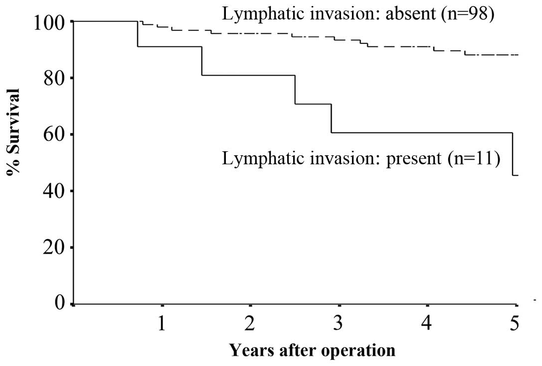
5-year disease-specific survival rate was significantly lower for
the group of patients with moderate to severe presence of lymphatic
invasion compared to that for the group with absent to mild
presence of lymphatic invasion (55 vs. 90%, P<0.01) (Fig. 1). Similarly, the 5-year
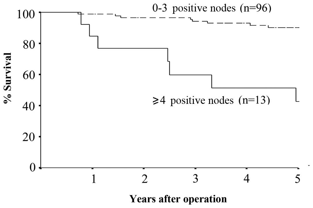
disease-specific survival rate was significantly lower for the
group with ≥4 positive micrometastatic nodes compared to that for
the group with 0–3 positive micrometastatic nodes (46 vs. 92%,
P<0.01) (Fig. 2). When a
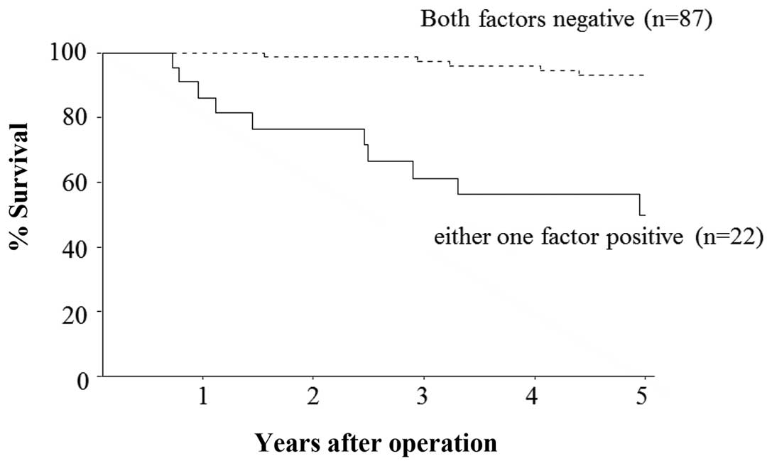
combination of two factors, lymphatic invasion and micrometastasis
was examined, the 5-year disease-specific survival rate for the
group of patients with either one positive factor was significantly
lower compared to that for the group with both factors negative (55
vs. 94%, P<0.01) (Fig. 3).
 | Table IUnivariate analysis for the 5-year
disease-specific survival in patients with stage II colorectal
cancer. |
Table I
Univariate analysis for the 5-year
disease-specific survival in patients with stage II colorectal
cancer.
| Factors | No. of patients | 5-year survival rate
(%) | P-value |
|---|
| Age (years) | | | |
| 0–70 | 73 | 79 | NS |
| ≥71 | 36 | 81 | |
| Gender | | | |
| Male | 61 | 85 | NS |
| Female | 48 | 88 | |
| Location of
tumor | | | |
| Right colon | 21 | 95 | NS |
| Left colon | 52 | 82 | |
| Rectum | 36 | 68 | |
| No. of resected lymph
nodes | | | |
| 0–11 | 43 | 81 | NS |
| ≥12 | 66 | 79 | |
| Bowel
obstruction | | | |
| Absent | 99 | 83 | <0.01 |
| Present | 10 | 50 | |
| Tumor size (cm) | | | |
| 0–4 | 29 | 90 | NS |
| >4 | 80 | 76 | |
| Depth of tumor
invasion | | | |
| T3 | 80 | 90 | NS |
| T4 | 29 | 76 | |
| Differentiation | | | |
| High | 68 | 84 | NS |
| Moderate | 36 | 75 | |
| Poor/mucinous | 5 | 60 | |
| Lymphatic
invasion | | | |
| Absent, mild | 98 | 90 | <0.01 |
| Moderate,
severe | 11 | 55 | |
| Venous invasion | | | |
| Absent | 80 | 89 | NS |
| Present | 29 | 79 | |
| Tumor budding | | | |
| Absent | 70 | 84 | NS |
| Present | 39 | 90 | |
| Peritumoral
lymphocytes | | | |
| Inconspicuous | 40 | 83 | NS |
| Conspicuous | 69 | 88 | |
| Tumor growth
pattern | | | |
| Expansive | 30 | 91 | NS |
| Infiltrative | 79 | 84 | |
| p53 | | | |
| Negative | 49 | 84 | NS |
| Positive | 60 | 88 | |
| CD10 | | | |
| Negative | 70 | 83 | NS |
| Positive | 39 | 92 | |
| Angiogenesis
(microvessel count) | | | |
| 0–50 | 90 | 80 | NS |
| >50 | 19 | 79 | |
| Ki-67 index | | | |
| Sparse | 94 | 78 | NS |
| Diffuse | 15 | 93 | |
| Lymph node
micrometastasis | | | |
| 0–3 positive
nodes | 96 | 92 | <0.01 |
| ≥4 positive
nodes | 13 | 46 | |
| Table IIMultivariate analysis for the 5-year
disease-specific survival in patients with stage II colorectal
cancer. |
Table II
Multivariate analysis for the 5-year
disease-specific survival in patients with stage II colorectal
cancer.
| Factors | HR | 95% CI | P-value |
|---|
| Lymphatic
invasion | 4.091 | 1.376–12.165 | 0.006 |
| Lymph node
micrometastasis | 3.704 | 1.458–9.406 | 0.011 |
Discussion
In the present study, 20% of the stage II colorectal
cancer patients presented with tumor recurrence or distant
metastasis during follow-up, after curative resection. A
multivariate analysis allowed us to define a subgroup of patients
at high risk of recurrence, which included those with lymph node
micrometastasis and those with lymphatic invasion. In addition,
these factors were significantly associated with the prognosis of
stage II colorectal cancer patients.
Recent advances in immunohistochemistry and
molecular biology suggest that molecular changes of the primary
tumor may serve as prognostic indicators for individual patients.
Several studies have attempted to identify the prognostic
biomarkers in patients with stage II or node-negative colorectal
cancer (2–7, 12,17,18).
Although several studies have been conducted on
lymph node micrometastasis in patients with colorectal cancer, the
significance of the presence of lymph node micrometastasis has been
a subject of debate (16,18–21).
Yasuda et al (16) reported
that micrometastasis in ≥4 lymph nodes and micrometastasis to N2 or
higher nodes were significantly correlated with postoperative
recurrence and prognosis in stage II colorectal cancer patients.
Bukholm et al (21)
reported that the presence of isolated tumor cells in the
mesenteric lymph nodes was independently associated with reduced
relative survival in patients with stage II colon cancer. Our study
also demonstrated that the number of lymph node micrometastases was
a more powerful indicator than the presence and level of lymph node
micrometastasis. Therefore, it is helpful to investigate the number
of lymph node micrometastases with immunohistochemistry in stage II
colorectal cancer patients.
The aim of adjuvant chemotherapy is the destruction
of microscopic metastases that may already be present and the
reduction of the risk of recurrence. Postoperative chemotherapy for
stage III colorectal cancer patients has been shown to improve
prognosis and is recommended as standard therapy (22,23).
However, the value of adjuvant chemotherapy for patients with stage
II colorectal cancer is controversial (24,25).
The International Multicentre Pooled Analysis of B2 Cancer Trials
(IMPACT B2) (26) and the
meta-analysis reported by Figueredo et al (27) did not demonstrate any improvement
in prognosis of stage II colon cancer patients treated with
adjuvant chemotherapy. However, the QUASAR study demonstrated a
significantly reduced recurrence rate and improved survival of
patients with stage II colorectal cancer in favour of the adjuvant
chemotherapy arm (28).
Although several large studies have investigated the
subject of adjuvant chemotherapy for stage II colorectal cancer
patients, the use of adjuvant chemotherapy for all stage II
colorectal cancer patients may be inappropriate and expensive
(29). Therefore, there is an
increasing need for accurate stratification of stage II colorectal
cancer patients in order to identify those at high-risk of
recurrence who may benefit from adjuvant chemotherapy.
Our data suggest that two factors, lymph node
micrometastasis and lymphatic invasion, should be included in the
high-risk group of patients with stage II colorectal cancer. Sirop
et al (30) reported
improved outcomes of micrometastasis after being considered as
high-risk disease and treated with chemotherapy in their pilot
study. These results suggest a trend in favour of adjuvant
chemotherapy in stage II colorectal cancer patients with high-risk
factors.
In conclusion, we demonstrated that each of the two
factors investigated, lymph node micrometastasis and lymphatic
invasion, carries independent prognostic significance with respect
to the 5-year disease-specific survival rates of patients with
stage II colorectal cancer. This finding may be useful in
identifying the high-risk patients for recurrence or metastasis
among stage II colorectal cancer patients. We recommend that stage
II colorectal cancer patients with lymph node micrometastasis and
lymphatic invasion be evaluated for the benefit of adjuvant
chemotherapy in the future, through further prospective randomized
control studies.
Acknowledgements
This study was supported in part by
the National Cancer Center Research and Development Fund
(23-A-19).
References
|
1.
|
Jemal A, Siegel R, Ward EA, et al: Cancer
statistics, 2009. CA Cancer J Clin. 59:225–249. 2009. View Article : Google Scholar
|
|
2.
|
Graziano F and Cascinu S: Prognostic
molecular markers for planning adjuvant chemotherapy trials in
Dukes’ B colorectal cancer patients: how much evidence is enough?
Ann Oncol. 14:1026–1038. 2003.PubMed/NCBI
|
|
3.
|
Allegra CJ, Paik S, Colangelo LH, et al:
Prognostic value of thymidylate synthase, Ki-67, and p53 in
patients with Dukes’ B and C colon cancer: a National Cancer
Institute-National Surgical Adjuvant Breast and Bowel Project
collaborative study. J Clin Oncol. 21:241–250. 2003.
|
|
4.
|
Garrity MM, Burgart LJ, Mahoney MR, et al:
Prognostic value of proliferation, apoptosis, defective DNA
mismatch repair, and p53 overexpression in patients with resected
Dukes’ B2 or C colon cancer: a North Central Cancer Treatment Group
Study. J Clin Oncol. 22:1572–1582. 2004.PubMed/NCBI
|
|
5.
|
Bhatavdekar JM, Patel DD, Chikhlikar PR,
et al: Molecular markers are predictors of recurrence and survival
in patients with Dukes B and Dukes C colorectal adenocarcinoma. Dis
Colon Rectum. 44:523–533. 2001. View Article : Google Scholar : PubMed/NCBI
|
|
6.
|
Gervaz P, Cerottini JP, Bouzourene H, et
al: Comparison of microsatellite instability and chromosomal
instability in predicting survival of patients with T3N0 colorectal
cancer. Surgery. 131:190–197. 2002. View Article : Google Scholar : PubMed/NCBI
|
|
7.
|
Sinicrope FA, Hart J, Hsu HA, et al:
Apoptotic and mitotic indices predict survival rates in lymph
node-negative colon carcinomas. Clin Cancer Res. 5:1793–1804.
1999.PubMed/NCBI
|
|
8.
|
Kim YH, Lee JH, Chun H, et al: Apoptosis
and its correlation with proliferative activity in rectal cancer. J
Surg Oncol. 79:236–242. 2002. View Article : Google Scholar : PubMed/NCBI
|
|
9.
|
Sadahiro S, Suzuki T, Maeda Y, et al:
Predictors of tumor downsizing and regression with preoperative
radiotherapy alone and with concomitant tegafur/uracil (UFT) for
resectable advanced rectal adenocarcinoma. Hepatogastroenterology.
54:1107–1112. 2007.
|
|
10.
|
Fujimoto Y, Nakanishi Y, Sekine S, et al:
CD10 expression in colorectal carcinoma correlates with liver
metastasis. Dis Colon Rectum. 48:1883–1889. 2005. View Article : Google Scholar : PubMed/NCBI
|
|
11.
|
Ogawa H, Iwaya K, Izumi M, et al:
Expression of CD10 by stromal cells during colorectal tumor
development. Hum Pathol. 33:806–811. 2002. View Article : Google Scholar : PubMed/NCBI
|
|
12.
|
Weber JC, Nakano H, Bachellier P, et al:
Is a proliferation index of cancer cells a reliable prognostic
factor after hepatectomy in patients with colorectal liver
metastases? Am J Surg. 182:81–88. 2001. View Article : Google Scholar : PubMed/NCBI
|
|
13.
|
Ueno H, Price AB, Wilkinson KH, et al: A
new prognostic staging system for rectal cancer. Ann Surg.
240:832–839. 2004. View Article : Google Scholar : PubMed/NCBI
|
|
14.
|
Jass JR, Love SB and Northover JM: A new
prognostic classification of rectal cancer. Lancet. 1:1303–1306.
1987. View Article : Google Scholar : PubMed/NCBI
|
|
15.
|
Oh-e H, Tanaka S, Kitadai Y, et al:
Angiogenesis at the site of deepest penetration predicts lymph node
metastasis of submucosal colorectal cancer. Dis Colon Rectum.
44:1129–1136. 2001. View Article : Google Scholar : PubMed/NCBI
|
|
16.
|
Yasuda K, Adachi Y, Shiraishi N, et al:
Pattern of lymph node micrometastasis and prognosis of patients
with colorectal cancer. Ann Surg Oncol. 8:300–304. 2001. View Article : Google Scholar : PubMed/NCBI
|
|
17.
|
Okuyama T, Nakamura T and Yamaguchi M:
Budding is useful to select high-risk patients in stage II
well-differentiated or moderately differentiated colon
adenocarcinoma. Dis Colon Rectum. 46:1400–1406. 2003. View Article : Google Scholar : PubMed/NCBI
|
|
18.
|
Feezor RJ, Copeland EM III and Hochwald
SN: Significance of micrometastases in colorectal cancer. Ann Surg
Oncol. 9:944–953. 2002. View Article : Google Scholar : PubMed/NCBI
|
|
19.
|
Palmqvist R, Sellberg P, Oberg A, et al:
Low tumour cell proliferation at the invasive margin is associated
with a poor prognosis in Dukes’ stage B colorectal cancers. Br J
Cancer. 79:577–581. 1999.PubMed/NCBI
|
|
20.
|
Noura S, Yamamoto H, Miyake Y, et al:
Immunohistochemical assessment of localization and frequency of
micrometastases in lymph nodes of colorectal cancer. Clin Cancer
Res. 8:759–767. 2002.PubMed/NCBI
|
|
21.
|
Bukholm IR, Bondi J, Wiik P, et al:
Presence of isolated tumour cells in mesenteric lymph nodes
predicts poor prognosis in patients with stage II colon cancer. Eur
J Surg Oncol. 29:862–866. 2003. View Article : Google Scholar : PubMed/NCBI
|
|
22.
|
O’Connell MJ, Mailliard JA, Kahn MJ, et
al: Controlled trial of fluorouracil and low-dose leucovorin given
6 months as postoperative adjuvant therapy for colon cancer. J Clin
Oncol. 15:246–250. 1997.PubMed/NCBI
|
|
23.
|
Barone C: Adjuvant chemotherapy of colon
cancer current strategies. Eur J Cancer. 6:60–63. 2008. View Article : Google Scholar
|
|
24.
|
Van Cutsem E and Costa F: Progress in the
adjuvant treatment of colon cancer. Has it influenced clinical
practice? JAMA. 7:2758–2760. 2005.PubMed/NCBI
|
|
25.
|
O’Connell MJ: Oxaliplatin or irinotecan as
adjuvant therapy for colon cancer: the results are in. J Clin
Oncol. 27:3082–3084. 2009.
|
|
26.
|
No authors listed:. Efficacy of adjuvant
fluorouracil and folinic acid in B2 colon cancer. International
Multicentre Pooled Analysis of B2 Colon Cancer Trials (IMPACT B2)
Investigators. J Clin Oncol. 17:1356–1363. 1999.
|
|
27.
|
Figueredo A, Charrette M, Maroun J, et al:
Adjuvant therapy for stage II colon cancer: a systematic review
from the Cancer Care Ontario Program in evidence-based care’s
gastrointestinal cancer disease site group. J Clin Oncol.
22:3395–3407. 2004.
|
|
28.
|
Quasar Collaborative Group; Gray R,
Barnwell J, McConkey C, Hills RK, Williams NS and Kerr DJ: Adjuvant
chemotherapy versus observation in patients with colorectal cancer:
a randomised study. Lancet. 370:2020–2029. 2007. View Article : Google Scholar : PubMed/NCBI
|
|
29.
|
Norum J: Adjuvant chemotherapy in Dukes’ B
and C colorectal cancer has only a minor influence on psychological
distress. Support Care Cancer. 5:318–321. 1997.
|
|
30.
|
Sirop S, Kanaan M, Korant A, et al:
Detection and prognostic impact of micrometastasis in colorectal
cancer. J Surg Oncol. 103:534–537. 2011. View Article : Google Scholar : PubMed/NCBI
|