Introduction
It is a well known fact that environmental factors
are as important as genetic factors in the carcinogenesis of
urothelial cancer (UC). Cigarette smoking, in particular, is a
major risk factor for cancer, with the risk of developing bladder
cancer in smokers being several-fold that in non-smokers (1). In addition to carcinogenesis,
cigarette smoking plays an important role in cancer progression and
the prognosis of patients with bladder cancer (2–4).
Although a similar role for smoking has been reported in patients
with upper tract (UT) UC, there are conflicting opinions regarding
the pathological significance and prognostic role of smoking status
in these patients (5–9). In addition, it was previously
suggested that the changes in the malignant potential of UTUC
induced by cigarette smoke were different from those in bladder
cancer (4). Therefore, a detailed
investigation on the effect of smoking is crucial for understanding
the fundamental characteristics of UTUC. However, the association
between smoking and changes in pathological characteristics and
molecular expression of cancer-related molecules in UTUC has not
been fully elucidated. Cigarette smoke affects numerous
pathological characteristics and molecules in vivo and in
vitro. In the field of oncology, exposure to cigarette smoke
may modify various cancer-related factors and molecules (10). In this study, we focused
specifically on the following invasion- and metastasis-related
factors: cancer cell proliferation, apoptosis, angiogenesis,
lymphangiogenesis and expression of cyclooxygenase (COX)-2,
vascular endothelial growth factors (VEGFs), matrix
metalloproteinases (MMPs) and urokinase-type plasminogen activator
(uPA), as these factors play important roles in UC (2, 11–15).
The effect of smoking on the carcinogenesis,
pathological characteristics and prognosis of UTUC has been
reported and reviewed in earlier studies (5–9).
Therefore, the main objective of this study was to elucidate the
association between the smoking status of patients and the
malignant aggressiveness of UTUC at the molecular level. Tumor
growth, invasion and metastasis affect the quality of life and
survival of patients with UTUC. Therefore, a detailed understanding
of cancer cell dissemination and recurrence are crucial for the
determination of appropriate observation and treatment strategies.
Our results may provide new information on smoking-induced changes
in the expression of cancer-related factors in patients with
UTUC.
Materials and methods
Patients and tissue samples
A total of 134 patients diagnosed with
non-metastatic UTUC were retrospectively investigated. Patients who
had received preoperative therapy and/or who had carcinoma in
situ (Cis) were excluded, due to difficulties in the evaluation
of cancer-related factors and semi-quantification of immunological
stainings. All the histological characteristics, including tumor
grade and pT stage, were determined using formalin-fixed and
paraffin-embedded specimens from open nephroureterectomy; cancer
stage and grade were assessed using the 2002 tumor-node-metastasis
(TNM) classification (16). The
cancer was classified into three grades, namely G1, G2 and G3,
according to the World Health Organization classification (17). A single pathologist performed all
the pathological examinations. The median duration of the follow-up
was 52 months (range, 2-250 months). The study protocol was
approved by the Human Ethics Review Committee of the Nagasaki
University Hospital.
Immunohistochemistry and evaluation of
cancer-related factors
Immunohistochemical staining was performed according
to our previous reports (11,
14, 18–21).
Briefly, 5-µm sections were deparaffinized stepwise in xylene and
rehydrated in graded solutions of ethanol. Antigen retrieval was
performed at 121˚C for 15 min (for the anti-Ki-67 antibody) or 95˚C
for 40 min (for all the other antibodies) in 0.01 M sodium citrate
buffer (pH 6.0). All the sections were then immersed in 3% hydrogen
peroxide for 30 min to block the endogenous peroxidase activity.
The primary anti-Ki-67 and anti-D2-40 antibodies were obtained from
Dako (Glostrup, Denmark); the anti-CD105 antibody was obtained from
Vector Laboratories (Burlingame, CA, USA); the anti-VEGF-A antibody
was obtained from Santa Cruz Biotechnology, Inc. (Santa Cruz, CA,
USA); the anti-COX-2 antibody was obtained from Immuno-Biological
Laboratories Co., Ltd. (Gunma, Japan); the anti-VEGF-C and
anti-VEGF-D antibodies were obtained from Zymed Laboratories, Inc.
(San Francisco, CA, USA); the anti-MMP-2 and anti-MMP-9 antibodies
were obtained from Daiichi Fine Chemical Co., Ltd. (Toyama, Japan);
and the anti-uPA antibody was obtained from American Diagnostica,
Inc. (Stamford, CT, USA). The sections were incubated overnight at
4˚C with the primary antibodies. Subsequently, the sections were
treated with DAKO EnVision ™ + Peroxidase (Dako, Carpinteria, CA,
USA) for 60 min using the labeled polymer method. The peroxidase
reaction was visualized using Liquid DAB Substrate kit (Zymed
Laboratories, Inc). The sections were counterstained with
hematoxylin, dehydrated stepwise through a graded alcohol series
and cleared in xylene prior to mounting. Consecutive sections from
each sample processed without the primary antibody were used as the
negative control. In addition, in situ labeling for
apoptosis was performed as previously described (18), using the ApopTag ® In
Situ Apoptosis Detection kit (Intergen Company, Purchase, NY, USA),
which is based on the terminal deoxynucleotidyl
transferase-mediated nick end labeling method.
Evaluation
All the analyses of immunohistochemically stained
sections were performed using light microscopy within a tumor area
containing ≥ 500 carcinoma cells. In this study, lymphatic vessel
density (LVD) was described in the peritumoral area according to
previous report (20). Two
investigators (S.W. and Y.M.), who were blinded to clinical data,
independently performed the semi-quantitative analyses and the
immunostaining interpretations. The disagreement rate for analyses
between the two investigators was <10% and results from both
investigators were averaged for the statistical analyses. The
presence of intratumoral lymp hatic vessel (iLV) was defined as at
least one D2-40 positive vessel clearly visible in the intratumoral
area (20). As regards the
expression of VEGF-A and -C, staining intensity was classified into
four grades as follows : absent, weak, moderate and strong; the
specimens were considered positive when >25% of the carcinoma
cells were clearly (moderately or strongly) stained for statistical
evaluation according to a previous report (11). The expression of MMP-2 and −9 was
also evaluated as previously reported (14). Briefly, the staining intensity was
graded as weak, moderate, or intense. In addition, the extent of
positive staining in the tumor area was graded as focal (≤ 10%),
regional (11-50%), or diffuse (≥ 50%). Finally, the staining
patterns of moderate and diffuse, intense and regional, or intense
and diffuse were considered to be positive for MMPs. For the
evaluation of uPA, staining intensity was scored as 1 (weak), 2
(moderate) or 3 (intense staining). In addition, the extent of
staining was scored as 1, 2 or 3 (<10, 10–50 or >50% of tumor
area stained, respectively). These two scores were summed up to
give each case a final score (immunoreactive score of 0–6) and uPA
was considered as positive when the immunoreactive score was 3–6
according to a previous report (21). Tumor sections stained with each
antibody were examined under a Nikon E-400 bright-field microscope
(Nikon, Tokyo, Japan) and images were captured using a digital
camera (DU100; Nikon) at ×200 magnification. For the assessment of
microvessel density (MVD) and LVD, 3–5 fields with the greatest
density of positively stained vessels (hot spots) were evaluated.
Values were defined as the number of positively stained vessels per
high-power field, as estimated from a computer-aided image analysis
(WinROOF version 6.4; Mitani, Fukui, Japan).
Statistical analyses
Normality was evaluated by normal distribution and
histograms for each variable. The results are expressed as mean ±
standard deviation (SD), unless otherwise stated. A Student's
t-test was performed for continuous variables. The Scheffé test was
used for multiple comparisons of data. Crude and adjusted effects
were estimated by logistic regression analysis and described as
odds ratios (ORs) with 95% confidence intervals, together with
P-values. For the survival analyses, variables that achieved
statistical significance (P<0.050) in the univariate analyses
were subsequently entered into a multivariate analysis using the
Cox proportional hazards model. All the statistical tests were
two-sided; statistical significance was set at P<0.050. All the
statistical analyses were performed on a personal computer using
the StatView program of the Windows statistical package, version
5.0 (Abacus Concepts, Berkeley, CA, USA).
Results
Smoking status and clinicopathological
characteristics
Our study population included patients who had never
smoked/non-smokers (n=54, 40.3%), former smokers (n=46, 34.3%) and
current smokers (n=34, 25.4%). The associations between the
clinicopathological characteristics of UTUC and the smoking status
of the patients are shown in Table
I. Age at diagnosis in former smokers tended to be higher
compared to that in other groups; however, this difference was not
statistically significant. The number of former or current smokers
was significantly higher (P<0.001) among male (39.2 and 32.4%,
respectively) compared to that among female patients (18.8 and 3.1
%, respectively). However, the pathological characteristics of UTUC
exhibited no significant differences according to smoking
status.
 | Table I.Association between
clinicopathological characteristics and smoking status. |
Table I.
Association between
clinicopathological characteristics and smoking status.
|
|
| Smoking status at
diagnosis, n (%) |
|---|
|
|
|
|
|---|
|
Characteristics | Total patient
number (n=134) | Never (n=54) | Former (n=46) | Current (n=34) |
|---|
| Mean age (SD) |
| 67.9 (11.8) | 70.1 (8.6) | 66.8 (10.8) |
| Gender |
|
Male | 102 | 29 (28.4) | 40 (39.2) | 33 (32.4) |
|
Female | 32 | 25 (78.1) | 6 (18.8) | 1 (3.1) |
|
P-value |
|
| <0.001 |
|
| Grade |
| 1 | 24 | 6 (25.0) | 11 (45.8) | 7 (29.2) |
| 2 | 62 | 24 (38. 8) | 19 (30.6) | 19 (30.6) |
| 3 | 48 | 24 (50.0) | 16 (33.3) | 8 (16.7) |
|
P-value |
|
| 0.200 |
|
| pT stage |
| Ta | 13 | 6 (46.2) | 4 (30.7) | 3 (23.1) |
| T1 | 43 | 18 (41.9) | 15 (34.9) | 10 (23. 2) |
| T2 | 24 | 7 (29. 2) | 9 (37.5) | 8 (33.3) |
| T3 | 44 | 18 (40.9) | 17 (38.6) | 9 (20.5) |
| T4 | 10 | 5 (50.0) | 1 (10.0) | 4 (40.0) |
|
P-value |
|
| 0.740 |
|
| Low
(pTa and T 1) | 56 | 24 (42.9) | 19 (33.9) | 13 (23.2) |
| High
(pT2-4) | 78 | 30 (38.5) | 27 (34.6) | 21 (26.9) |
|
P-value |
|
| 0.845 |
|
Smoking status and cancer-related
factors
The associations between the smoking status of the
patients and the cancer-related factors are shown in Table II. The proliferation index (PI) as
well as the apoptotic index appeared to be positively correlated
with smoking status; however, the correlation was not statistically
significant. However, the mean/SD level of LVD in current smokers
(40.9/12.9) was significantly higher (P=0.034) compared to that in
patients who had never smoked (34.4/10.6). In addition, smoking
status was correlated with the presence of iLV (P=0.010) and the
expression of VEGF-D (P=0.011), COX-2 (P=0.032) and MMP-9
(P=0.032). With respect to the expression of VEGF-A, VEGF-C and
MMP-2, although the positively stained ratio in current smokers was
higher compared to that in those who had never smoked or former
smokers, the differences were not statistically significant.
Similarly, smoking status did not appear to be associated with MVD
or uPA expression.
| Table II.Association between cancer-related
factors and smoking status. |
Table II.
Association between cancer-related
factors and smoking status.
|
| Smoking status |
|
|---|
|
|
|
|
|---|
| Factors | Never (n=54) | Former (n=46) | Current (n=34) | P-value |
|---|
| Proliferation index
(mean/SD) | 20.1/10.5 | 21.2/10.3 | 22.7/11.7 | NS |
| Apoptotic index
(mean/SD) | 1.5/0.7 | 1.6/0.8 | 1.7/0.9 | NS |
| Microvessel density
(mean/SD) | 31.0/16.4 | 27.0/13.2 | 32.1/19.6 | NS |
| Lymphatic vessel
density (mean/SD) | 34.4/10.6 | 39.6/10.8 | 40.9/12.9 | 0.034a |
| Intratumoral lymph
vessels, n (%) |
|
|
| 0.010 |
|
Absent | 51 (94.4) | 37 (80.4) | 24 (70.5) |
|
|
Present | 3 (5.6) | 9 (19.6) | 10 (29.5) |
|
| VEGF-A expression,
n (%) |
|
|
| 0.542 |
|
Negative | 30 (55.6) | 22 (47.8) | 15 (44.1) |
|
|
Positive | 24 (44.4) | 24 (52.2) | 19 (55.9) |
|
| VEGF-C expression,
n (%) |
|
|
| 0.779 |
|
Negative | 29 (53.7) | 22 (47.8) | 16 (47.1) |
|
|
Positive | 25 (46.3) | 24 (52.2) | 18 (52.9) |
|
| VEGF-D expression,
n (%) |
|
|
| 0.011 |
|
Negative | 47 (47.0) | 33 (33.0) | 20 (20.0) |
|
| Positive | 7 (20.6) | 13 (38.2) | 14 (41.2) |
|
| COX-2 expression, n
(%) |
|
|
| 0.032 |
|
Negative | 34 (63.0) | 21 (45.7) | 12 (35.3) |
|
|
Positive | 20 (37.0) | 25 (54.3) | 22 (64.7) |
|
| MMP-2 expression, n
(%) |
|
|
| 0.359 |
|
Negative | 33 (61.1) | 23 (50.0) | 16 (47.1) |
|
|
Positive | 21 (38.9) | 23 (50.0) | 18 (52.9) |
|
| MMP-9 expression, n
(%) |
|
|
| 0.032 |
|
Negative | 35 (64.8) | 21 (45.7) | 13 (38.2) |
|
|
Positive | 19 (35.2) | 25 (54.3) | 21 (61.8) |
|
| uPA expression, n
(%) |
|
|
| 0.995 |
|
Negative | 24 (44.4) | 20 (43.5) | 15 (44.1) |
|
|
Positive | 30 (55.6) | 26 (56.5) | 19 (55.9) |
We next investigated the independent correlation
between the smoking status and the cancer-related factors using a
multivariate analysis model that included pathological
characteristics. As shown in Table
III, former smoking was independently associated with LVD (OR=
1.04, P=0.024), the presence of iLV (OR=5.1, P=0.027) and VEGF-D
expression (OR=3.0, P=0.043), but not with the expression of COX-2
or MMP-9. However, current smoking was independently associated
with all these factors (Table
III).
| Table III.Correlation of different factors with
smoking by multivariate analys i sa. |
Table III.
Correlation of different factors with
smoking by multivariate analys i sa.
| Factors | OR | 95% CI | P-value |
|---|
| In former
smokers |
|
|
|
| LV
density | 1.04 | 1.01-1.1 | 0.024 |
|
Presence of iLV | 5.1 | 1.2-21.7 | 0.027 |
| VEGF-D:
positive | 3.0 | 1.03-8.4 | 0.043 |
| COX-2:
positive | 2.2 | 0.9-5.1 | 0.079 |
| MMP-9:
positive | 2.0 | 0.9-4.7 | 0.112 |
| In current
smokers |
|
|
|
| LV
density | 1.1 | 1.01-1.1 | 0.011 |
|
Presence of iLV | 9.7 | 2.2-42.3 | 0.003 |
| VEGF-D:
positive | 5.3 | 1.8-15.5 | 0.003 |
| COX-2:
positive | 3.3 | 1.3-8.7 | 0.014 |
| MMP-9:
positive | 3.5 | 1.3-9.0 | 0.011 |
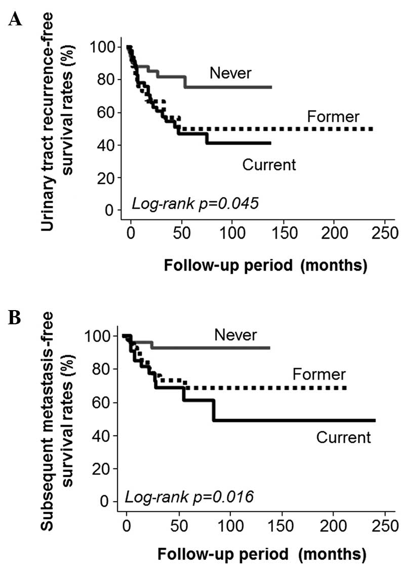
Survival analyses
The Kaplan-Meier survival curves demonstrated that
the smoking status of a patient was a significant predictor of the
recurrence of urinary tract cancer (log-rank P=0.045; Fig. 1A) and subsequent metastasis
(log-rank P=0.016; Fig. 1B), but
not of cause-specific survival (log-rank P=0.514). Based on these
results, the roles of various cancer-related factors in the
recurrence of urinary tract carcinoma and subsequent metastasis
were evaluated and are shown in Table
IV. The Cox proportional hazard analyses indicated that the
presence of iLV [hazard ratio (HR)=2.5, P=0.007] and the expression
of COX-2 (HR=2.1, P=0.018) and MMP-9 (HR=2.0, P=0.026), but not
LVD, were significantly associated with the recurrence of urinary
tract cancer. However, similar analyses revealed that they were all
significantly associated with subsequent metastasis (Table IV).
| Table IV.Cox proportional hazard analyses of
smoking-related factors. |
Table IV.
Cox proportional hazard analyses of
smoking-related factors.
|
|
|
| Positive
expression |
|---|
|
|
|
|
|
|---|
| Factors | LVD | Presence of
iLV | COX-2 | MMP-9 |
|---|
| For urinary tract
recurrence |
|
|
|
|
| Hazard
ratio | 1.02 | 2.5 | 2.1 | 2.0 |
| 95%
CI | 0.99-1.05 | 1.3-4.8 | 1.1-3.9 | 1.1-3.6 |
|
P-value | 0.087 | 0.007 | 0.018 | 0.026 |
| For subsequent
metastasis |
|
|
|
|
| Hazard
ratio | 1.05 | 5.6 | 8.3 | 3.5 |
| 95%
CI | 1.01-1.1 | 2.5-12.3 | 2.5-27.9 | 1.5-8.4 |
|
P-value | 0.002 | <0.001 | <0.001 | 0.005 |
Discussion
The present study demonstrated that smoking leads to
lymphangiogenesis and increased expression levels of COX-2 and
MMP-9 in patients with UTUC. Furthermore, these changes may play
important roles in the recurrence of cancer in the urinary tract
and in subsequent metastasis following nephroureterectomy, as these
two molecules have been reported to be associated with recurrence
and prognosis in patients with UC (14, 22,
23). Although smoking is a major
causative risk factor and a significant predictor of outcome in
patients with UC, including UTUC (5, 9),
the smoking-induced changes in the malignant behavior and
expression of cancer-related factors have not yet been fully
elucidated. To the best of our knowledge, this is the first study
to describe the association between smoking status and the
aggressiveness of cancer at the molecular level in human UTUC
tissues.
The expression levels of VEGF-D, COX-2 and MMP-9,
which are invasion- and metastasis-related molecules, were found to
be significantly associated with smoking status in our study
population. In bladder cancer tissues, a recent study reported that
VEGF-D overexpression was associated with a high frequency of lymph
node metastasis and poor survival in bladder cancer (24). Interestingly, this study also
demonstrated that VEGF-C expression did not play such significant
pathological and prognostic roles. We hypothesized that this
phenomenon was the case not only in bladder cancer, but also in
UTUC. There is little information on the association between COX-2
expression and UTUC according to smoking status. However, in an
in vitro study using bladder cancer cell lines, cigarette
smoke extract induced COX-2 expression in a time-dependent manner
(25). Similar findings were also
reported in human bladder cancer tissues (3, 26).
Thus, various studies suggested that smoking mediates bladder
cancer aggressiveness by regulating the expression of COX-2
(2). Based on these previously
published reports and the results of the present study, we suggest
that a similar positive correlation may exist in patients with UTUC
and that it may represent one of the important mechanisms
underlying the smoking-related malignant behavior of cancer cells.
It is known that smoking induces polymorphism and expression of
MMP-9 in bladder cancer (27);
however, there is no report on the effect of smoking on MMP-9
expression in human UTUC tissues. In other types of cancer,
cigarette smoke extract induces MMP-9 expression in vitro
(28, 29). Of note, exposure to cigarette smoke
also increases the expression levels of COX-2 in lung cancer tissue
(28). Furthermore, in patients
with cardiovascular diseases, several investigators have observed
that smoking increases the expression level of MMP-9, but not that
of MMP-2 (30, 31). Therefore, we hypothesized that
smoking may stimulate cancer cell invasion and metastasis by
inducing an increase in the expression levels of COX-2 and MMP-9 in
patients with UTUC.
Several studies previously reported that the
upregulation of lymphangiogenesis and lymphovascular invasion are
crucial steps in cancer cell dissemination in patients with UTUC
(20, 32). However, there is little information
regarding the effect of smoking status on lymphangiogenesis in
patients with UTUC. In patients with lung cancer, smoking was found
to be associated with high expression levels of interleukin-17 and
was hypothesized to play an important role in metastasis by
promoting lymphangiogenesis (33).
In addition, although the association between lymphangiogenesis and
the expression of COX-2 or MMP-9 in UTUC is not fully understood,
these two molecules have been reported to stimulate
lymphangiogenesis in other types of cancer (34, 35). On the basis of these facts, we
hypothesized that smoking induces cancer cell dissemination by
stimulating lymphangiogenesis via the upregulation of COX-2 and
MMP-9 expression in patients with urothelial carcinoma of the upper
urinary tract (UC-UUT).
Our results demonstrated that the smoking status of
patients exhibited no significant correlation with either cell
proliferation or apoptosis. Similarly, smoking status was reported
to have no significant correlation with PI in bladder cancer
(36). However, a previous study
suggested that smoking status is significantly associated with
apoptosis in patients with bladder cancer (37). By contrast, other studies reported
that the expression of apoptosis-related molecules, such as p53 and
caspase-3, were not associated with smoking intensity in several
cancers, including UC (3, 38). We suggest that the effect of
smoking on cancer cell proliferation and apoptosis in patients with
UC-UUT is limited.
VEGF-A is one of most potent pro-angiogenic
molecules and its expression is known to be closely associated with
MVD in several types of solid tumors. In addition, VEGF-A
expression is a useful predictive factor of prognosis in patients
with UC. Therefore, we hypothesized that, among UTUC patients,
VEGF-A expression in current smokers may be significantly higher
compared to that in non-smokers. However, various studies have
demonstrated that VEGF-A is not invariably associated with smoking
in UC. In fact, in an earlier study on bladder cancer, it was
observed that cancer patients who smoked exhibited higher
expression levels of VEGF-A compared to non-smokers; however, the
difference between the two levels was not statistically significant
(39). Similarly, several studies
reported that there is no significant association between VEGF-A
expression and smoking intensity in bladder cancer patients
(3, 37). In addition, VEGF-A expression was
not affected by the smoking status of patients with various
disorders, including other smoking-related pathological conditions
(38, 40). Therefore, we suggest that there may
be no correlation between smoking and VEGF-A expression in patients
with UTUC. In addition to VEGF-A, smoking has been reported to
increase the activity and expression of VEGF-C under several
pathological conditions, including cancer (41, 42). We previously reported that
lymphangiogenesis is a crucial process in cancer cell dissemination
in patients with UTUC (20).
Additionally, the present study demonstrated that smoking
stimulated lymphangiogenesis in UC-UUT patients. Therefore, we
hypothesized that VEGF-C expression was significantly associated
with the smoking status. However, such an association was not found
in our study population.
Our results demonstrated that the smoking status of
the patients was not associated with any of the pathological
characteristics of UTUC, including muscle invasion and cancer
grade. Initially, we hypothesized that the smoking status was
closely associated with malignant potential in patients with UTUC.
Indeed, we had previously reported that lymphangiogenesis and the
expression of COX-2 and MMP-9 significantly affected malignant
potential and tumor aggressiveness in patients with UTUC (14, 20). Furthermore, other previous reports
demonstrated that the smoking status of these patients was
significantly associated with pT stage and grade (6–8).
However, there are conflicting opinions regarding these
associations. For example, several studies reported that current
smokers were significantly more likely to have a low pT stage and a
low-grade cancer compared to non-smokers and former smokers
(6, 7). Conversely, a previous study suggested
that former and current smokers exhibit a higher pT stage and grade
compared to non-smokers (8).
In addition to the correlation with pathological
characteristics, although we observed that a higher intensity of
smoking was a worse prognostic factor for cancer recurrence and
metastasis following radical surgery, it was not found to be
significantly associated with cause-specific survival in our study
population. Several studies demonstrated that smoking intensity is
a significant predictor for prognosis and survival in patients with
UTUC (6, 7). However, a recent review demonstrated
that there is a lack of consensus regarding the association between
smoking status and cancer outcome, including cancer recurrence,
progression and survival, in UTUC patients following
nephroureterectomy (9).
Thus, there is no general agreement regarding the
pathological significance and the prognostic role of smoking in
patients with UTUC. Several investigators have suggested that this
discrepancy may be attributed to the differences in study
methodology, including the study population, and in the definition
of high pT stage and grade. For example, in this study, to ensure
the accurate evaluation of cancer-related factors and molecules, we
excluded patients with Cis. In addition, we excluded patients with
lymph node metastasis in order to accurately determine the
association between smoking status and cancer-related factors.
In previous studies on the pathological role of
smoking status, high pT stage was defined as pT2-4 (7) or pT3/4 (6) and tumor grade was divided into three
(G1, G2 and G3) (7) or two (low
and high) groups (6, 8). In addition, differences in smoking
intensity and length of time after smoking cessation may result in
such a discrepancy (8). Therefore,
the differences in methodology, the diversity in patients'
background, reliability of immunohistochemistry and accuracy of
smoking status determination are potential limitations of such
studies, including the present study. Furthermore, the effects of
smoking on the malignant behavior and outcome of UTUC are complex.
A previous study demonstrated that the recurrence-free survival
rate was lower in current smokers compared to that in non-smokers
(7), while a different study
reported that smoking did not increase the risk of recurrence
(6).
We were unable to explain the reasons why smoking
status was associated with recurrence and metastasis following
radical surgery, despite the fact that it was not associated with
pathological characteristics at diagnosis. Therefore, detailed
studies on the changes of pathological characteristics and on the
effects of smoking at the molecular level are essential for a more
accurate understanding of the effect of smoking on patients with
UTUC. Although the small number of patients was a limitation of the
present study, we emphasize that our findings may provide important
information for urologists and medical oncologists.
In conclusion, our results demonstrated that the
smoking status of the patients was associated with a recurrence of
cancer in the urinary tract and subsequent metastasis following
nephroureterectomy in patients with UC-UUT. Smoking-induced
lymphangiogenesis and expression of COX-2 and MMP-9 appeared to be
associated with such a prognosis. In addition to being carcinogenic
and responsible for the stimulation of cancer-related factors,
smoking is well known to be associated with other disadvantages,
such as poor nutritional status and immune dysfunction. A complex
mechanism appears to be associated with the smoking-related
malignant behavior of cancer cells in UC-UUT. Therefore, larger and
more detailed studies are required to ascertain appropriate
observation and treatment strategies on the basis of the smoking
status of patients with UC-UUT.
Acknowledgements
This study was supported in part by a Grant-in-Aid
from the Japan Society for the Promotion of Science (to Yasuyoshi
Miyata).
References
|
1
|
Strope SA and Montie JE: The causal role
of cigarette smoking in bladder cancer initiation and progression,
and the role of urologists in smoking cessation. J Urol. 180:31–37.
2008. View Article : Google Scholar : PubMed/NCBI
|
|
2
|
Huang RY and Chen GG: Cigarette smoking,
cyclooxygenase-2 pathway and cancer. Biochim Biophys Acta.
1815:158–169. 2011.PubMed/NCBI
|
|
3
|
Mitra AP, Castelao JE, Hawes D, et al:
Combination of molecular alternations and smoking intensity
predicts bladder cancer outcome: a report from the Los Angeles
Cancer Surveillance Program. Cancer. 119:756–765. 2013. View Article : Google Scholar : PubMed/NCBI
|
|
4
|
Wang YH, Yeh SD, Wu MM, et al: Comparing
the joint effect of arsenic exposure, cigarette smoking and risk
genotypes of vascular endothelial growth factor on upper urinary
tract urothelial carcinoma and bladder cancer. J Hazard Mater.
262:1139–1146. 2013. View Article : Google Scholar : PubMed/NCBI
|
|
5
|
Jensen OM, Knudsen JB, McLaughlin JK and
Sørensen BL: The Copenhagen case-control study of renal pelvis and
ureter cancer: role of smoking and occupational exposures. Int J
Cancer. 41:557–561. 1988. View Article : Google Scholar : PubMed/NCBI
|
|
6
|
Ehdaie B, Furberg H, Zabor EC, et al:
Impact of smoking status at diagnosis on disease recurrence and
death in upper tract urothelial carcinoma. BJU Int. 111:589–595.
2013. View Article : Google Scholar : PubMed/NCBI
|
|
7
|
Hagiwara M, Kikuchi E, Tanaka N, et al:
Impact of smoking status on bladder tumor recurrence after radical
nephroureterectomy for upper tract urothelial carcinoma. J Urol.
189:2062–2068. 2013. View Article : Google Scholar : PubMed/NCBI
|
|
8
|
Rink M, Xylinas E, Margulis V, et al:
Impact of smoking on oncologic outcomes of upper tract urothelial
carcinoma after radical nephroureterectomy. Eur Urol. 63:1082–1090.
2013. View Article : Google Scholar : PubMed/NCBI
|
|
9
|
Crivelli JJ, Xylians E, Kluth LA, Rieken
M, Rink M and Shariat SF: Effect of smoking on outcomes of
urothelial carcinoma: a systematic review of the literature. Eur
Urol. 65:742–754. 2014. View Article : Google Scholar : PubMed/NCBI
|
|
10
|
Chen RJ, Chang LW, Lin P and Wang YJ:
Epigenetic effects and molecular mechanisms of tumorigenesis
induced by cigarette smoke: an overview. J Oncol. 2011:6549312011.
View Article : Google Scholar : PubMed/NCBI
|
|
11
|
Miyata Y, Kanda S, Ohba K, et al:
Lymphangiogenesis and angiogenesis in bladder cancer: prognostic
implications and regulation by vascular endothelial growth
factors-A, -C, and -D. Clin Cancer Res. 12:800–806. 2006.
View Article : Google Scholar : PubMed/NCBI
|
|
12
|
Bolenz C and Lotan Y: Translational
research in bladder cancer: from molecular pathogenesis to useful
tissue biomarkers. Cancer Biol Ther. 10:407–415. 2010. View Article : Google Scholar : PubMed/NCBI
|
|
13
|
Birrane G, Li H, Yang S, Tachado SD and
Seng S: Cigarette smoke induces nuclear translocation of heme
oxygenase 1 (HO-1) in prostate cancer cells: Nuclear HO-1 promotes
vascular endothelial growth factor secretion. Int J Oncol.
42:1919–1928. 2013.PubMed/NCBI
|
|
14
|
Miyata Y, Kanda S, Nomata K, Hayashida Y
and Kanetake H: Expression of metalloproteinase-2,
metalloproteinase-9 and tissue inhibitor of metalloproteinase-1 in
transitional cell carcinoma of the upper urinary tract: correlation
with tumor stage and survival. Urology. 63:602–608. 2004.
View Article : Google Scholar : PubMed/NCBI
|
|
15
|
Bhuvarahamurthy V, Schroeder J, Denkert C,
et al: In situ gene expression of urokinase-type plasminogen
activator and its receptor in transitional cell carcinoma of the
human bladder. Oncol Rep. 12:909–913. 2004.PubMed/NCBI
|
|
16
|
AJCC, . Renal pelvis and ureter. In:
American Joint Committee on Cancer. AJCC Cancer Staging Manual.
6th. New York, NY: Springer; pp. 329–334. 2002
|
|
17
|
Mostofi FK, Sobin LH and Torloni H:
Histological typing of urinary bladder tumorsInternational
Histological Classification of Tumors, No. 10. Geneva: World Health
Organization; 1973
|
|
18
|
Miyata Y, Kanda S, Sakai H, Hakariya T and
Kanetake H: Relationship between changes in prostate cancer cell
proliferation, apoptotic index, and expression of apoptosis-related
proteins by neoadjuvant hormonal therapy and duration of such
treatment. Urology. 65:1238–1243. 2005. View Article : Google Scholar : PubMed/NCBI
|
|
19
|
Miyata Y, Sagara Y, Watanabe S, et al:
CD105 is a more appropriate marker for evaluating angiogenesis in
urothelial cancer of the upper urinary tract than CD31 or CD34.
Virchows Arch. 463:673–679. 2013. View Article : Google Scholar : PubMed/NCBI
|
|
20
|
Miyata Y, Kanda S, Ohba K, et al: Tumor
lymphangiogenesis in transitional cell carcinoma of the upper
urinary tract: association with clinicopathological features and
prognosis. J Urol. 176:348–353. 2006. View Article : Google Scholar : PubMed/NCBI
|
|
21
|
Ohba K, Miyata Y, Kanda S, Koga S, Hayashi
T and Kanetake H: Expression of urokinase-type plasminogen
activator, urokinase-type plasminogen activator receptor and
plasminogen activator inhibitors in patients with renal cell
carcinoma: correlation with tumor associated macrophage and
prognosis. J Urol. 174:461–465. 2005. View Article : Google Scholar : PubMed/NCBI
|
|
22
|
Oku S, Higashi M, Imazono Y, et al:
Overexpression of cyclooxygenase-2 in high-grade human transitional
cell carcinoma of the upper urinary tract. BJU Int. 91:109–114.
2003. View Article : Google Scholar : PubMed/NCBI
|
|
23
|
Ishida M, Mikami S, Kikuchi E, et al:
Activation of the aryl hydrocarbon receptor pathway enhances cancer
cell invasion by upregulating the MMP expression and is associated
with poor prognosis in upper urinary tract urothelial cancer.
Carcinogenesis. 31:287–295. 2010. View Article : Google Scholar : PubMed/NCBI
|
|
24
|
von Hardenberg J, Martini T, Knauer A, et
al: Expression and predictive value of lymph-specific markers in
urothelial carcinoma of the bladder. Urol Oncol. 32:54.e9–17. 2014.
View Article : Google Scholar
|
|
25
|
Kamat AM, Sethi G and Aggarwal BB:
Curcumin potentiates the apoptotic effects of chemotherapeutic
agents and cytokines though down-regulation of nuclear factor-kappa
B and nuclear factor-kappa B-regulated gene products in
IFN-alpha-sensitive and IFN-alpha-resistant human bladder cancer
cells. Mol Cancer Ther. 6:1022–1030. 2007. View Article : Google Scholar : PubMed/NCBI
|
|
26
|
Badawi AF, Habib SL, Mohammed MA, Abadi AA
and Michael MS: Influence of cigarette smoking on prostaglandin
synthesis and cyclooxygenase-2 gene expression in human urinary
bladder cancer. Cancer Invest. 20:651–656. 2002. View Article : Google Scholar : PubMed/NCBI
|
|
27
|
Srivastava P, Mandhani A, Kapoor R and
Mittal RD: Role of MMP-3 and MMP-9 and their haplotypes in risk of
bladder cancer in North Indian cohort. Ann Surg Oncol.
17:3068–3075. 2010. View Article : Google Scholar : PubMed/NCBI
|
|
28
|
Shishodia S, Potdar P, Gairola CG and
Aggarwal BB: Curcumin (diferuloylmethane) down-regulates cigarette
smoke-induced NF-kappa B activation through inhibition of Ikappa
Balpha kinase in human lung epithelial cells: correlation with
suppression of COX-2, MMP-9 and cyclin D1. Carcinogenesis.
24:1269–1279. 2003. View Article : Google Scholar : PubMed/NCBI
|
|
29
|
Ye YN, Wu WK, Shin VY and Cho CH: A
mechanistic study of colon cancer growth promoted by cigarette
smoke extract. Eur J Pharmacol. 519:52–57. 2005. View Article : Google Scholar : PubMed/NCBI
|
|
30
|
Dellalibera-Joviliano R, Jacob-Ferreira
AL, Joviliano EE, Tanus-Santos JE and Evora PR: Imbalanced matrix
metalloproteinase-9 and tissue inhibitor of metalloproteinase-1
activities in patients with thromboangiitis obliterans. Vasc Med.
17:73–78. 2012. View Article : Google Scholar : PubMed/NCBI
|
|
31
|
Sivaraman SK, Zachariah G and Annamala P:
Effect of smoking on metalloproteinases (MMPs) activity in patients
with acute myocardial infarction (AMI). J Clin Diagn Res. 8:27–30.
2014.PubMed/NCBI
|
|
32
|
Ku JH, Byun SS, Jeong H, Kwak C, Kim HH
and Lee SE: Lymphovascular invasion as a prognostic factor in the
upper urinary tract urothelial carcinoma: a systematic review and
meta-analysis. Eur J Cancer. 49:2665–2680. 2013. View Article : Google Scholar : PubMed/NCBI
|
|
33
|
Chen X, Wan J, Liu J, et al: Increased
IL-17-producing cells correlate with poor survival and
lymphangiogenesis in NSCLC patients. Lung Cancer. 69:348–354. 2010.
View Article : Google Scholar : PubMed/NCBI
|
|
34
|
Yoo YA, Kang MH, Lee HJ, et al: Sonic
hedgehog pathway promotes metastasis and lymphangiogenesis via
activation of Akt, EMT, and MMP-9 pathway in gastric cancer. Cancer
Res. 71:7061–7070. 2011. View Article : Google Scholar : PubMed/NCBI
|
|
35
|
Morita Y, Hata K, Nakanishi M, Nishisho T,
Yura Y and Yoneda T: Cyclooxygenase-2 promotes tumor
lymphangiogenesis and lymph node metastasis in oral squamous cell
carcinoma. Int J Oncol. 41:885–892. 2012.PubMed/NCBI
|
|
36
|
Lenz P, Pfeiffer R, Baris D, et al:
Cell-cycle control in urothelial carcinoma: large-scale tissue
array analysis of tumor tissue from Maine and Vermont. Cancer
Epidemiol Biomarkers Prev. 21:1555–1564. 2012. View Article : Google Scholar : PubMed/NCBI
|
|
37
|
Rahmani A, Alzohairy M, Khadri H, Mandal
AK and Rizvi MA: Expressional evaluation of vascular endothelial
growth factor (VEGF) protein in urinary bladder carcinoma patients
exposed to cigarette smoke. Int J Clin Exp Pathol. 5:195–202.
2012.PubMed/NCBI
|
|
38
|
Scheidt JH, Yurgel LS, Romanini J,
Cherubini K, de Figueiredo MA and Salum FG: Oral squamous cell
carcinoma from users and nonusers of tobacco and alcohol:
clinicopathologic features and immunoreactivity of VEGF, caspase-3,
and p 53. Appl Immunohistochem Mol Morphol. 21:148–153.
2013.PubMed/NCBI
|
|
39
|
Quentin T, Schlott T, Korabiowska M, et
al: Alteration of the vascular endothelial growth factor and
angiopoietins-1 and −2 pathways in transitional cell carcinomas of
the urinary bladder associated with tumor progression. Anticancer
Res. 24:2745–2756. 2004.PubMed/NCBI
|
|
40
|
Mondul AM, Rager HC, Kopp W, Virtamo J and
Albanes D: Supplementation with alpha-tocophenol or beta-carotene
reduces serum concentrations of vascular endothelial growth
factor-D, but not -A or -C, in male smokers. J Nutl. 141:2030–2034.
2011. View Article : Google Scholar
|
|
41
|
Lane D, Gray EA, Mathur RS and Mathur SP:
Up-regulation of vascular endothelial growth factor-C by nicotine
in cervical cancer cell lines. Am J Reprod Immunol. 53:153–158.
2005. View Article : Google Scholar : PubMed/NCBI
|
|
42
|
Inamine M, Nagai Y, Mitsuhashi A, et al:
Cigarette smoke stimulates VEGF-C expression in cervical
intraepithelial neoplasia (CIN) 1 and 2 lesions. Int J Clin Oncol.
17:498–504. 2012. View Article : Google Scholar : PubMed/NCBI
|