Thymoma exhibiting spontaneous regression in size, pleural effusion and serum cytokeratin fragment level: A case report

  • Authors:
    • Kenta Furuya
    • Kazutoshi Isobe
    • Go Sano
    • Kyohei Kaburaki
    • Kyoko Gocho
    • Fumiaki Ishida
    • Naoshi Kikuchi
    • Keishi Sugino
    • Susumu Sakamoto
    • Yujiro Takai
    • Hajime Otsuka
    • Yoshinobu Hata
    • Akira Iyoda
    • Megumi Wakayama
    • Kazutoshi Shibuya
    • Sakae Homma
  • View Affiliations

  • Published online on: June 22, 2015     https://doi.org/10.3892/mco.2015.583
  • Pages: 1058-1062
Metrics: Total Views: 0 (Spandidos Publications: | PMC Statistics: )
Total PDF Downloads: 0 (Spandidos Publications: | PMC Statistics: )


Abstract

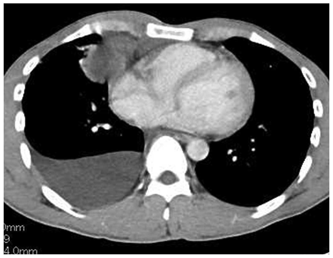
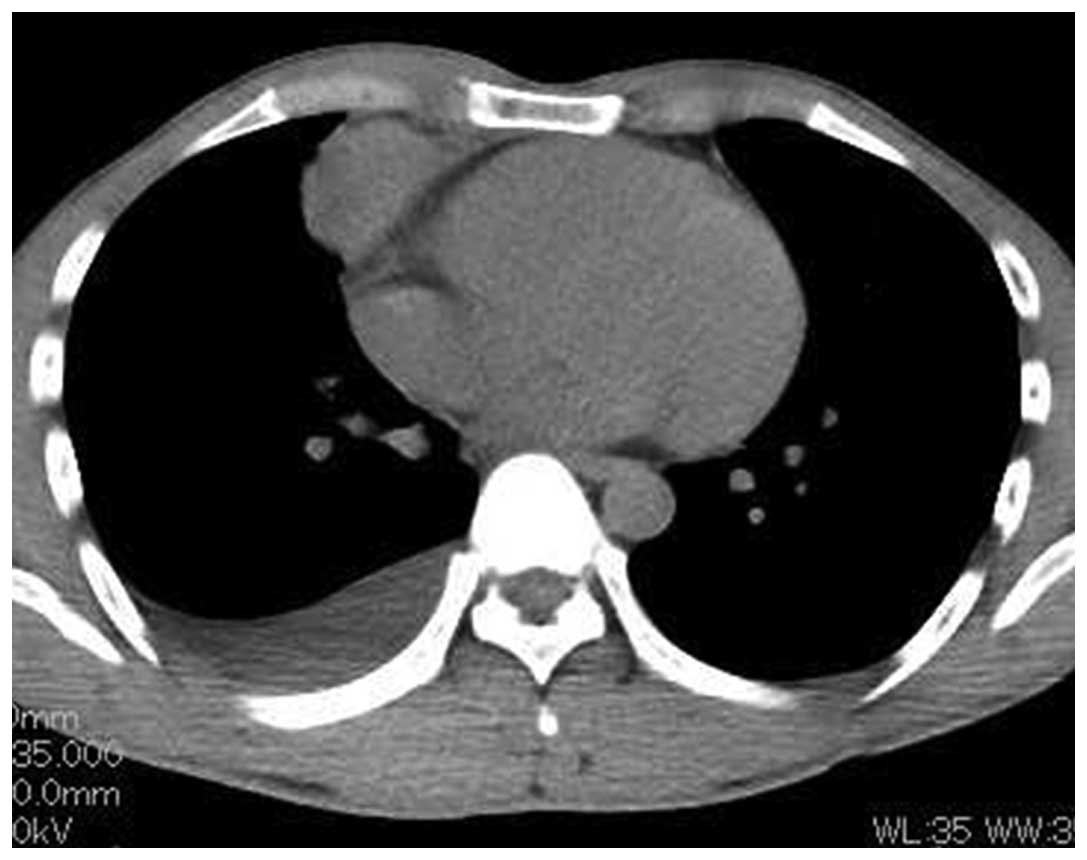
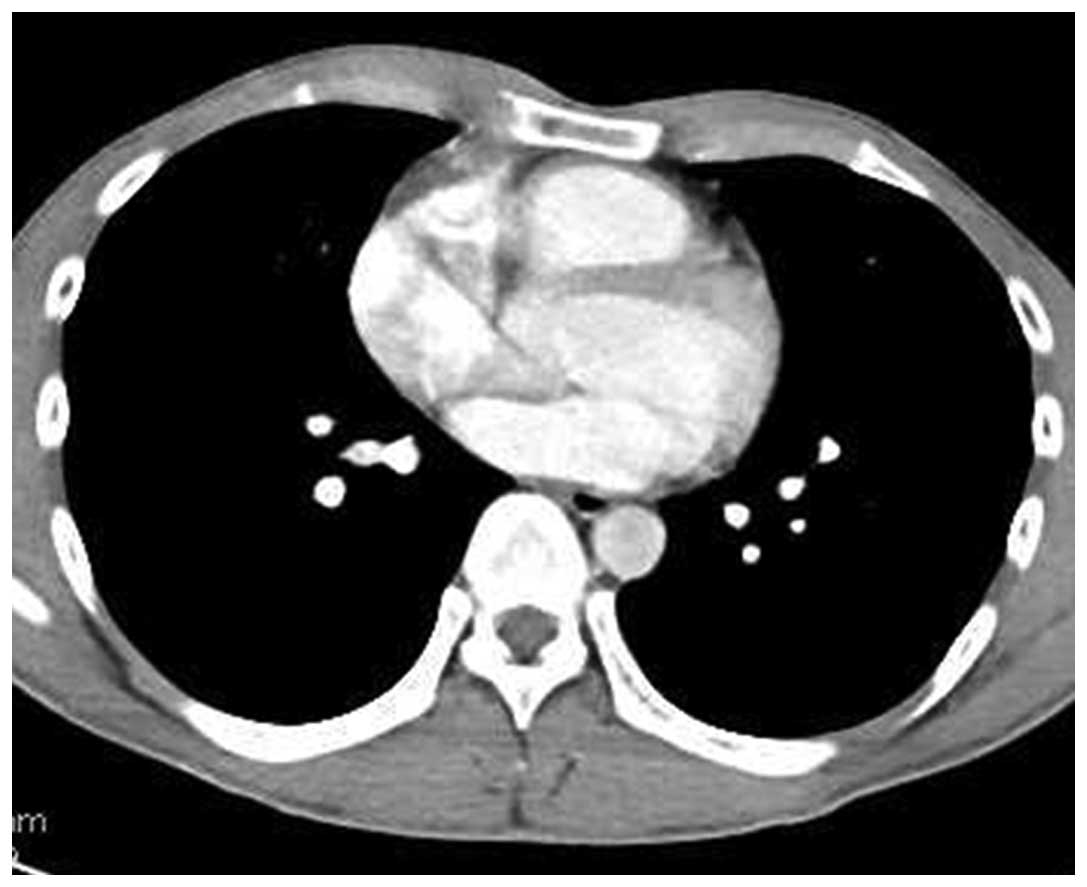
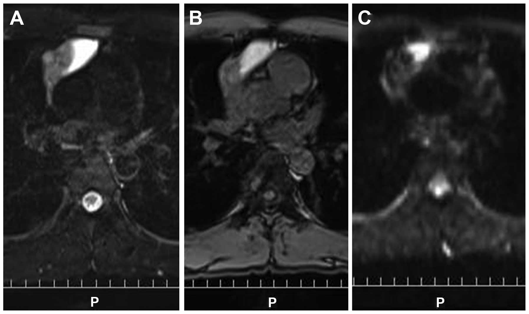
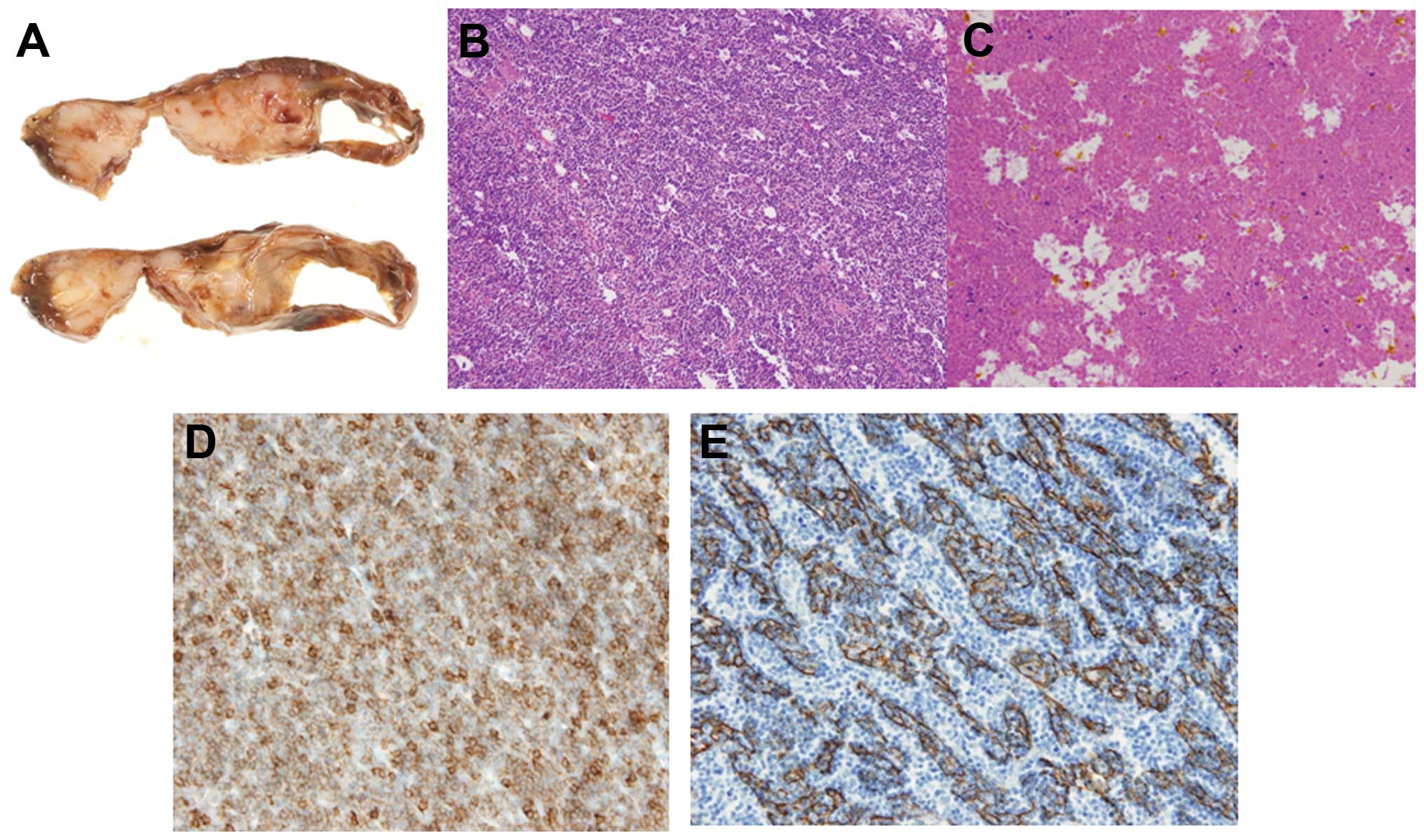
A 30-year-old man was admitted to Toho University Omori Medical Center for assessment of right chest pain and fever. Chest computed tomography (CT) revealed an anterior mediastinal tumor sized 11.0x6.0x5.0 cm, with right pleural effusion. The laboratory analysis revealed elevated white blood cell count (11,000/µl), C‑reactive protein (4.1 mg/dl) and cytokeratin fragment (CYFRA; 12.7 ng/ml; normal, <2 ng/ml). The level of CYFRA in the pleural effusion was also markedly elevated (143 ng/ml). On the first day after admission (6 days after the initial CT), there was a mild regression on CT (10.0x5.5x4.4 cm; reduction rate, 26.7%), with decrease of the pleural effusion volume. A CT‑guided needle biopsy was performed, but the findings were not conclusive, as most of the tissue was necrotic. Seven days later (13 days after the initial CT), a CT revealed further regression (9.5x5.4x4.2 cm; reduction rate, 34.7%) with disappearance of the pleural effusion. The patient was followed up on an outpatient basis. At 35 days after the initial CT, the tumor continued to shrink without treatment (8.0x3.6x3.0 cm; reduction rate, 73.8%) and the serum CYFRA level had decreased to 0.8 ng/ml, although it had not returned to normal levels. At 62 days after the initial CT, the patient underwent surgical resection. The resected specimen was diagnosed as thymoma (World Health Organization type B2; Masaoka classification, stage II), with prominent degeneration and necrosis. One possible cause of the spontaneous regression may be increased internal pressure, probably associated with rapid tumor growth, leading to massive necrosis with resulting chest pain, inflammatory reaction with pleural effusion and subsequent tumor regression. The serum CYFRA level may be a useful marker for the evaluation of the clinical course of thymoma with extensive necrosis.
View Figures
View References

Related Articles

Journal Cover

September-2015
Volume 3 Issue 5

Print ISSN: 2049-9450
Online ISSN:2049-9469

Sign up for eToc alerts

Recommend to Library

Copy and paste a formatted citation
x
Spandidos Publications style
Furuya K, Isobe K, Sano G, Kaburaki K, Gocho K, Ishida F, Kikuchi N, Sugino K, Sakamoto S, Takai Y, Takai Y, et al: Thymoma exhibiting spontaneous regression in size, pleural effusion and serum cytokeratin fragment level: A case report. Mol Clin Oncol 3: 1058-1062, 2015
APA
Furuya, K., Isobe, K., Sano, G., Kaburaki, K., Gocho, K., Ishida, F. ... Homma, S. (2015). Thymoma exhibiting spontaneous regression in size, pleural effusion and serum cytokeratin fragment level: A case report. Molecular and Clinical Oncology, 3, 1058-1062. https://doi.org/10.3892/mco.2015.583
MLA
Furuya, K., Isobe, K., Sano, G., Kaburaki, K., Gocho, K., Ishida, F., Kikuchi, N., Sugino, K., Sakamoto, S., Takai, Y., Otsuka, H., Hata, Y., Iyoda, A., Wakayama, M., Shibuya, K., Homma, S."Thymoma exhibiting spontaneous regression in size, pleural effusion and serum cytokeratin fragment level: A case report". Molecular and Clinical Oncology 3.5 (2015): 1058-1062.
Chicago
Furuya, K., Isobe, K., Sano, G., Kaburaki, K., Gocho, K., Ishida, F., Kikuchi, N., Sugino, K., Sakamoto, S., Takai, Y., Otsuka, H., Hata, Y., Iyoda, A., Wakayama, M., Shibuya, K., Homma, S."Thymoma exhibiting spontaneous regression in size, pleural effusion and serum cytokeratin fragment level: A case report". Molecular and Clinical Oncology 3, no. 5 (2015): 1058-1062. https://doi.org/10.3892/mco.2015.583