Cervical cancer (CC) is the four most common cancer
in women with an estimate of 604,000 new cases and 342,000 deaths
per year worldwide (1). Most of
the new cases (85%) and deaths (90%) occur in low- and
middle-income countries, where CC is the third most common cancer
among women. According to GLOBOCAN 2020, CC is the second most
common cancer in Mexico with 9,439 new cases and 4,335 deaths per
year. The Federation of Gynecology and Obstetrics (FIGO) staging
system classifies CC in four stages, I-IV, which in turn are
subdivided in various subtypes; this classification is mainly based
on surgery, pathologic analysis and imaging (2).
The expression of certain circulating biomolecules
is modified when a disease is established, having a great potential
to detect and predict the disease as well as to identify the
response to different treatments (3-5).
For CC, there are several studies showing biomolecules, proteins,
non-coding RNAs (ncRNAs), and circulating DNA (cDNA) with a great
potential to be useful biomarkers for diagnosis, prognosis, and to
determine the response to treatments currently used in medical
attention for CC. Although certain of these protein biomarkers may
also function as biomarkers for other HPV-related cancers, previous
studies showed that the combined use of these molecules increases
their potential as biomarkers for this disease (6-8).
Although these biomarkers have a sensitivity very similar to that
of colposcopy and p16 (Table I)
(9-29),
a significant advantage is that it is a minimally invasive
methodology, having a very important impact on the number of women
who would be screened with this test. The aim of the present study
was to review the literature related to these potential biomarkers,
emphasizing improved results as biomarkers when these molecules
have been analyzed in combination.
The information was searched in Pubmed and in
academic Google. The criteria followed to search the literature
were the following: Circulating biomarkers, CC, ncRNAs and cDNA. In
addition, biomarkers for HPV-associated cancers and public spending
in Mexico for the treatment of CC were searched. The inclusion
criterion was full access to the reviewed articles. Those articles
that could not be accessed were excluded from the review.
The search of circulating proteins as potential
biomarkers in pathologies such as cancer has been addressed for
several decades. The objective is clear, to diagnose and monitor
the diverse types of cancer in a minimally invasive way; however,
this search has not been easy and even when there are proteins
currently used in the medical attention, their sensitivity (the
ability to detect a disease in patients in whom the disease is
actually present) and specificity (the ability to rule out the
disease in patients in whom the disease is actually absent) is
relatively low (9). To date, the
most common circulating cancer marker proteins used in medical
attention for distinct types of cancer are: i) The squamous cell
carcinoma antigen (SCC-A), ii) The carcinoembryonic antigen (CEA),
iii) The α-fetoprotein, iv) The β-subunit of human chorionic
gonadotropin, v) Lactate dehydrogenase and vi) The cancer antigen
125(10). Regarding CC, massive
analyses have revealed groups of circulating proteins
differentially expressed in this disease with a great potential to
be used as biomarkers. Notably, the use of two or more of these
proteins has been revealed to considerably increase their
sensitivity and specificity (Table
II) (30,31).
In the 1970s, the SCC-A was identified by using the
hybridoma technique in SCC of human uterine cervix (11). SCC-A is a serpin that comprises two
nearly identical proteins (45 kDa), SCC-1 and SCC-2, which possess
unique proteinase inhibitory properties (10,11).
SCC-1 exerts an anti-apoptotic action through the inhibition of
chymotrypsin and cathepsin L. The mechanism of protection of tumor
cells from apoptosis involves the inhibition of the caspase-3
activity and/or upstream proteases. SCC-2 inhibits cathepsin G and
mast cell chymase, thus protecting epithelial cells from these
proteases-induced inflammation (32).
Increased serum SCC-A levels were observed in more
advanced SCC stages (in 28-88% of the patients) allowing the use of
SCC-A as diagnostic and prognostic biomarker for this cancer
subtype (30-32).
Differences in the percentage of SCC-A detection were attributed to
various factors, such as the histological grade and the cutoff in
the SCC-A serum concentration. Although numerous years have passed
since its discovery, the clinical use of SCC-A remains under
debate, for the increase on its expression has been reported in
patients with SCC of the esophagus, lung, head, neck, and in anal
canal and uterine cervix, as well as in patients with several
non-malignant skin lesions, such as pemphigus and renal failure.
Regarding this, the exposure to TNF-α significantly increased the
production of SCC-A in normal human epidermal keratinocytes
(33).
In addition to SCC-A, other potential circulating
biomarkers have been identified. Mitsuhashi et al (21) described that the serum YKL-40 level
was elevated in both SCC and adenocarcinoma. YLK-40 is a
glycoprotein member of the glycosyl hydrolase 18 family; it is
secreted by active macrophages, chondrocytes, neutrophils and
synovial cells. Recent studies suggested that YLK-40 plays a role
in the inflammation process and tissue remodeling (34-36).
This molecule appears to be a favorable CC biomarker in both SSC
and adenocarcinoma subtypes, and it appears to be more specific
than SCC-A and CA125. Previous findings demonstrated that serum
YKL-40 level is increased in several solid tumors with a variety of
histological types (37,38). This protein is a biomarker
associated with inflammation and, despite this, it could be a
correlation between the C-reactive protein (CRP) and
YKL-40(39). YKL-40 serum appears
to be more a non-specific biomarker of inflammation, since its
expression was higher than that of CRP, allowing to discriminate
patients with CC from tumor-free individuals. In addition, YKL-40
appears to be an improved serum biomarker for adenocarcinomas
detection than CA-125 exhibiting 78 and 68% sensitivity for all
grades and for stage I tumors, respectively (21). Although it does not appear to be an
ideal biomarker due to its relative low sensitivity to detect CC,
the receiver operating characteristic (ROC) and area under a ROC
curve (AUC) analysis revealed that YKL-40 discriminates healthy
individuals from patients with CC. Similarly, the YKL-40 levels
were identified to be a poor prognostic variable for relapse of the
disease (40).
It is well documented that activation of
Macrophage-Colony Stimulating Factor (M-CSF) and vascular
endothelial growth factor (VEGF) is involved in the pathogenesis
and spread of distinct types of cancer, including CC. Regarding
this, Sidorkiewicz et al (24) examined the M-CSF and VEGF plasma
levels and compared them with those of CA-125 and SCC-A in three
groups of patients: i) The CC group (patients with either SCC or
adenocarcinoma), ii) The cervical dysplasia group and iii) The
control group. The median levels of M-CSF and VEGF as well as those
of CA-125 and SCC-A were significantly different in the three
groups relative to the control group. The sensitivity and
specificity for VEGF and SCC-Ag were of 82 and 76%, and 81 and 74%,
respectively. In the adenocarcinoma group, the VEGF sensitivity and
specificity were respectively of 87 and 76% (24). The results indicated a possible
clinical applicability for these proteins and a relatively high
diagnostic power for the M-CSF, VEGF, CA-125 and SCC-Ag
combination. Similarly, the combined analysis of α-Actinin 4
(ACTN4) and SSC-A is a promising serological examination for CC
detection. ACTN4 plays an essential role in regulating cellular
signaling pathways correlated with various types of cancer
progression and poor patient prognosis, involved in the invasion
and metastasis of colorectal, pancreatic and ovarian cancer. Its
principal function is by regulating cell invasion due to its
participation in the epithelial-to-mesenchymal transition; however,
it is also involved in controlling the cancer stem cell properties
and chemoresistance in CC. Zhu et al (43) demonstrated the circulating and
tumor ACTN4 overexpression in patients with CIN3 or more advanced
stages. In addition, the ACTN4 mRNA was also overexpressed in CC
tissues and in tissues with advanced FIGO stages, larger tumor
sizes, and positive lymph node metastasis. In conclusion, ACTN4, in
combination with SCC-Ag, is a potential biomarker for the diagnosis
and prognosis of patients with CIN3 or more advanced stages.
Summarizing, different circulating proteins are
differentially expressed in patients with CC relative to
individuals without cancer, having a great potential to be used in
clinical diagnosis and even more when two or more proteins are
analyzed in a combined manner. Importantly, not only circulating
proteins have been identified as biomarkers for CC but also
microRNAs (miRNAs) and long non-coding RNAs (lncRNAs), as well as
cDNA.
ncRNAs are RNA molecules, which are not translated
into a protein, including transfer RNAs, ribosomal RNAs, small
non-coding RNAs (snRNAs) and lncRNAs (44). snRNAs and lncRNAs regulate numerous
biological functions and their expression is finely regulated at
different stages of development of organisms to fulfill very
particular functions (45,46). Numerous lines of evidence indicate
their involvement in cancer, specifically in CC, and their
differential expression, mainly in blood, has been related to
diagnosis, prognosis, and treatment response of patients with CC,
as described below.
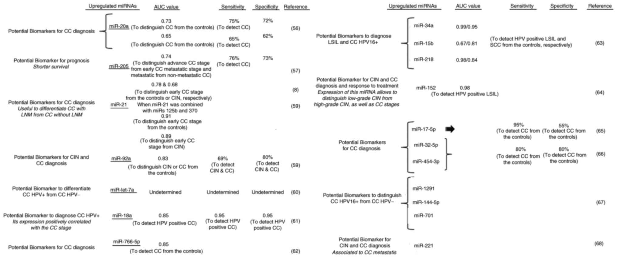
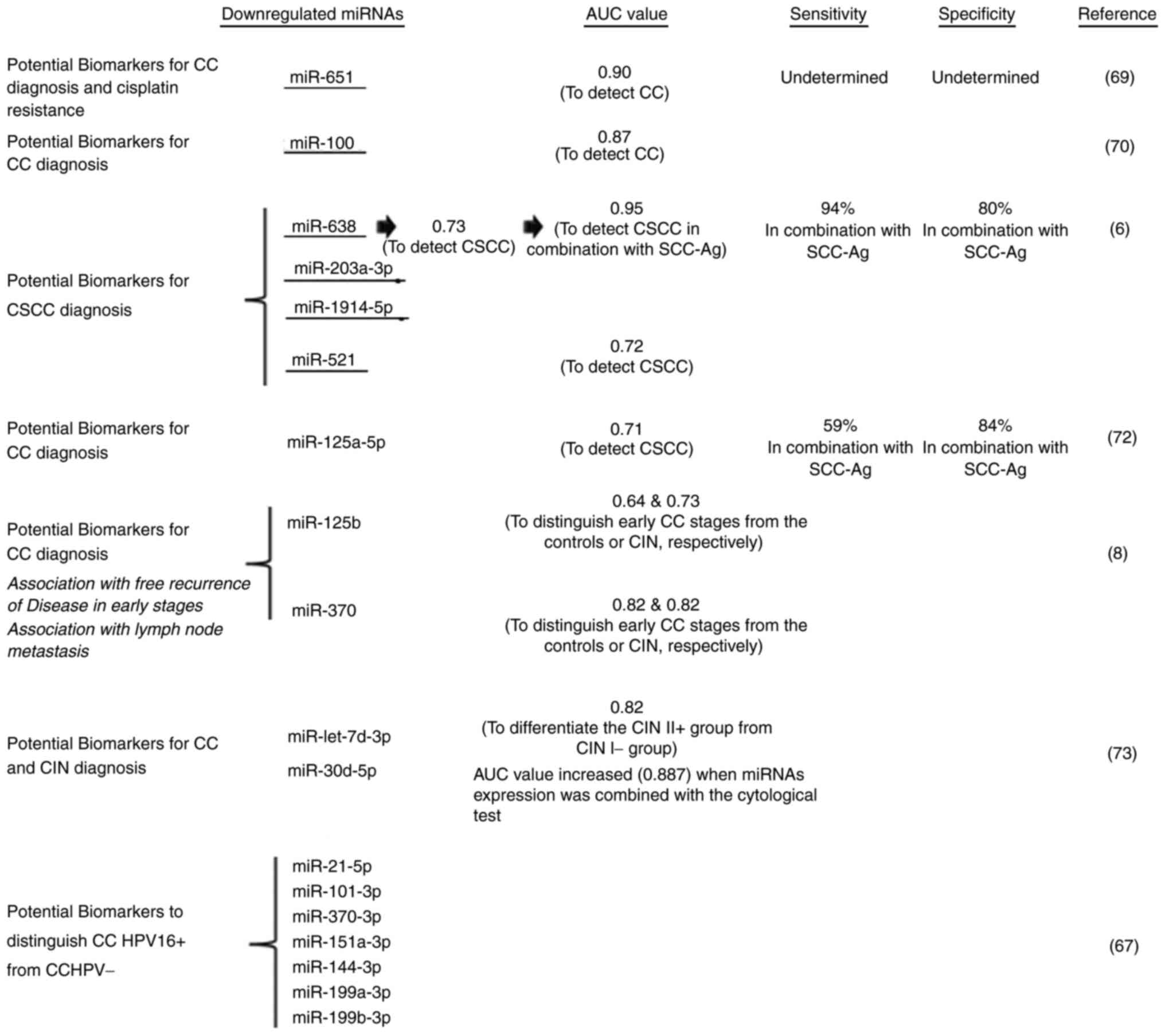
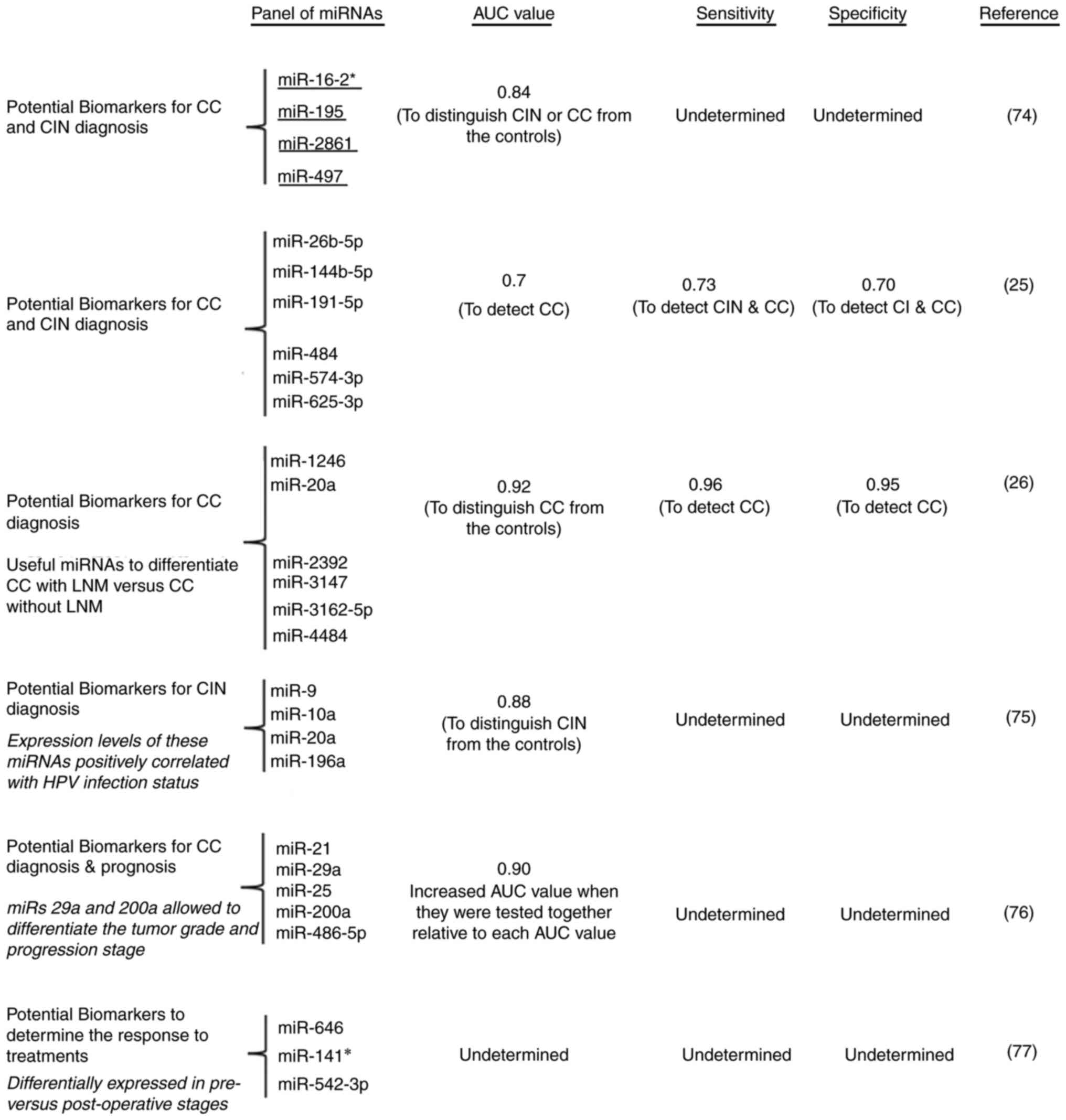
To date, numerous circulating miRNAs have been
identified as possible biomarkers for low-grade squamous
intraepithelial lesions (LSIL), CIN and CC and most of them
demonstrate higher AUC values and a higher sensitivity and
specificity than proteins. Notably, both sensitivity and
specificity to detect CC by miRNAs is relatively high and they were
considerably increased when analyzed in combination (6-8,54-72)
(Fig. 1, Fig. 2 and Fig. 3). The majority of miRNAs have the
potential to be biomarkers for CIN and CC diagnosis and only
certain for prognosis (Fig. 3).
MiRs 34a and 218 are particularly important, since they allow to
distinguish LSIL and CC HPV16+ from healthy women
(60) (Fig. 1). Their clinical use in LSIL
detection-in a minimally invasive form-would have a huge impact on
public health in countries where CC remains a public health
problem, such as Mexico.
A very interesting study showed the miR-221-3p
enrichment in exosomes, which were secreted by CSCC and captured by
human lymphatic endothelial cells (HLECs), resulted in their
migration promotion, tube formation, lymphanogenesis induction and
LN metastasis in CSCC patients. These processes appear to be
regulated, at least partly, by targeting vasohibin-1, leading to
the ERK/AKT pathway activation in HLECs (65).
To date, there are several circulating miRNAs that
can be used for the LSIL, CIN and CC diagnosis and certain of them
are specific to differentiate HPV+ CC from the negative
one. Importantly, certain miRNAs render it possible to identify CC
metastasis to the lymph nodes.
LncRNAs are ≥200 nt RNAs with very complex secondary
structures and a myriad of cellular functions: i) Maintaining the
chromatin structure and regulating gene transcription (73), ii) They are molecular scaffolds for
several factors involved in transcription control (74) and iii) They are miRNAs sponges
(75). These type of RNAs have
been detected in body fluids and have been associated with cancer,
including CC; however, the information regarding circulating
lncRNAs participating in CC is very scarce. Sun et al
(27) found that lncRNAs HOTAIR,
PVT1, XLOC_000303 and AL592284 are overexpressed in the blood serum
of patients with CC when compared with the controls. The analysis
of these four lncRNAs together improved the AUC value: 0.875. In a
similar way, the analysis of the overexpressed lncRNAs CCAT2,
LINC01133 and LINC00511 by including the SCC-A, increased the AUC
value to 0.94(28). Meanwhile, the
expression of HOTAIR was increased in patients with CC relative to
the controls and this correlated with numerous clinical aspects as
well as with tumor recurrence and short overall survival (76). The identification of more
circulating lncRNAs as potential CC biomarkers will definitely have
a noticeable impact on the diagnosis, prognosis and response to
treatments of this type of cancer. In addition, the discovery of
the functions performed by these RNAs in time shall allow the
identification of therapeutic targets in the future.
There is a close relationship between gynecological
cancers and alterations in hormone-mediated regulatory pathways,
modifying gene expression. Most information is related to protein
biomarkers and some of them were regulated by estrogens in tissues:
SCC-Ag, C-125, VEGFA and ACTN4, and only circulating HMGB1 and
YKL-40 were regulated by estrogens (77-81).
Notably, the circulating miR-21 expression was downregulated by
estrogens (82).
To the best of our knowledge, for the remaining
biomarkers there is no evidence indicating changes on their
circulating expression in response to estrogens; however, this does
not mean that their expression cannot be regulated by this hormone,
since most of them show a close relationship with the expression of
estrogen receptors.
Previous studies showed that HPV-cDNA detection
positively correlates with low-grade cervical lesions (LGCL) and
high-grade cervical lesions (HGCL), CIN and CC, tumor grade, and
with the genomic HPV insertion, which is associated with a poor
patient's prognosis (91-98)
(Table III). Importantly,
Rungkamoltip et al (29)
showed a 100% sensitivity and 88% specificity to detect E7 HPV16/18
cfDNA by using the amplification-by recombinase polymerase-in
combination with lateral Flow strip. In addition to cDNA, the
presence of specific cDNA mutations allows to detect CC from
healthy individuals and positively correlates with disease
progression and with a shorter progression-free survival, as well
as with the overall survival of patients with metastatic relapse CC
compared with the controls (patients without any detectable of
these mutations) (99).
Very importantly, cDNA detection allows to
differentiate among LGCL and HGCL, and CC from patients with
non-HPV dependent CC as well as from individuals without CC. LGCL
detection by means of cDNA is very promising since the early
detection of this type of lesions can significantly prevent their
progression to cancer. In addition, cDNA concentration appears to
be useful for monitoring treatment response and patient's
prognosis.
Making a prediction of which mechanisms will be
regulated by specific circulating molecules-only based on their
canonical functions-is a high-risk task. This is because the few
studies that have focused on studying the action of circulating
molecules have elucidated mechanisms of action somewhat unexpected
and that have nothing to do with the previously reported
effects.
In general, it is known that exogenous miRNAs
regulate gene expression through the canonical pathway, involving
the binding to their target mRNA; however, Fabbri et al
(100) revealed that miRs-21 and
-29a function by a different mechanism. The aforementioned study
demonstrated the interaction of these miRNAs with Toll-like
receptor (TLR) 7 and TRL8 in cells from the immune system. Notably,
the binding of miRs-21 and -29a to these receptors triggered a
prometastatic inflammatory response, resulting in the tumor growth
induction and metastasis. Thus, this is the first indication that
miRNAs function as paracrine agonists of TLRs to regulate tumor
environment.
Based on canonical functions reported for the
proteins proposed as biomarkers for CC, KEGG analysis showed their
involvement mainly in the control of signaling pathways, such as
PI3K-Akt, HIF-1, and Rap1, among others (Table IV). The cellular processes that
may be modified by changes in these signaling pathways were cell
proliferation, adhesion, migration, cytoskeleton remodeling and
gene expression regulation (Table
IV).
Changes on expression of miRNAs and/or on their
function could alter numerous signaling pathways and cellular
processes, which is related to their ability to regulate several
mRNAs in the same cell. Secretion of miRNAs to blood serum and
target organ recognition could mainly alter proteoglycans in
cancer, pathways in cancer, renal cell carcinoma, viral
carcinogenesis, among others; these signaling pathways control
cellular processes related with hallmarks of cancer (Table V).
Numerous functional studies are necessary to know
the mechanism(s) of action of each of these molecules separately
and/or together, and what ‘benefit’ the tumor obtains by releasing
these molecules.
The aforementioned biomarkers are postulated as a
favorable tool for both the detection of cervical lesions and
cancer; however, various studies have shown the expression of some
of these biomarkers in other types of HPV-related cancers. Their
expression has been detected mainly at the tumor level and to a
markedly lesser extent as circulating molecules. SCC-A is the most
studied biomarker and is overexpressed in distinct squamous cell
cancers, including the following: Esophagus, lung, head, neck, and
in anal canal, and uterine cervix, as well as in vulvar and penile
carcinoma (101-105).
Meanwhile, YKL-40 has demonstrated high tissue levels in anal
carcinoma and it has been detected in the plasma of patients with
esophageal cancer (106) and
CA-125 showed relatively high circulating levels in vulvar
carcinoma (107). In addition,
CA-125 had an increased expression in oropharyngeal cancer
(108) and relatively high VEGFA
expression levels were detected in vulvar carcinoma (109) and oropharyngeal cancer (110). Although PIGF was detected in
oropharyngeal cancer, its expression was not related with the
malignancy of this cancer type (111).
In the case of ncRNAs, to the best of our knowledge
only miR-205 and HOTAIR have been related with head and neck
(112), and oropharyngeal cancers
(113), as well as with
cervicovaginal lavage specimens, respectively (114).
Even though the Mexican Institute of Social Security
has a substantial cost for the detection, follow-up and treatment
of CC, this cancer type remains the second most common in Mexican
women after breast cancer. A previous study carried out by
Granados-García et al (17)
revealed the high cost of evaluating a patient's condition
regarding CC by cytology, colposcopy, biopsies, and pathology, as
well as by diagnostic tests and treatments for cervical
intraepithelial neoplasia grade II and III (CIN 2/3) and CC. The
aforementioned study identified that the cost to perform 2.7
million cytology tests was nearly 38 million dollars, representing
26.1% of the total program cost (145.4 million). False negatives
account for nearly 43% of the program costs. According to the
aforementioned results, it was concluded that the low sensitivity
of the cytology test generates high rates of false negatives,
resulting in high institutional costs from the treatment of
undetected CC cases.
In accordance with the aforementioned studies, the
establishment of a panel of biomarkers with high sensitivity and
specificity would be a great molecular tool to improve the
diagnosis and treatment of women with LGCLs, HGCLs and CC. An
increase in the detection of women with LGCLs and HGCLs-by this
panel-may decrease the number of women progressing towards CC.
From the authors' point of view, the biomarkers that
would have a greater impact on women and health sector are those
detecting both LGCLs and HGCLs. Early detection of these type of
lesions would allow early treatment of women and this would
considerably decrease the progression towards cancer. At this point
it is important to mention that the use of this biomarker panel
will increase the power of detection, prognosis and response to
treatments (Table VI).
As observed, early CC detection is a crucial factor
to effectively treat low-grade CC lesions and thus avoid the
transition to cancer; therefore, the establishment of molecular
tools that allow performing this task is imperative. Currently,
there are diverse biomolecules-particularly ncRNAs-which have a
high sensitivity and specificity to detect LGCLs as well as CC,
thus the establishment of these biomarkers for their use in the
clinical studies to detect LGCLs and CC is crucial. In addition,
the biomolecules that enable us to know the response to treatments
is also very important and, in the same way, it should be part of
the molecular tools used in the medical attention.
Collectively, the biomarkers found to date have
great potential to be used as clinically useful biomarkers for
detection and response to treatments. Further studies are needed to
establish which are the ones that will best support the medical
attention.
Not applicable.
Funding: The present study was supported by Proyectos-ATSO.
Not applicable.
RREG searched and organized the information and
wrote the manuscript. MGS reviewed the last version of the
manuscript. MAVF reviewed and corrected the information of the last
version of the manuscript. All authors read and approved the final
version of the manuscript.
Not applicable.
Not applicable.
The authors declare that they have no competing
interests.
|
1
|
Sung H, Ferlay J, Siegel RL, Laversanne M,
Soerjomataram I, Jemal A and Bray F: Global cancer statistics 2020:
GLOBOCAN estimates of incidence and mortality worldwide for 36
cancers in 185 countries. CA Cancer J Clin. 71:209–249.
2021.PubMed/NCBI View Article : Google Scholar
|
|
2
|
Lee SI and Atri M: 2018 FIGO staging
system for uterine cervical cancer: Enter cross-sectional imaging.
Radiology. 292:15–24. 2019.PubMed/NCBI View Article : Google Scholar
|
|
3
|
Cho H, Oh CK, Cha J, Chung JI, Byun SS,
Hong SK, Chung JS and Han KH: Association of serum
prostate-specific antigen (PSA) level and circulating tumor
cell-based PSA mRNA in prostate cancer. Prostate Int. 10:14–20.
2022.PubMed/NCBI View Article : Google Scholar
|
|
4
|
Nieder C, Dalhaug A and Mannsåker B:
Established serum biomarkers are prognostic factors in patients
with oligometastatic cancer and brain involvement. In Vivo.
36:801–805. 2022.PubMed/NCBI View Article : Google Scholar
|
|
5
|
López-Aguilar JE, Velázquez-Flores MA,
Simón-Martínez LA, Ávila-Miranda R, Rodríguez-Florido MA and
Ruiz-Esparza Garrido R: Circulating microRNAs as biomarkers for
pediatric astrocytomas. Arch Med Res. 48:323–332. 2017.PubMed/NCBI View Article : Google Scholar
|
|
6
|
Zheng S, Li R, Liang J, Wen Z, Huang X, Du
X, Dong S, Zhu K, Chen X, Liu D, et al: Serum miR-638 combined with
squamous cell carcinoma-related antigen as potential screening
biomarkers for cervical squamous cell carcinoma. Genet Test Mol
Biomarkers. 24:188–194. 2020.PubMed/NCBI View Article : Google Scholar
|
|
7
|
Jia W, Wu Y, Zhang Q, Gao GE, Zhang C and
Xiang Y: Expression profile of circulating microRNAs as a promising
fingerprint for cervical cancer diagnosis and monitoring. Mol Clin
Oncol. 3:851–858. 2015.PubMed/NCBI View Article : Google Scholar
|
|
8
|
Qiu H, Liang D, Liu L, Xiang Q, Yi Z and
Ji Y: A novel circulating MiRNA-based signature for the diagnosis
and prognosis prediction of early-stage cervical cancer. Technol
Cancer Res Treat. 19(1533033820970667)2020.PubMed/NCBI View Article : Google Scholar
|
|
9
|
ACCP. Pap smears: An important but
imperfect method. Cervical Cancer Prevention Fact Sheet, 2002.
|
|
10
|
Milla Villeda RH, Alvarado Zaldívar G,
Sánchez Anguiano LF, Barrera Tovar M and Vázquez Arreola I:
Colposcopy and cervical biopsy in patients with routine
Papanicolaou smear. Ginecol Obstet Mex. 65:235–238. 1997.PubMed/NCBI
|
|
11
|
Chu E, Bratwaite O, Gonzalez I, Campo Z
and De León RG: Diagnosis of human papillomavirus by Papanicolau
and PCR in a group of adolescents and young women. Instituto
conmemorativo gorgas de estudios de la salud, centro de
investigación en reproducción humana. URL: Microsoft
Word-2087_2002_doc(gorgas.gob.pa).
|
|
12
|
Flores-Juárez DJ and García-González GA:
Usefulness of Papanicolau for cervical dysplasia in patients HPV
positive with CD4 <200 cell/mm3. Escuela de Estudios de
Postgrado de la Facultad de Ciencias Médicas Maestría en
Ginecología y Obstetricia Tesis para obtener el grado de Maestras
en Ciencias en Ginecología y Obstetricia, pp1-45, 2014. URL:
https://biblioteca.medicina.usac.edu.gt/tesis/post/2014/071.pdf.
|
|
13
|
Terrádez Raro JJ, Coloma Colomer F,
Navarro Conde P and Gasull Ibáñez J: Cervical cancer screening in
public health system in Valencia community and Pap test limit. Rev
ESP Patol. 38:3–7. 2005.
|
|
14
|
Dzul-Rosado KR, Puerto-Solís M and
González Losa MR: Cáncer cervicouterino: Métodos actuales para su
detección. Rev Biomed. 15:233–241. 2004.
|
|
15
|
Vlastos AT, Richards-Kortum R, Zuluaga A
and Follen M: New approaches to cervical cancer screening.
Contemporary Ob/Gyn. 47:87–107. 2002.
|
|
16
|
Andrade AZ, Zaragoza JZ, Blanco BR and
Marañón RT: Evaluación del papanicolaou y la colposcopia en el
diagnóstico de la infección por el virus del papiloma humano. Rev
Fac Med UNAM. 44:5–7. 2001.
|
|
17
|
Granados-García V, Flores YN, Pérez R,
Rudolph SE, Lazcano-Ponce E and Salmerón J: Cost of the cervical
cancer screening program at the mexican social security institute.
Salud Publica Mex. 56:502–510. 2014.PubMed/NCBI View Article : Google Scholar
|
|
18
|
Solares LF, Álvarez AM, García-Echeverría
AM and Velasco MJ: Diagnóstico citológico de ASCUS. Identificación
del riesgo para displasia cervical mediante test del virus del
papiloma humano. Clin Invest Ginecol Obstet. 32:50–53. 2005.
|
|
19
|
Zhang Q, Kuhn L, Denny LA, De Souza M,
Taylor S and Wright TC Jr: Impact of utilizing p16INK4A
immunohistochemistry on estimated performance of three cervical
cancer screening tests. Int J Cancer. 120:351–356. 2007.PubMed/NCBI View Article : Google Scholar
|
|
20
|
Mattosinho de Castro Ferraz Mda G, Nicolau
SM, Stávale JN, Focchi J, Castelo A, Dôres GB, Mielzynska-Lohnas I,
Lorincz A and Rodrigues de Lima G: Cervical biopsy-based comparison
of a new liquid-based thin-layer preparation with conventional Pap
smears. Diagn Cytopathol. 30:220–226. 2004.PubMed/NCBI View
Article : Google Scholar
|
|
21
|
Mitsuhashi A, Matsui H, Usui H, Nagai Y,
Tate S, Unno Y, Hirashiki K, Seki K and Shozu M: Serum YKL-40 as a
marker for cervical adenocarcinoma. Ann Oncol. 20:71–77.
2009.PubMed/NCBI View Article : Google Scholar
|
|
22
|
Xu G, Fan W, Wang F, Lu H, Xing X, Zhang R
and Jiang P: CTHRC1 as a novel biomarker in the diagnosis of
cervical squamous cell carcinoma. Int J Clin Exp Pathol.
11:847–854. 2018.PubMed/NCBI
|
|
23
|
Yang SH, Wang XL, Cai J and Wang SH:
Diagnostic value of circulating PIGF in combination with Flt-1 in
early cervical cancer. Curr Med Sci. 40:973–978. 2020.PubMed/NCBI View Article : Google Scholar
|
|
24
|
Sidorkiewicz I, Zbucka-Krętowska M, Zaręba
K, Lubowicka E, Zajkowska M, Szmitkowski M, Gacuta E and Ławicki S:
Plasma levels of M-CSF and VEGF in laboratory diagnostics and
differentiation of selected histological types of cervical cancers.
BMC Cancer. 19(398)2019.PubMed/NCBI View Article : Google Scholar
|
|
25
|
Ning R, Meng S, Wang L, Jia Y, Tang F, Sun
H, Zhang Z, Zhang C, Fan X, Xiao B, et al: 6 Circulating miRNAs can
be used as non-invasive biomarkers for the detection of cervical
lesions. J Cancer. 12:5106–5113. 2021.PubMed/NCBI View Article : Google Scholar
|
|
26
|
Chen J, Yao D, Li Y, Chen H, He C, Ding N,
Lu Y, Ou T, Zhao S, Li L and Long F: Serum microRNA expression
levels can predict lymph node metastasis in patients with
early-stage cervical squamous cell carcinoma. Int J Mol Med.
32:557–567. 2013.PubMed/NCBI View Article : Google Scholar
|
|
27
|
Sun W, Wang L, Zhao D, Wang P, Li Y and
Wang S: Four circulating long non-coding RNAs Act as biomarkers for
predicting cervical cancer. Gynecol Obstet Invest. 83:533–539.
2018.PubMed/NCBI View Article : Google Scholar
|
|
28
|
Wang WJ, Wang D, Zhao M, Sun XJ, Li Y, Lin
H, Che YQ and Huang CZ: Serum lncRNAs (CCAT2, LINC01133, LINC00511)
with squamous cell carcinoma antigen panel as novel non-invasive
biomarkers for detection of cervical squamous carcinoma. Cancer
Manag Res. 12:9495–9502. 2020.PubMed/NCBI View Article : Google Scholar
|
|
29
|
Rungkamoltip P, Temisak S, Piboonprai K,
Japrung D, Thangsunan P, Chanpanitkitchot S, Chaowawanit W,
Chandeying N, Tangjitgamol S and lempridee T: Rapid and
ultrasensitive detection of circulating human papillomavirus E7
cell-free DNA as a cervical cancer biomarker. Exp Biol Med
(Maywood). 246:654–666. 2021.PubMed/NCBI View Article : Google Scholar
|
|
30
|
Gadducci A, Tana R, Cosio S and Genazzani
AR: The serum assay of tumour markers in the prognostic evaluation,
treatment monitoring and follow-up of patients with cervical
cancer: A review of the literature. Crit Rev Oncol Hematol.
66:10–20. 2008.PubMed/NCBI View Article : Google Scholar
|
|
31
|
Duffy MJ: Evidence for the clinical use of
tumor markers. Ann Clin Biochem. 41:370–377. 2004.PubMed/NCBI View Article : Google Scholar
|
|
32
|
Uemera Y, Pak SC, Luke C, Cataltepec S,
Tsu C, Schick C, Kamachi Y, Pomeroy SL, Perlmutter DH and Silverman
GA: Circulating serpin tumor markers SCCA1 and SCCA2 are not
actively secreted but reside in the cytosol of squamous carcinoma
cells. Int J Cancer. 89:368–377. 2000.PubMed/NCBI View Article : Google Scholar
|
|
33
|
Numa F, Takeda O, Nakata M, Nawata S,
Tsunaga N, Hirabayashi K, Suminami Y, Kato H and Hamanaka S: Tumor
necrosis factor-alpha stimulates the production of squamous cell
carcinoma antigen in normal squamous cells. Tumour Biol. 17:97–101.
1996.PubMed/NCBI View Article : Google Scholar
|
|
34
|
Liposits G, Skuladottir H, Ryg J, Winther
SB, Möller S, Hofsli E, Shah CH, Poulsen LØ, Berglund Å, Qvortrup
C, et al: The prognostic value of pre-treatment circulating
biomarkers of systemic inflammation (CRP, dNLR, YKL-40, and IL-6)
in vulnerable older patients with metastatic colorectal cancer
receiving palliative chemotherapy-the randomized NORDIC9-study. J
Clin Med. 11(5603)2022.PubMed/NCBI View Article : Google Scholar
|
|
35
|
Dolin TG, Christensen IJ, Lund CM, Bojesen
SE, Lykke J, Nielsen DL, Larsen JS and Johansen JS: Preoperative
plasma vitamin D in patients with localized colorectal cancer:
Age-dependent association with inflammation, postoperative
complications, and survival. Eur J Surg Oncol.
(S0748-7983(22)00651-5)2022.PubMed/NCBI View Article : Google Scholar : (Epub ahead of
print).
|
|
36
|
Rathcke CN and Vestergaard H: YKL-40, a
new inflammatory marker with relation to insulin resistance and
with a role in endothelial dysfunction and atherosclerosis. Inflamm
Res. 55:221–227. 2006.PubMed/NCBI View Article : Google Scholar
|
|
37
|
Bao J, Ouyang Y, Qiao L, He J, Liu F, Wang
Y, Miao L, Fu A, Lou Z, Zang Q, et al: Serum CHI3L1 as a biomarker
for non-invasive diagnosis of liver fibrosis. Discov Med. 33:41–49.
2022.PubMed/NCBI
|
|
38
|
Shi M, Ge Q, Wang X, Diao W, Yang B, Sun
S, Wang G, Liu T, Chan AML, Gao Z, et al: Functional analysis of
the short splicing variant encoded by CHI3L1/YKL-40 in
glioblastoma. Front Oncol. 12(910728)2022.PubMed/NCBI View Article : Google Scholar
|
|
39
|
Videmark AN, Christensen IJ, Feltoft CL,
Villadsen M, Borg FH, Jørgensen BM, Bojesen SE, Kistorp C,
Ugleholdt R and Johansen JS: Combined plasma C-reactive protein,
interleukin 6 and YKL-40 for detection of cancer and prognosis in
patients with serious nonspecific symptoms and signs of cancer.
Cancer Med: Nov 28, 2022 (Epub ahead of print).
|
|
40
|
Hermunen K, Soveri LM, Boisen MK, Mustonen
HK, Dehlendorff C, Haglund CH, Johansen JS and Osterlund P:
Postoperative serum CA19-9, YKL-40, CRP and IL-6 in combination
with CEA as prognostic markers for recurrence and survival in
colorectal cancer. Acta Oncol. 59:1416–1423. 2020.PubMed/NCBI View Article : Google Scholar
|
|
41
|
Sheng X, Du X, Zhang X, Li D, Lu C, Li Q,
Ma Z, Song Q and Wang C: Clinical value of serum HMGB1 levels in
early detection of recurrent squamous cell carcinoma of uterine
cervix: Comparison with serum SCCA, CYFRA21-1, and CEA levels.
Croat Med J. 50:455–464. 2009.PubMed/NCBI View Article : Google Scholar
|
|
42
|
Chen Y, Xiong X, Wang Y, Zhao J, Shi H,
Zhang H, Wang Y, Wei Y, Xue W and Zhang J: Proteomic screening for
serum biomarkers for cervical cancer and their clinical
significance. Med Sci Monit. 25:288–297. 2019.PubMed/NCBI View Article : Google Scholar
|
|
43
|
Zhu B, Dong B, Hong S, Wang M, Dai W,
Zheng Q, Wu D and Cao Y: Combined detection of ACTN4 and SCC-Ag is
a promising serological biomarker for cervical intraepithelial
neoplasia 3 or worse: A case-control study. Risk Manag Health
Policy. 13:2677–2687. 2020.PubMed/NCBI View Article : Google Scholar
|
|
44
|
Lander ES, Linton LM, Birren B, Nusbaum C,
Zody MC, Baldwin J, Devon K, Dewar K, Doyle M, FitzHugh W, et al:
Initial sequencing and analysis of the human genome. Nature.
409:860–921. 2001.PubMed/NCBI View Article : Google Scholar
|
|
45
|
Wang F, Sun H, Li K, Yang K, Xiang Y and
Tian X: CircRASSF2 promotes IGF1R and osteosarcoma metastasis via
sponging miR-6838-5p. Ann Transl Med. 10(11)2022.PubMed/NCBI View Article : Google Scholar
|
|
46
|
Gupta A, Vats A, Ghosal A, Mandal K,
Sarkar R, Bhattacharya I, Das S, Pal R and Majumdar SS:
Follicle-stimulating hormone-mediated decline in miR-92a-3p
expression in pubertal mice Sertoli cells is crucial for germ cell
differentiation and fertility. Cell Mol Life Sci.
79(136)2022.PubMed/NCBI View Article : Google Scholar
|
|
47
|
Bartel DP: Metazoan MicroRNAs. Cell.
173:20–51. 2018.PubMed/NCBI View Article : Google Scholar
|
|
48
|
Hansen TB, Venø MT, Jensen TI, Schaefer A,
Damgaard CK and Kjems J: Argonaute-associated short introns are a
novel class of gene regulators. Nat Commun. 7(11538)2016.PubMed/NCBI View Article : Google Scholar
|
|
49
|
Vincent K, Pichler M, Lee GW and Ling H:
MicroRNAs, genomic instability and cancer. Int J Mol Sci.
15:14475–14491. 2014.PubMed/NCBI View Article : Google Scholar
|
|
50
|
Fabian MR, Sonenberg N and Filipowicz W:
Regulation of mRNA translation and stability by microRNAs. Annu Rev
Biochem. 79:351–379. 2010.PubMed/NCBI View Article : Google Scholar
|
|
51
|
Liu H, Lei C, He Q, Pan Z, Xiao D and Tao
Y: Nuclear functions of mammalian MicroRNAs in gene regulation,
immunity and cancer. Mol Cancer. 17(64)2018.PubMed/NCBI View Article : Google Scholar
|
|
52
|
Leucci E, Patella F, Waage J, Holmstrøm K,
Lindow M, Porse B, Kauppinen S and Lund AH: microRNA-9 targets the
long non-coding RNA MALAT1 for degradation in the nucleus. Sci Rep.
3(2535)2013.PubMed/NCBI View Article : Google Scholar
|
|
53
|
Zitzer NC, Garzon R and Ranganathan P:
Toll-like receptor stimulation by MicroRNAs in acute graft-vs-host
disease. Front Immunol. 9(2561)2018.PubMed/NCBI View Article : Google Scholar
|
|
54
|
Zhao S, Yao D, Chen J and Ding N:
Circulating miRNA-20a and miRNA-203 for screening lymph node
metastasis in early stage cervical cancer. Genet Test Mol
Biomarkers. 17:631–636. 2013.PubMed/NCBI View Article : Google Scholar
|
|
55
|
Ma Q, Wan G, Wang S, Yang W, Zhang J and
Yao X: Serum microRNA-205 as a novel biomarker for cervical cancer
patients. Cancer Cell Int. 14(81)2014.PubMed/NCBI View Article : Google Scholar
|
|
56
|
Kong Q, Tang Z, Xiang F, Jiang J, Yue H,
Wu R and Kang X: Diagnostic value of serum hsa-mir-92a in patients
with cervical cancer. Clin Lab. 63:335–340. 2017.PubMed/NCBI View Article : Google Scholar
|
|
57
|
Hoelzle CR, Arnoult S, Borém CRM, Ottone
M, de Magalhaes KCSF, da Silva IL and Simoes RT: MicroRNA levels in
cervical cancer samples and relationship with lesion grade and HPV
infection. Microrna. 10:139–145. 2021.PubMed/NCBI View Article : Google Scholar
|
|
58
|
Zhang Y, Qiu S, Guo Y, Zhang J, Wu X and
Hong G: Diagnostic value of vaginal microecology, serum miR-18a,
and PD-L1 for identifying HPV-positive cervical cancer. Technol
Cancer Res Treat. 20(1533033821995281)2021.PubMed/NCBI View Article : Google Scholar
|
|
59
|
Cai Y, Zhang K, Cao L, Sun H and Wang H:
Inhibition of microrna-766-5p attenuates the development of
cervical cancer through regulating SCAI. Technol Cancer Res Treat.
19(1533033820980081)2020.PubMed/NCBI View Article : Google Scholar
|
|
60
|
Ocadiz-Delgado R, Lizcano-Meneses S,
Trejo-Vazquez JA, Conde-Perezprina JC, Garrido-Palmas F,
Alvarez-Rios E, García-Villa E, Ruiz G, Illades-Aguiar B,
Leyva-Vázquez MA, et al: Circulating miR-15b, miR-34a and miR-218
as promising novel early low-invasive biomarkers of cervical
carcinogenesis. APMIS. 129:70–79. 2021.PubMed/NCBI View Article : Google Scholar
|
|
61
|
Yang D and Zhang Q: miR-152 may function
as an early diagnostic and prognostic biomarker in patients with
cervical intraepithelial neoplasia and patients with cervical
cancer. Oncol Lett. 17:5693–5698. 2019.PubMed/NCBI View Article : Google Scholar
|
|
62
|
Ding N, Lu Y, Zhu SL, Zhao S, Chen JY, He
CJ, Ren F and Yao DS: MiR-17 promotes cervical squamous cell
tumorigenesis and metastasis by targeting E2F1. Int J Clin Exp
Pathol. 9:10224–10232. 2016.
|
|
63
|
Shukla V, Varghese VK, Kabekkodu SP,
Mallya S, Chakrabarty S, Jayaram P, Pandey D, Banerjee S, Sharan K
and Satyamoorthy K: Enumeration of deregulated miRNAs in liquid and
tissue biopsies of cervical cancer. Gynecol Oncol. 155:135–143.
2019.PubMed/NCBI View Article : Google Scholar
|
|
64
|
Pulati N, Zhang Z, Gulimilamu A, Qi X and
Yang J: HPV16+ -miRNAs in cervical cancer and the
anti-tumor role played by miR-5701. J Gene Med.
21(e3126)2019.PubMed/NCBI View Article : Google Scholar
|
|
65
|
Zhou CF, Ma J, Huang L, Yi HY, Zhang YM,
Wu XG, Yan RM, Liang L, Zhong M, Yu YH, et al: Cervical squamous
cell carcinoma-secreted exosomal miR-221-3p promotes
lymphangiogenesis and lymphatic metastasis by targeting VASH1.
Oncogene. 38:1256–1268. 2019.PubMed/NCBI View Article : Google Scholar
|
|
66
|
Zhu X, Long L, Xiao H and He X:
Cancer-derived exosomal miR-651 as a diagnostic marker restrains
cisplatin resistance and directly targets ATG3 for cervical cancer.
Dis Markers. 2021(1544784)2021.PubMed/NCBI View Article : Google Scholar
|
|
67
|
Yamanaka Z, Sasaki T, Yamanaka A, Kato K
and Nishi H: Circulating and tissue miR-100 acts as a potential
diagnostic biomarker for cervical cancer. Cancer Biomark.
32:551–558. 2021.PubMed/NCBI View Article : Google Scholar
|
|
68
|
Lv A, Tu Z, Huang Y, Lu W and Xie B:
Circulating exosomal miR-125a-5p as a novel biomarker for cervical
cancer. Oncol Lett. 21(54)2021.PubMed/NCBI View Article : Google Scholar
|
|
69
|
Zheng M, Hou L, Ma Y, Zhou L, Wang F,
Cheng B, Wang W, Lu B, Liu P, Lu W and Lu Y: Exosomal let-7d-3p and
miR-30d-5p as diagnostic biomarkers for non-invasive screening of
cervical cancer and its precursors. Mol Cancer.
18(76)2019.PubMed/NCBI View Article : Google Scholar
|
|
70
|
Zhang Y, Zhang D, Wang F, Xu D, Guo Y and
Cui W: Serum miRNAs panel (miR-16-2*, miR-195, miR-2861, miR-497)
as novel non-invasive biomarkers for detection of cervical cancer.
Sci Rep. 5(17942)2015.PubMed/NCBI View Article : Google Scholar
|
|
71
|
Xin F, Liu P and Ma CF: A circulating
serum miRNA panel as early detection biomarkers of cervical
intraepithelial neoplasia. Eur Rev Med Pharmacol Sci. 20:4846–4851.
2016.PubMed/NCBI
|
|
72
|
Wang WT, Zhao YN, Yan JX, Weng MY, Wang Y,
Chen YQ and Hong SJ: Differentially expressed microRNAs in the
serum of cervical squamous cell carcinoma patients before and after
surgery. J Hematol Oncol. 7(6)2014.PubMed/NCBI View Article : Google Scholar
|
|
73
|
Subhash S, Mishra K, Akhade VS, Kanduri M,
Mondal T and Kanduri C: H3K4me2 and WDR5 enriched chromatin
interacting long non-coding RNAs maintain transcriptionally
competent chromatin at divergent transcriptional units. Nucleic
Acids Res. 46:9384–9400. 2018.PubMed/NCBI View Article : Google Scholar
|
|
74
|
Montero JJ, López-Silanes I, Megías D, F
Fraga M, Castells-García Á and Blasco MA: TERRA recruitment of
polycomb to telomeres is essential for histone trymethylation marks
at telomeric heterochromatin. Nat Commun. 9(1548)2018.PubMed/NCBI View Article : Google Scholar
|
|
75
|
Kallen AN, Zhou XB, Xu J, Qiao C, Ma J,
Yan L, Lu L, Liu C, Yi JS, Zhang H, et al: The imprinted H19 lncRNA
antagonizes let-7 microRNAs. Mol Cell. 52:101–112. 2013.PubMed/NCBI View Article : Google Scholar
|
|
76
|
Li J, Wang Y, Yu J, Dong R and Qiu H: A
high level of circulating HOTAIR is associated with progression and
poor prognosis of cervical cancer. Tumour Biol. 36:1661–1665.
2015.PubMed/NCBI View Article : Google Scholar
|
|
77
|
Catanzaro JM, Guerriero JL, Liu J, Ullman
E, Sheshadri N, Chen JJ and Zong WX: Elevated expression of
squamous cell carcinoma antigen (SCCA) is associated with human
breast carcinoma. PLoS One. 6(e19096)2011.PubMed/NCBI View Article : Google Scholar
|
|
78
|
Qiao D, Qin X, Yang H, Liu X, Liu L, Liu S
and Jia Z: Estradiol mediates the interaction of LINC01541 and
miR-429 to promote angiogenesis of G1/G2 endometrioid
adenocarcinoma in-vitro: A pilot study. Front Oncol.
12(951573)2022.PubMed/NCBI View Article : Google Scholar
|
|
79
|
Lu Z, Zhang Y, Yan X, Chen Y, Tao X, Wang
J, Jia N, Lyu T, Wang J, Ding J, et al: Estrogen stimulates the
invasion of ovarian cancer cells via activation of the PI3K/AKT
pathway and regulation of its downstream targets E-cadherin and
α-actinin-4. Mol Med Rep. 10:2433–2440. 2014.PubMed/NCBI View Article : Google Scholar
|
|
80
|
Liu L, Liu S, Luo H, Chen C, Zhang X, He L
and Tu G: GPR30-mediated HMGB1 upregulation in CAFs induces
autophagy and tamoxifen resistance in ERα-positive breast cancer
cells. Aging (Albany NY). 13:16178–16197. 2021.PubMed/NCBI View Article : Google Scholar
|
|
81
|
Johansen JS, Brasso K, Iversen P, Teisner
B, Garnero P, Price PA and Christensen IJ: Changes of biochemical
markers of bone turnover and YKL-40 following hormonal treatment
for metastatic prostate cancer are related to survival. Clin Cancer
Res. 13:3244–3249. 2007.PubMed/NCBI View Article : Google Scholar
|
|
82
|
Hu X, Wang Q, Zhao H, Wu W, Zhao Q, Jiang
R, Liu J, Wang L and Yuan P: Role of miR-21-5p/FilGAP axis in
estradiol alleviating the progression of monocrotaline-induced
pulmonary hypertension. Animal Model Exp Med. 5:217–226.
2022.PubMed/NCBI View Article : Google Scholar
|
|
83
|
Mandel P and Métais P: Les acides
nucléiques du plasma sanguin chez l'homme. C R Seances Soc Biol Ses
Fil. 142:241–243. 1948.PubMed/NCBI
|
|
84
|
Leon SA, Shapiro B, Sklaroff DM and Yaros
MJ: Free DNA in the serum of cancer patients and the effect of
therapy. Cancer Res. 37:646–650. 1977.PubMed/NCBI
|
|
85
|
Schwarzenbach H, Hoon DSB and Pantel K:
Cell-free nucleic acids as biomarkers in cancer patients. Nat Rev
Cancer. 11:426–437. 2011.PubMed/NCBI View Article : Google Scholar
|
|
86
|
Bettegowda C, Sausen M, Leary RJ, Kinde I,
Wang Y, Agrawal N, Bartlett BR, Wang H, Luber B, Alani RM, et al:
Detection of circulating tumor DNA in early- and late-stage human
malignancies. Sci Transl Med. 6(224ra24)2014.PubMed/NCBI View Article : Google Scholar
|
|
87
|
Kustanovich A, Schwartz R, Peretz T and
Grinshpun A: Life and death of circulating cell-free DNA. Cancer
Biol Ther. 20:1057–1067. 2019.PubMed/NCBI View Article : Google Scholar
|
|
88
|
Wang W, Kong P, Ma G, Li L, Zhu J, Xia T,
Xie H, Zhou W and Wang S: Characterization of the release and
biological significance of cell-free DNA from breast cancer cell
lines. Oncotarget. 8:43180–43191. 2017.PubMed/NCBI View Article : Google Scholar
|
|
89
|
Trejo-Becerril C, Pérez-Cárdenas E,
Taja-Chayeb L, Anker P, Herrera-Goepfert R, Medina-Velázquez LA,
Hidalgo-Miranda A, Pérez-Montiel D, Chávez-Blanco A, Cruz-Velázquez
J, et al: Cancer progression mediated by horizontal gene transfer
in an in vivo model. PLoS One. 7(e52754)2012.PubMed/NCBI View Article : Google Scholar
|
|
90
|
Marsman G, Zeerleder S and Luken BM:
Extracellular histones, cell-free DNA, or nucleosomes: Differences
in immunostimulation. Cell Death Dis. 7(e2518)2016.PubMed/NCBI View Article : Google Scholar
|
|
91
|
Takahashi A, Okada R, Nagao K, Kawamata Y,
Hanyu A, Yoshimoto S, Takasugi M, Watanabe S, Kanemaki MT, Obuse C
and Hara E: Exosomes maintain cellular homeostasis by excreting
harmful DNA from cells. Nat Commun. 8(15287)2017.PubMed/NCBI View Article : Google Scholar
|
|
92
|
Pornthanakasem W, Shotelersuk K,
Termrungruanglert W, Voravud N, Niruthisard S and Mutirangura A:
Human papillomavirus DNA in plasma of patients with cervical
cancer. BMC Cancer. 1(2)2001.PubMed/NCBI View Article : Google Scholar
|
|
93
|
Guan T, Guo XY, Ye CL and Jiang YH:
Analysis of circulating DNA level in the plasma of cervical cancer
patients. Nan Fang Yi Ke Da Xue Xue Bao. 28:1663–1664, 1667.
2008.PubMed/NCBI(In Chinese).
|
|
94
|
Yang HJ, Liu VWS, Tsang PCK, Yip AMW, Tam
KF, Wong LC, Ng TY and Ngan HYS: Quantification of human
papillomavirus DNA in the plasma of patients with cervical cancer.
Int J Gynecol Cancer. 14:903–910. 2004.PubMed/NCBI View Article : Google Scholar
|
|
95
|
Venezuela RF, Kiguen AX, Frutos MC and
Cuffini CG: Circulation of human papillomavirus (HPV) genotypes in
women from Córdoba, Argentina, with squamous intraepithelial
lesions. Rev Inst Med Trop Sao Paulo. 54:11–16. 2012.PubMed/NCBI View Article : Google Scholar
|
|
96
|
Campitelli M, Jeannot E, Peter M,
Lappartient E, Saada S, de la Rochefordière A, Fourchotte V, Alran
S, Petrow P, Cottu P, et al: Human papillomavirus mutational
insertion: Specific marker of circulating tumor DNA in cervical
cancer patients. PLoS One. 78(e43393)2012.PubMed/NCBI View Article : Google Scholar
|
|
97
|
Han K, Leung E, Barbera L, Barnes E, Croke
J, Di Grappa MA, Fyles A, Metser U, Milosevic M, Pintilie M, et al:
Circulating human papillomavirus DNA as a biomarker of response in
patients with locally advanced cervical cancer treated with
definitive chemoradiation. JCO Precis Oncol. 2:1–8. 2018.PubMed/NCBI View Article : Google Scholar
|
|
98
|
Cabel L, C Bonneau C, Bernard-Tessier A,
Héquet D, Tran-Perennial C, Bataillon G, Rouzier R, Féron JG,
Fourchotte V, Le Brun JF, et al: HPV ctDNA detection of high-risk
HPV types during chemoradiotherapy for locally advanced cervical
cancer. ESMO Open. 6(100154)2021.PubMed/NCBI View Article : Google Scholar
|
|
99
|
Tian X, Ge D, Zhang F, Zhang B, Bai W, Xu
X, Li Z, Cao Y, Li P, Zou K and Zou L: Dynamic analysis of
circulating tumor DNA to predict prognosis and monitor therapeutic
response in metastatic relapsed cervical cancer. Int J Cancer.
148:921–931. 2021.PubMed/NCBI View Article : Google Scholar
|
|
100
|
Fabbri M, Paone A, Calore F, Galli R,
Gaudio E, Santhanam R, Lovat F, Fadda P, Mao C, Nuovo GJ, et al:
MicroRNAs bind to Toll-like receptors to induce prometastatic
inflammatory response. Proc Natl Acad Sci USA. 109:E2110–E2116.
2012.PubMed/NCBI View Article : Google Scholar
|
|
101
|
Quillien V, Raoul JL, Laurent JF, Meunier
B and Le Prise E: Comparison of Cyfra 21-1, TPA and SCC tumor
markers in esophageal squamous cell carcinoma. Oncol Rep.
5:1561–1565. 1998.PubMed/NCBI View Article : Google Scholar
|
|
102
|
Bi H, Yin L, Fang W, Song S, Wu S and Shen
J: Association of CEA, NSE, CYFRA 21-1, SCC-Ag, and ProGRP with
clinicopathological characteristics and chemotherapeutic outcomes
of lung cancer. Lab Med: lmac122, 2022 (Epub ahead of print).
|
|
103
|
Fatica EM, Larson BJ, Algeciras-Schimnich
A and Bornhorst JA: Performance characteristics of the BRAHMS
KRYPTOR automated squamous cell carcinoma antigen assay. J Immunol
Methods. 504(113257)2022.PubMed/NCBI View Article : Google Scholar
|
|
104
|
Petrelli NJ, Shaw N, Bhargava A, Daufeldt
J, Herrera L, Stulc JP, Sischy B and Mittelman A: Squamous cell
carcinoma antigen as a marker for squamous cell carcinoma of the
anal canal. J Clin Oncol. 6:782–785. 1988.PubMed/NCBI View Article : Google Scholar
|
|
105
|
Kommu S, Hadway P and Watkin N: Squamous
cell carcinoma antigen as a biomarker for penile cancer. BJU Int.
95:478–479. 2005.PubMed/NCBI View Article : Google Scholar
|
|
106
|
Huang R, Yao J, Ding X, Liu J, Fan C, Duan
H and Ye H: Plasma YKL-40: A potential biomarker for tumor invasion
in esophageal cancer. Clin Lab. 66:2020.PubMed/NCBI View Article : Google Scholar
|
|
107
|
Tomas C, Risteli J, Risteli L, Stenback F
and Kauppila A: Measurement of epithelial and stromal changes in
vulvar carcinoma-a clinical, biochemical and immunohistochemical
study. Int J Oncol. 7:101–105. 1995.PubMed/NCBI
|
|
108
|
Kannan A, Hertweck KL, Philley JV, Wells
RB and Dasgupta S: Genetic mutation and exosome signature of human
papilloma virus associated oropharyngeal cancer. Sci Rep.
7(46102)2017.PubMed/NCBI View Article : Google Scholar
|
|
109
|
Zhang T, Liu Q, Yu M, Lan Y and Zhou J:
Expression profiles reveal involvement of VEGF, IGF1, BIRC5, and
MMP1 in vulvar carcinogenesis. Technol Cancer Res Treat.
20(15330338211004922)2021.PubMed/NCBI View Article : Google Scholar
|
|
110
|
Ramqvist T, Näsman A, Franzén B, Bersani
C, Alexeyenko A, Becker S, Haeggblom L, Kolev A, Dalianis T and
Munck-Wikland E: Protein expression in tonsillar and base of tongue
cancer and in relation to human papillomavirus (HPV) and clinical
outcome. Int J Mol Sci. 19(978)2018.PubMed/NCBI View Article : Google Scholar
|
|
111
|
Lassig AAD, Joseph AM, Lindgren BR and
Yueh B: Association of oral cavity and oropharyngeal cancer
biomarkers in surgical drain fluid with patient outcomes. JAMA
Otolaryngol Head Neck Surg. 143:670–678. 2017.PubMed/NCBI View Article : Google Scholar
|
|
112
|
Emmett SE, Stark MS, Pandeya N, Panizza B,
Whiteman DC and Antonsson A: MicroRNA expression is associated with
human papillomavirus status and prognosis in mucosal head and neck
squamous cell carcinomas. Oral Oncol. 113(105136)2021.PubMed/NCBI View Article : Google Scholar
|
|
113
|
Weiss BG, Anczykowski MZ, Ihler F,
Bertlich M, Spiegel JL, Haubner F, Canis M, Küffer S, Hess J, Unger
K, et al: MicroRNA-182-5p and microRNA-205-5p as potential
biomarkers for prognostic stratification of p16-positive
oropharyngeal squamous cell carcinoma. Cancer Biomark. 33:331–347.
2022.PubMed/NCBI View Article : Google Scholar
|
|
114
|
Zhang J, Liu SC, Luo XH, Tao GX, Guan M,
Yuan H and Hu DK: Exosomal long noncoding RNAs are differentially
expressed in the cervicovaginal lavage samples of cervical cancer
patients. J Clin Lab Anal. 30:1116–1121. 2016.PubMed/NCBI View Article : Google Scholar
|