Spandidos Publications Logo
  • About
    • About Spandidos
    • Aims and Scopes
    • Abstracting and Indexing
    • Editorial Policies
    • Reprints and Permissions
    • Job Opportunities
    • Terms and Conditions
    • Contact
  • Journals
    • All Journals
    • Oncology Letters
      • Oncology Letters
      • Information for Authors
      • Editorial Policies
      • Editorial Board
      • Aims and Scope
      • Abstracting and Indexing
      • Bibliographic Information
      • Archive
    • International Journal of Oncology
      • International Journal of Oncology
      • Information for Authors
      • Editorial Policies
      • Editorial Board
      • Aims and Scope
      • Abstracting and Indexing
      • Bibliographic Information
      • Archive
    • Molecular and Clinical Oncology
      • Molecular and Clinical Oncology
      • Information for Authors
      • Editorial Policies
      • Editorial Board
      • Aims and Scope
      • Abstracting and Indexing
      • Bibliographic Information
      • Archive
    • Experimental and Therapeutic Medicine
      • Experimental and Therapeutic Medicine
      • Information for Authors
      • Editorial Policies
      • Editorial Board
      • Aims and Scope
      • Abstracting and Indexing
      • Bibliographic Information
      • Archive
    • International Journal of Molecular Medicine
      • International Journal of Molecular Medicine
      • Information for Authors
      • Editorial Policies
      • Editorial Board
      • Aims and Scope
      • Abstracting and Indexing
      • Bibliographic Information
      • Archive
    • Biomedical Reports
      • Biomedical Reports
      • Information for Authors
      • Editorial Policies
      • Editorial Board
      • Aims and Scope
      • Abstracting and Indexing
      • Bibliographic Information
      • Archive
    • Oncology Reports
      • Oncology Reports
      • Information for Authors
      • Editorial Policies
      • Editorial Board
      • Aims and Scope
      • Abstracting and Indexing
      • Bibliographic Information
      • Archive
    • Molecular Medicine Reports
      • Molecular Medicine Reports
      • Information for Authors
      • Editorial Policies
      • Editorial Board
      • Aims and Scope
      • Abstracting and Indexing
      • Bibliographic Information
      • Archive
    • World Academy of Sciences Journal
      • World Academy of Sciences Journal
      • Information for Authors
      • Editorial Policies
      • Editorial Board
      • Aims and Scope
      • Abstracting and Indexing
      • Bibliographic Information
      • Archive
    • International Journal of Functional Nutrition
      • International Journal of Functional Nutrition
      • Information for Authors
      • Editorial Policies
      • Editorial Board
      • Aims and Scope
      • Abstracting and Indexing
      • Bibliographic Information
      • Archive
    • International Journal of Epigenetics
      • International Journal of Epigenetics
      • Information for Authors
      • Editorial Policies
      • Editorial Board
      • Aims and Scope
      • Abstracting and Indexing
      • Bibliographic Information
      • Archive
    • Medicine International
      • Medicine International
      • Information for Authors
      • Editorial Policies
      • Editorial Board
      • Aims and Scope
      • Abstracting and Indexing
      • Bibliographic Information
      • Archive
  • Articles
  • Information
    • Information for Authors
    • Information for Reviewers
    • Information for Librarians
    • Information for Advertisers
    • Conferences
  • Language Editing
Spandidos Publications Logo
  • About
    • About Spandidos
    • Aims and Scopes
    • Abstracting and Indexing
    • Editorial Policies
    • Reprints and Permissions
    • Job Opportunities
    • Terms and Conditions
    • Contact
  • Journals
    • All Journals
    • Biomedical Reports
      • Information for Authors
      • Editorial Policies
      • Editorial Board
      • Aims and Scope
      • Abstracting and Indexing
      • Bibliographic Information
      • Archive
    • Experimental and Therapeutic Medicine
      • Information for Authors
      • Editorial Policies
      • Editorial Board
      • Aims and Scope
      • Abstracting and Indexing
      • Bibliographic Information
      • Archive
    • International Journal of Epigenetics
      • Information for Authors
      • Editorial Policies
      • Editorial Board
      • Aims and Scope
      • Abstracting and Indexing
      • Bibliographic Information
      • Archive
    • International Journal of Functional Nutrition
      • Information for Authors
      • Editorial Policies
      • Editorial Board
      • Aims and Scope
      • Abstracting and Indexing
      • Bibliographic Information
      • Archive
    • International Journal of Molecular Medicine
      • Information for Authors
      • Editorial Policies
      • Editorial Board
      • Aims and Scope
      • Abstracting and Indexing
      • Bibliographic Information
      • Archive
    • International Journal of Oncology
      • Information for Authors
      • Editorial Policies
      • Editorial Board
      • Aims and Scope
      • Abstracting and Indexing
      • Bibliographic Information
      • Archive
    • Medicine International
      • Information for Authors
      • Editorial Policies
      • Editorial Board
      • Aims and Scope
      • Abstracting and Indexing
      • Bibliographic Information
      • Archive
    • Molecular and Clinical Oncology
      • Information for Authors
      • Editorial Policies
      • Editorial Board
      • Aims and Scope
      • Abstracting and Indexing
      • Bibliographic Information
      • Archive
    • Molecular Medicine Reports
      • Information for Authors
      • Editorial Policies
      • Editorial Board
      • Aims and Scope
      • Abstracting and Indexing
      • Bibliographic Information
      • Archive
    • Oncology Letters
      • Information for Authors
      • Editorial Policies
      • Editorial Board
      • Aims and Scope
      • Abstracting and Indexing
      • Bibliographic Information
      • Archive
    • Oncology Reports
      • Information for Authors
      • Editorial Policies
      • Editorial Board
      • Aims and Scope
      • Abstracting and Indexing
      • Bibliographic Information
      • Archive
    • World Academy of Sciences Journal
      • Information for Authors
      • Editorial Policies
      • Editorial Board
      • Aims and Scope
      • Abstracting and Indexing
      • Bibliographic Information
      • Archive
  • Articles
  • Information
    • For Authors
    • For Reviewers
    • For Librarians
    • For Advertisers
    • Conferences
  • Language Editing
Login Register Submit
  • This site uses cookies
  • You can change your cookie settings at any time by following the instructions in our Cookie Policy. To find out more, you may read our Privacy Policy.

    I agree
Search articles by DOI, keyword, author or affiliation
Search
Advanced Search
presentation
Molecular and Clinical Oncology
Join Editorial Board Propose a Special Issue
Print ISSN: 2049-9450 Online ISSN: 2049-9469
Journal Cover
May-2023 Volume 18 Issue 5

Full Size Image

Sign up for eToc alerts
Recommend to Library

Journals

International Journal of Molecular Medicine

International Journal of Molecular Medicine

International Journal of Molecular Medicine is an international journal devoted to molecular mechanisms of human disease.

International Journal of Oncology

International Journal of Oncology

International Journal of Oncology is an international journal devoted to oncology research and cancer treatment.

Molecular Medicine Reports

Molecular Medicine Reports

Covers molecular medicine topics such as pharmacology, pathology, genetics, neuroscience, infectious diseases, molecular cardiology, and molecular surgery.

Oncology Reports

Oncology Reports

Oncology Reports is an international journal devoted to fundamental and applied research in Oncology.

Experimental and Therapeutic Medicine

Experimental and Therapeutic Medicine

Experimental and Therapeutic Medicine is an international journal devoted to laboratory and clinical medicine.

Oncology Letters

Oncology Letters

Oncology Letters is an international journal devoted to Experimental and Clinical Oncology.

Biomedical Reports

Biomedical Reports

Explores a wide range of biological and medical fields, including pharmacology, genetics, microbiology, neuroscience, and molecular cardiology.

Molecular and Clinical Oncology

Molecular and Clinical Oncology

International journal addressing all aspects of oncology research, from tumorigenesis and oncogenes to chemotherapy and metastasis.

World Academy of Sciences Journal

World Academy of Sciences Journal

Multidisciplinary open-access journal spanning biochemistry, genetics, neuroscience, environmental health, and synthetic biology.

International Journal of Functional Nutrition

International Journal of Functional Nutrition

Open-access journal combining biochemistry, pharmacology, immunology, and genetics to advance health through functional nutrition.

International Journal of Epigenetics

International Journal of Epigenetics

Publishes open-access research on using epigenetics to advance understanding and treatment of human disease.

Medicine International

Medicine International

An International Open Access Journal Devoted to General Medicine.

Journal Cover
May-2023 Volume 18 Issue 5

Full Size Image

Sign up for eToc alerts
Recommend to Library

  • Article
  • Citations
    • Cite This Article
    • Download Citation
    • Create Citation Alert
    • Remove Citation Alert
    • Cited By
  • Similar Articles
    • Related Articles (in Spandidos Publications)
    • Similar Articles (Google Scholar)
    • Similar Articles (PubMed)
  • Download PDF
  • Download XML
  • View XML
Case Report Open Access

Hemangioblastoma of the medulla oblongata that caused isolated fourth ventricle after stereotactic radiosurgery: A case report

  • Authors:
    • Yuya Hama
    • Takahiro Sasaki
    • Toshikazu Yamoto
    • Junya Fukai
    • Hiroki Nishibayashi
    • Naoyuki Nakao
  • View Affiliations / Copyright

    Affiliations: Department of Neurological Surgery, School of Medicine, Wakayama Medical University, Wakayama, Wakayama 641‑8510, Japan
    Copyright: © Hama et al. This is an open access article distributed under the terms of Creative Commons Attribution License.
  • Article Number: 37
    |
    Published online on: March 14, 2023
       https://doi.org/10.3892/mco.2023.2633
  • Expand metrics +
Metrics: Total Views: 0 (Spandidos Publications: | PMC Statistics: )
Metrics: Total PDF Downloads: 0 (Spandidos Publications: | PMC Statistics: )
Cited By (CrossRef): 0 citations Loading Articles...

This article is mentioned in:


Abstract

Isolated fourth ventricle is a rare complication following shunt insertion of the lateral ventricles for hydrocephalus. The present report describes a rare case of a hemangioblastoma of the medulla oblongata that caused isolated fourth ventricle due to intraventricular deposition of fibrin. A 34‑year‑old man presented with headache a month before admission. Magnetic resonance imaging indicated multiple tumors in the medulla oblongata and the bilateral cerebellar hemisphere with surrounding edema, and the patient was diagnosed with hemangioblastoma. The patient began to develop progressive headache and nausea after stereotactic radiosurgery, and computed tomography showed obstructive hydrocephalus. Endoscopic third ventriculostomy was performed, and the intraoperative view of this showed that the walls of the lateral and third ventricles were covered with a white membrane‑like substance. Endoscopic third ventriculostomy and then ventriculoperitoneal shunt did not improve the hydrocephalus. The patient's consciousness deteriorated due to isolated fourth ventricle and upward herniation. The patient underwent posterior fossa craniotomy and the tumor in the medulla oblongata was removed via a telovelar approach. Intraoperatively, the fourth ventricle was filled with a white membrane‑like substance, which was surgically removed and pathologically diagnosed as fibrin. The patient's consciousness and obstructive hydrocephalus improved after surgery. The present case suggests that isolated fourth ventricle may occur after VP shunt placement for the hydrocephalus with hyperproteinorachia.

Introduction

Isolated fourth ventricle (IFV) is a rare complication following shunt replacement during treatment for post-hemorrhagic, post-infective, post-inflammatory, and congenital hydrocephalus (1). IFV is most commonly seen in infancy with a history of prematurity following ventriculoperitoneal shunt for post-hemorrhagic hydrocephalus. IFV occurs in post-hemorrhagic or post-infective hydrocephalus, causing ependymal inflammation (2,3). Obstruction of the aqueduct and fourth ventricle outlet results in progressive dilation of the fourth ventricle followed by compression of the brainstem and the cerebellar parenchyma (1). Dilation of the fourth ventricle elevates infratentorial pressure and compression of the brain stem, and this can result in eye movement disorder, ataxia, and impaired consciousness. As it is an unusual and complicated disease, it may be missed on initial diagnosis. T2-weighted sagittal MRI to assess dilation of the fourth ventricle and obstruction of the aqueduct or fourth ventricular outlet is accurate for diagnosis of IFV (3). The development of clinical and radiographic features of IFV is slowly progressive, often remaining asymptomatic for months or years. When clinical symptoms develop and radiographic deterioration is detected, urgent operative intervention is necessitated. Several treatment options have been reported, including suboccipital craniectomy and outlet fenestration, fourth ventricular shunting procedures, and endoscopic procedures (3,4). Here, we report a rare case of a hemangioblastoma of the medulla oblongata that caused IFV due to intraventricular deposition of fibrin after stereotactic radiosurgery.

Case report

A 34-year-old man was referred to our outpatient department with intermittent headache from a month previously. Physical examination on admission revealed no neurological deficit. 
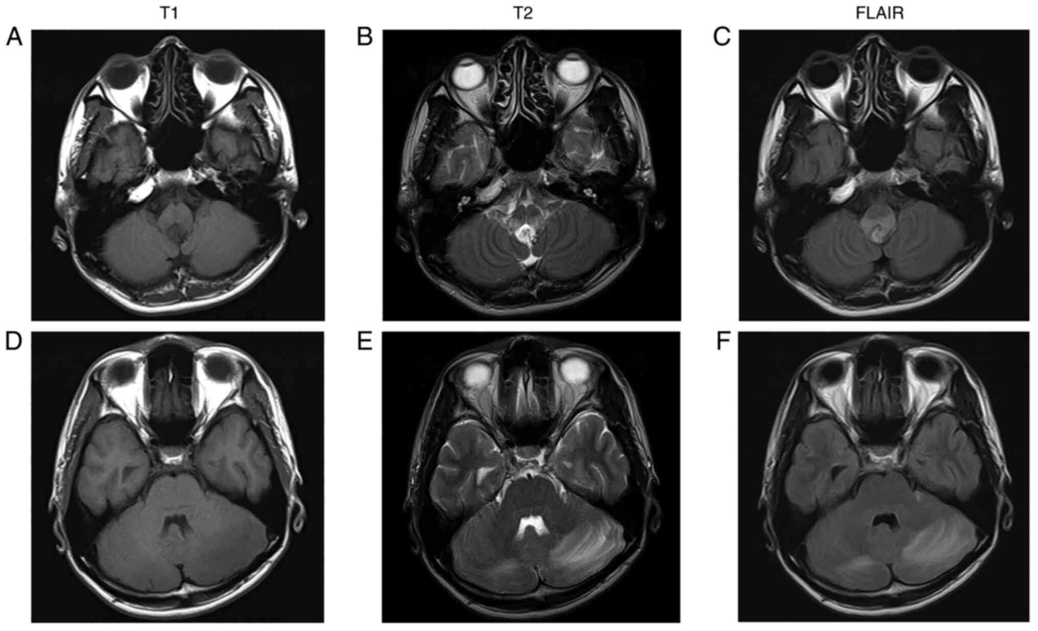
Magnetic resonance imaging (MRI) indicated a mass in the dorsal medulla oblongata and bilateral cerebellar edema (Fig. 1A-F). Post-contrast T1-imaging showed a 15 mm mass in the dorsal medulla oblongata and multiple masses <10 mm in bilateral cerebellar hemispheres that were 
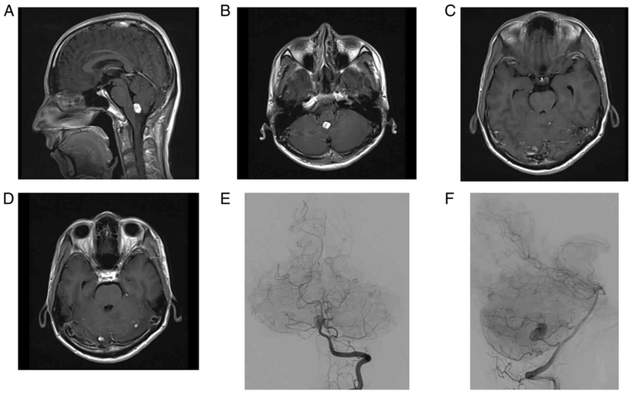
strongly enhanced with gadolinium (Fig. 2A-D). Cerebral angiography showed that the mass in the dorsal medulla oblongata received a blood supply from the left posterior inferior cerebellar artery (Fig. 2E and F). Systemic computed tomography (CT) showed a right renal tumor, multiple pancreatic cysts, and cystadenoma of the epididymis. The patient's family history was negative for von Hippel-Lindau (VHL) syndrome, and therefore he was diagnosed with solitary VHL syndrome clinically. With consideration of the operative risk, he elected to undergo stereotactic radiosurgery for multiple hemangioblastomas. Stereotactic radiosurgery (SRS) was performed with a dose of 18 Gy for the tumor of the dorsal medulla oblongata and 20 Gy for the other tumors. He 
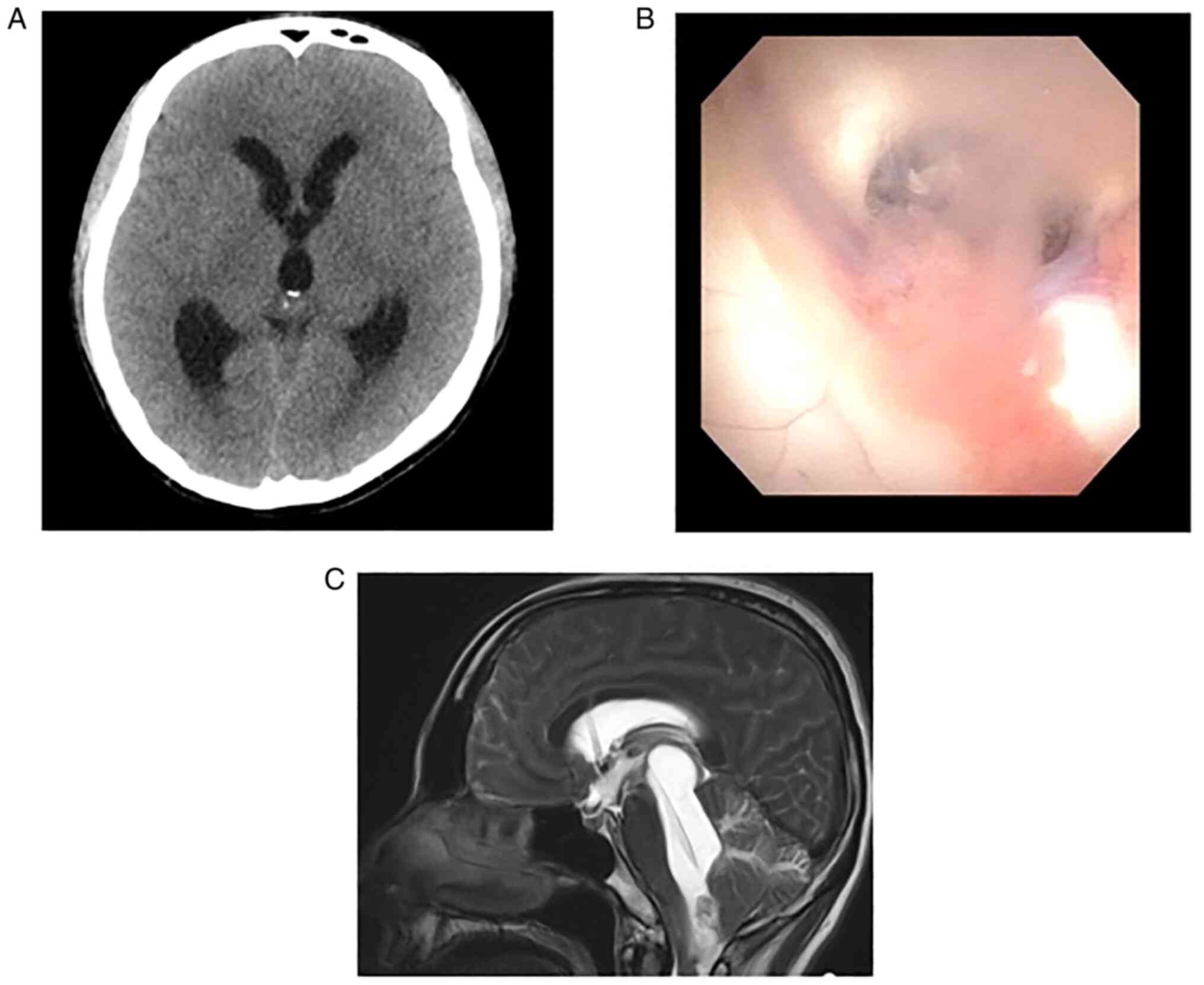
began to develop progressive headache and nausea three days after SRS, and CT showed obstructive hydrocephalus (Fig. 3A). We performed endoscopic third ventriculostomy (ETV), the endoscopic view of which showed turbid cerebrospinal fluid (CSF) and that the walls of the lateral and third ventricle were covered with white membrane-like substance (Fig. 3B). The protein level of CSF was 760 mg/dl. Subsequently, a ventriculoperitoneal (VP) shunt was placed a week after ETV because there was no improvement of 
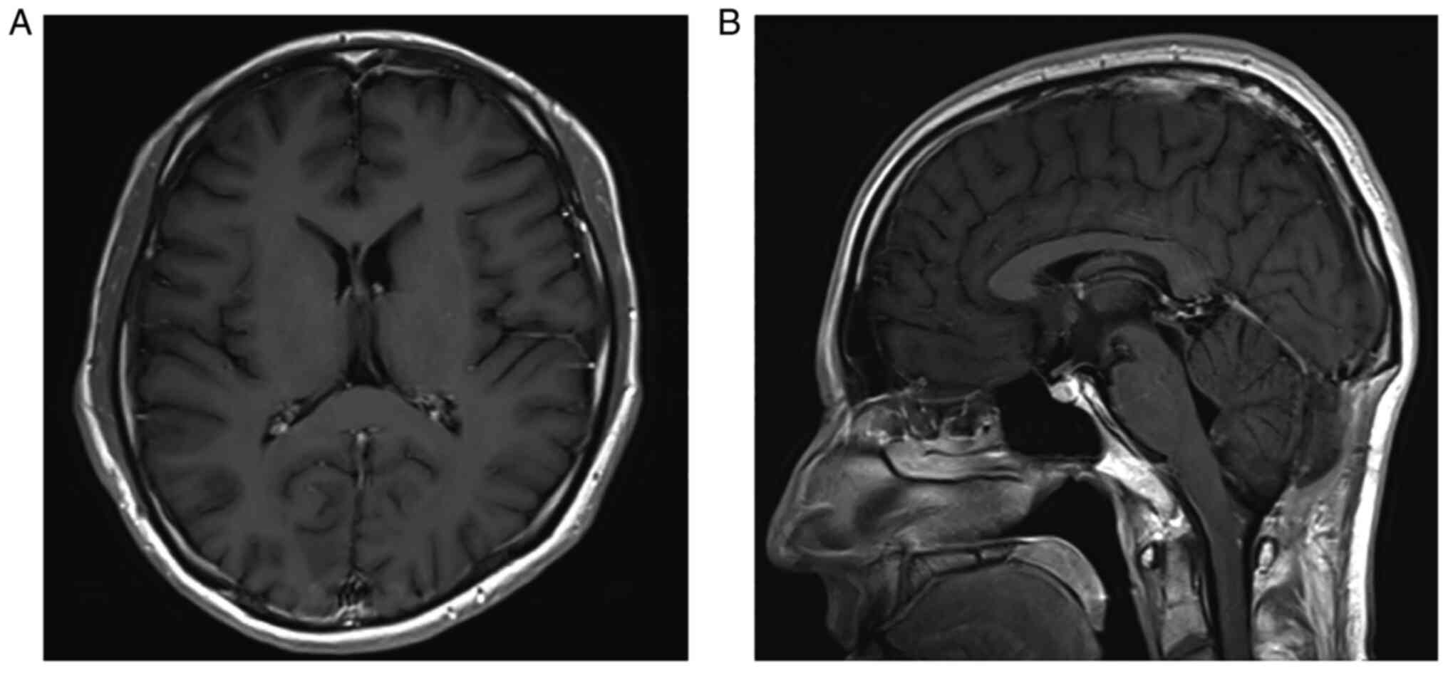
hydrocephalus after ETV. However, the patient's consciousness deteriorated gradually and the sagittal view of T2 weighted image showed isolated fourth ventricle and upward herniation 2 weeks after the VP shunt (Fig. 3C). The 
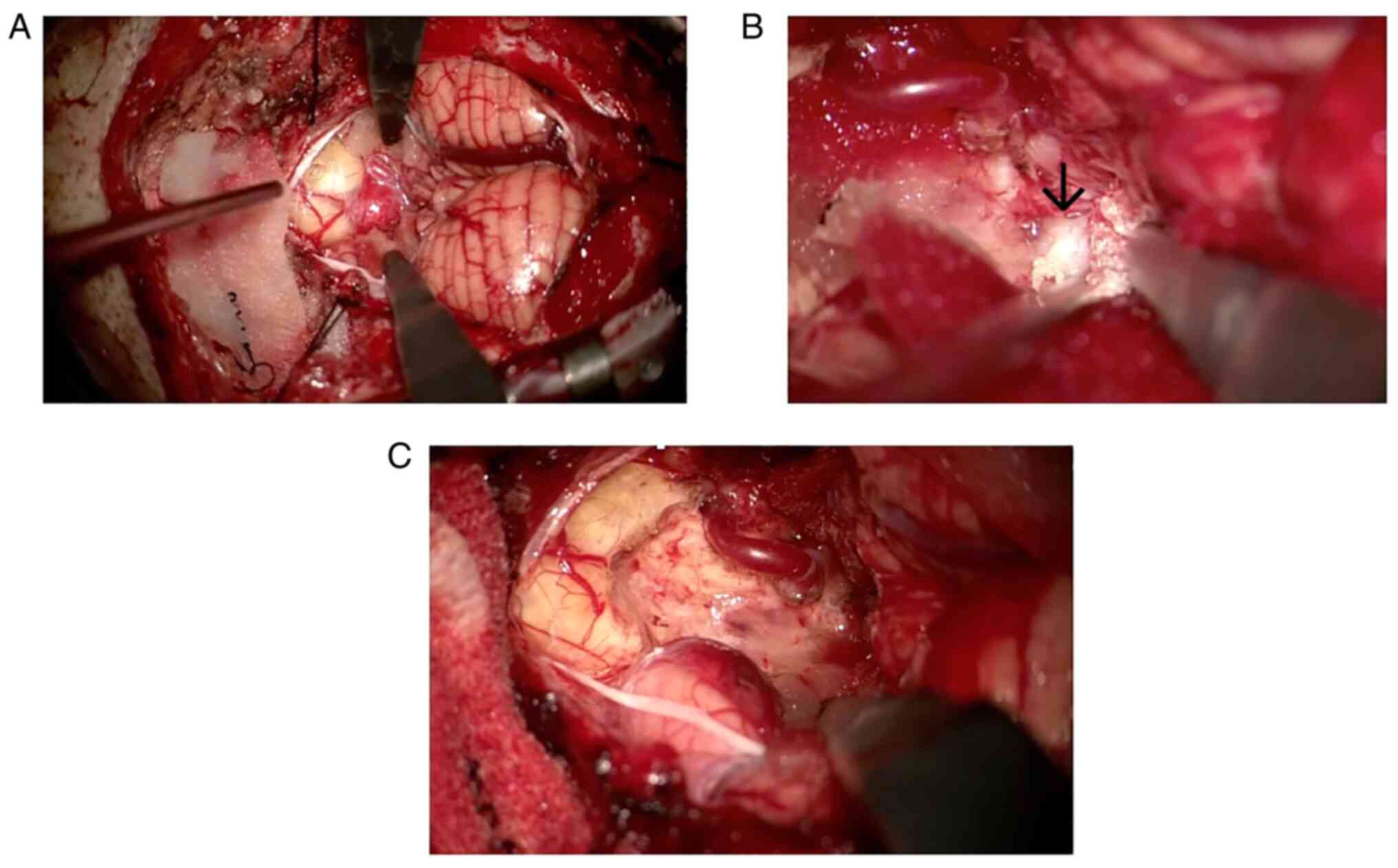
tumor in the medulla oblongata was emergently removed via posterior fossa craniotomy and telovelar approach. The fourth ventricle was filled with a white membrane-like substance, which was all surgically removed (Fig. 4A-C). We 
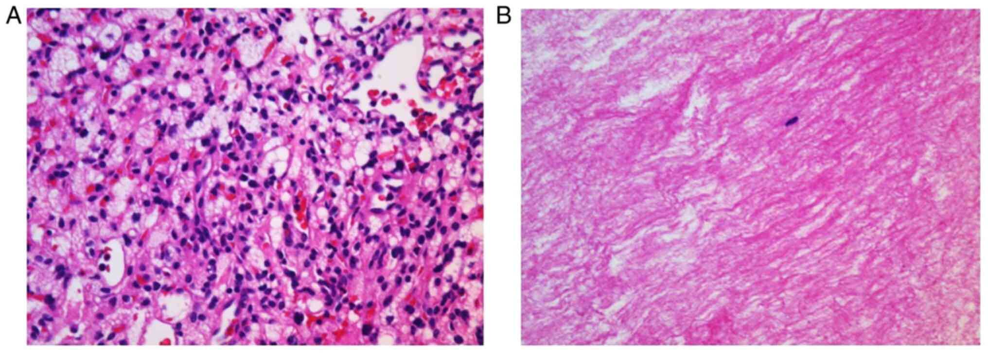
performed hematoxylin and eosin staining of the surgical specimen, following a protocol that included deparaffinization, rehydration, hematoxylin staining, eosin staining, and dehydration. Hematoxylin and eosin staining showed stromal cells with abundant vacuolated or lightly eosinophilic cytoplasm and histopathological diagnosis was hemangioblastoma (Fig. 5A). Pathologically, the white membrane-like substance was shown to consist of fibrin (Fig. 5B). The patient's consciousness and obstructive hydrocephalus improved after surgery (Fig. 6). Vocal cord paralysis occurred after surgery and he required tracheotomy, but the paralysis improved within a few weeks. Postoperative CSF protein level decreased to within normal range. The patient was discharged to his home three weeks after surgery with vertical diplopia as the only remaining symptom. Next generation sequence using peripheral was outsourced to SRL, Inc., Tokyo, Japan. The direct sequencing revealed his VHL variant mutation (NM_000551.3: c.464-2A>G). This variant is registered in ClinVar as pathogenic (Accession:VCV000223222.13). At the two-year follow-up, he was working in the same occupation as before the onset, and there had been no recurrence of the tumor.

Figure 1

Preoperative non-contrast magnetic resonance imaging. Axial views showed a mass in the dorsal medulla oblongata [(A) T1-weighted imaging, (B) T2-weighted imaging and (C) FLAIR] and bilateral cerebellar edema [(D) T1-weighted imaging, (E) T2-weighted imaging and (F) FLAIR]. FLAIR, fluid attenuated inversion recovery.

Figure 2

Post-contrast T1-imaging and cerebral angiography. Post-contrast T1-imaging showed multiple masses strongly enhanced with gadolinium [(A) sagittal view; (B) axial view of the medulla oblongata, (C) axial view of the midbrain and (D) axial view of the pons]. Cerebral angiography showed nodular stains from the left posterior inferior cerebellar artery [(E) anteroposterior view; (F) lateral view].

Figure 3

CT after stereotactic radiosurgery, surgical view of endoscopic third ventriculostomy, CT and T2-weighted imaging after VP shunt. (A) CT after stereotactic radiosurgery showed hydrocephalus. (B) Surgical view of endoscopic third ventriculostomy showed that a white membrane-like substance occludes the foramen of Monroe. (C) T2-weighted imaging after VP shunt showed upward herniation due to isolated fourth ventricle. VP, ventriculoperitoneal.

Figure 4

Surgical views of posterior cranial fossa craniotomy and telovelar approach. The surgical view showed (A) a tumor in the dorsal medulla oblongata and (B) a white membrane-like substance that occluded the foramen of Magendie (arrow). (C) The tumor and the white membrane-like substance were completely excised.

Figure 5

Photomicrographs of the surgical specimen. (A) Photomicrographs of the tumor stained with hematoxylin and eosin showed stromal cells with abundant vacuolated or lightly eosinophilic cytoplasm (magnification, x400). (B) The white membrane-like substance consisted of fibrin (magnification, x100).

Figure 6

Postoperative contrast-enhanced T1-weighted images. (A) Axial view and (B) sagittal view showing the tumor in the dorsal medulla oblongata was excised and improvement of the isolated fourth ventricle.

Discussion

IFV is an unusual type of obstructive hydrocephalus, which is characterized by the disproportionately enlarged fourth ventricle with caudal and rostral obstruction. IFV has often been observed after shunt treatment in patients with a history of ependymal inflammation from infection, or after hemorrhage in children (1-3). Dilation of the fourth ventricle elevates infratentorial pressure and compression of the brain stem, and this can result in eye movement disorder, ataxia, and impaired consciousness. IFV can be diagnosed by T2-weighted sagittal MRI to assess obstruction of the aqueduct or fourth ventricular outlet (4). Conventionally, patients with IFV have been managed by fourth ventricle shunt placement or fenestration of the occluded outlet foramen via posterior fossa craniotomy (3). With the development of neuroendoscopic surgery, patients with IFV have also been treated with endoscopic procedures, including aqueductoplasty and aqueductal stenting (4,5).

In our patient, hydrocephalus with hyperproteinorachia presented after SRS for hemangioblastoma of the medulla oblongata, and VP shunt was performed, but IFV developed. The tumor in the medulla oblongata was removed via posterior fossa craniotomy and telovelar approach. The fourth ventricle was filled with a white membrane-like substance, which was surgically removed. Pathologically, the white membrane-like substance consisted of eosinophilic fibrous matrix without atypical cells, which was shown to be fibrin. This is the first known case report of hemangioblastoma of the medulla oblongata causing IFV due to intraventricular precipitation of fibrin. Tumor removal and opening of the fourth ventricle resulted in improvement of the hydrocephalus and the protein level of CSF decreased after surgical treatment. We did not place a fourth ventricle shunt because the fourth ventricle was filled with fibrin and the shunt was expected to become occluded.

The mechanism behind hemangioblastoma cyst formation remains unclear. Intra-tumoral cysts have recently been suggested to result from vascular leakage and liquefaction of tumor cells (6). In one study, proteomic analysis indicated that the hemangioblastoma cyst fluid contained serum proteins (6). In this case, proteins secreted by the tumor flowed into the fourth ventricle and the fibrin deposition likely occluded the outflow of CSF from the fourth ventricle, leading to presentation of IFV. Our case suggests that IFV may occur after VP shunt placement for the hydrocephalus with hyperproteinorachia.

Resection of symptomatic hemangioblastomas may be curative, but SRS can be applied for small, multiple, high-surgical-risk hemangioblastomas. A retrospective international study of SRS for hemangioblastoma indicated good local tumor control and less adverse radiation effect, but a small number of patients with hemangioblastomas treated with SRS required additional surgical treatment (7). In this case, SRS was performed in consideration of small multiple lesions and the operative risk of lower cranial nerve palsy. However, SRS aggravated the hydrocephalus and required tumor removal. Careful treatment selection and follow-up after SRS are therefore important.

In conclusion, we presented a rare case of a hemangioblastoma of the medulla oblongata that caused IFV due to intraventricular deposition of fibrin. IFV may occur after VP shunt placement for hydrocephalus with hyperproteinorachia.

Acknowledgements

The authors would like to thank Mr. Benjamin Phillis (Clinical Study Support Center, Wakayama Medical University, Wakayama, Japan) for proofreading and editing.

Funding

Funding: No funding was received.

Availability of data and materials

The datasets used and/or analyzed during the current study are available from the corresponding author on reasonable request.

Authors' contributions

YH, TS, TY, JF, HN and NN contributed to the study conception and design. YH and TS wrote the final manuscript and acquired all data. YH and TS confirmed the authenticity of all the raw data. All authors read and approved the final manuscript.

Ethics approval and consent to participate

Not applicable.

Patient consent for publication

The featured patient provided written informed consent for the publication of the data and images of his case.

Competing interests

The authors declare that they have no competing interests.

References

1 

Mohanty A: Endoscopic options in the management of isolated fourth ventricles. Case report. J Neurosurg Pediatr. 103:73–78. 2005.PubMed/NCBI View Article : Google Scholar

2 

Ali K, Nannapaneni R and Hamandi K: The isolated fourth ventricle. BMJ Case Rep. 2013(bcr2013008791)2013.PubMed/NCBI View Article : Google Scholar

3 

Dauda HA and Sale D: Trapped fourth ventricle: A case report and review of literature. Int J Surg Case Rep. 80(105638)2021.PubMed/NCBI View Article : Google Scholar

4 

Panagopoulos D, Karydakis P and Themistocleous M: The entity of the trapped fourth ventricle: A review of its history, pathophysiology, and treatment options. Brain Circ. 7:147–158. 2021.PubMed/NCBI View Article : Google Scholar

5 

Imperato A, Almaguer Ascencio LM, Ruggiero C, Spennato P, Di Martino G, Aliberti F, Mirone G and Cinalli G: Endoscopic aqueductoplasty and stenting in the treatment of isolated fourth ventricle in children: 20-Year institutional experience. Childs Nerv Syst. 37:1587–1596. 2021.PubMed/NCBI View Article : Google Scholar

6 

Gläsker S, Vortmeyer AO, Lonser RR, Lubensky IA, Okamoto H, Xia JB, Li J, Milne E, Kowalak JA, Oldfield EH and Zhuang Z: Proteomic analysis of hemangioblastoma cyst fluid. Cancer Biol Ther. 5:549–553. 2006.PubMed/NCBI View Article : Google Scholar

7 

Kano H, Shuto T, Iwai Y, Sheehan J, Yamamoto M, McBride HL, Sato M, Serizawa T, Yomo S, Moriki A, et al: Stereotactic radiosurgery for intracranial hemangioblastomas: A retrospective international outcome study. J Neurosurg. 122:1469–1478. 2015.PubMed/NCBI View Article : Google Scholar

Related Articles

  • Abstract
  • View
  • Download
  • Twitter
Copy and paste a formatted citation
Spandidos Publications style
Hama Y, Sasaki T, Yamoto T, Fukai J, Nishibayashi H and Nakao N: Hemangioblastoma of the medulla oblongata that caused isolated fourth ventricle after stereotactic radiosurgery: A case report. Mol Clin Oncol 18: 37, 2023.
APA
Hama, Y., Sasaki, T., Yamoto, T., Fukai, J., Nishibayashi, H., & Nakao, N. (2023). Hemangioblastoma of the medulla oblongata that caused isolated fourth ventricle after stereotactic radiosurgery: A case report. Molecular and Clinical Oncology, 18, 37. https://doi.org/10.3892/mco.2023.2633
MLA
Hama, Y., Sasaki, T., Yamoto, T., Fukai, J., Nishibayashi, H., Nakao, N."Hemangioblastoma of the medulla oblongata that caused isolated fourth ventricle after stereotactic radiosurgery: A case report". Molecular and Clinical Oncology 18.5 (2023): 37.
Chicago
Hama, Y., Sasaki, T., Yamoto, T., Fukai, J., Nishibayashi, H., Nakao, N."Hemangioblastoma of the medulla oblongata that caused isolated fourth ventricle after stereotactic radiosurgery: A case report". Molecular and Clinical Oncology 18, no. 5 (2023): 37. https://doi.org/10.3892/mco.2023.2633
Copy and paste a formatted citation
x
Spandidos Publications style
Hama Y, Sasaki T, Yamoto T, Fukai J, Nishibayashi H and Nakao N: Hemangioblastoma of the medulla oblongata that caused isolated fourth ventricle after stereotactic radiosurgery: A case report. Mol Clin Oncol 18: 37, 2023.
APA
Hama, Y., Sasaki, T., Yamoto, T., Fukai, J., Nishibayashi, H., & Nakao, N. (2023). Hemangioblastoma of the medulla oblongata that caused isolated fourth ventricle after stereotactic radiosurgery: A case report. Molecular and Clinical Oncology, 18, 37. https://doi.org/10.3892/mco.2023.2633
MLA
Hama, Y., Sasaki, T., Yamoto, T., Fukai, J., Nishibayashi, H., Nakao, N."Hemangioblastoma of the medulla oblongata that caused isolated fourth ventricle after stereotactic radiosurgery: A case report". Molecular and Clinical Oncology 18.5 (2023): 37.
Chicago
Hama, Y., Sasaki, T., Yamoto, T., Fukai, J., Nishibayashi, H., Nakao, N."Hemangioblastoma of the medulla oblongata that caused isolated fourth ventricle after stereotactic radiosurgery: A case report". Molecular and Clinical Oncology 18, no. 5 (2023): 37. https://doi.org/10.3892/mco.2023.2633
Follow us
  • Twitter
  • LinkedIn
  • Facebook
About
  • Spandidos Publications
  • Careers
  • Cookie Policy
  • Privacy Policy
How can we help?
  • Help
  • Live Chat
  • Contact
  • Email to our Support Team